The Journal of Advanced Prosthodontics 163
This research was supported by Small and medium business administration (SMBA) and Korea medical devices industrial coop. association (KMDICA) in the Korean Government Fund.
Clinical outcome of immediately and early loaded implants with laser treated surface:
a 3-year retrospective study
Richard Leesungbok1, Jin-Ho Seo1, Sung-Am Cho2*
1Department of Biomaterials & Prosthodontics, Kyung Hee University Hospital at Gangdong, School of Dentistry, Kyung Hee University, Seoul, Republic of Korea
2Department of Prosthodontics, School of Dentistry, Kyung-Pook National University, Daegu, Republic of Korea
PURPOSE. The marginal bone loss of implants with laser treated surface was investigated after six weeks of loading after implant installation to the mandible molar area. MATERIALS AND METHODS. A total of 23 implants were placed in the edentulous molar area of the mandible: 13 implants were immediately loaded and 10 implants were early loaded. The implants used were made of titanium grade 23, screw shaped, 4.2 mm in diameter, and 10 mm in length. Patients were evaluated with resonance frequency analysis at implant fixture installation and 1, 2 (final prosthesis installation), 3, 5, 8, and 14 months later. X-rays were taken at 2 months after fixture installation and 1, 2, 3 years after to measure the marginal bone loss. RESULTS. The mean ISQ value measured at the implant installation was over 70 at all-time points. The average of marginal bone loss was average 0.33 mm. CONCLUSION. Immediate implant loading for laser treated implants would be possible.
[J Adv Prosthodont 2018;10:163-6]
KEYWORDS: Laser treated implant; Marginal bone loss; Immediate loading
https://doi.org/10.4047/jap.2018.10.2.163
https://jap.or.kr J Adv Prosthodont 2018;10:163-6
INTRODUCTION
The main purposes of implant surface treatment are to increase the surface area to obtain a higher mechanical fixa- tion between bone and implant immediately after insertion,1 to provide a surface structure that can maintain a blood clot well,2 and to provide a surface form that can promote the process of bone healing.3 SLActive technique involves forming surface roughness using large grit with the diameter
of 250 - 500 µm after sandblasting and etching by hydro- chloric acid and sulfuric acid, then washing in a nitrogen state.4
This surface forms a hydroxyl layer and has a high sur- face energy as a result of contact with water, and increases the ideal contact between the implant and the surrounding factors.4 The activated surface is preserved and stocked in a physiological saline solution to provide to dental clinics.5 The chlorine ions, as anions, and hydroxyl ions are com- bined to protect the activated surface from air and prevent hydrocarbon binding.6-8 Based on previous studies, it had been found that these surface properties significantly increased bone to implant contact and resulted in an accel- erated healing process of osseointegration during the early stage. This effect leads to enhanced stability of the implant and aids in healing during the critical early stages.9-11
According to a recent study, laser treated surface implants help improve the osseointegration process.12 As a unique surface, this method of treating implants prevents contami- nation with extraneous factors and has a high degree of sur- face purity, resulting in excellent surface roughness. That is, the entire laser treated surface of the implant has a porous structure that is pure and not contaminated. This porous structure increases the surface roughness and, as a result,
Corresponding author:
Sung-Am Cho
Department of Prosthodontics, School of Dentistry, Kyung-Pook National University, #2177 Dalgubeoldae-ro, Jung-gu, Daegu 41940, Republic of Korea Tel. +82536007672: e-mail, [email protected]
Received October 28, 2017 / Last Revision March 29, 2018 / Accepted April 16, 2018
© 2018 The Korean Academy of Prosthodontics
This is an Open Access article distributed under the terms of the Creative Commons Attribution Non-Commercial License (http://creativecommons.
org/licenses/by-nc/3.0) which permits unrestricted non-commercial use, distribution, and reproduction in any medium, provided the original work is properly cited.
pISSN 2005-7806, eISSN 2005-7814
164
enhances the strength of osseointegration.13,14
However, there has not yet been a clinical study on the immediate and early loading of implants with laser treated surfaces, which had excellent osseointegration in an animal study.15 Therefore, we applied the technology of laser treat- ment to an implant surface and conducted clinical trials to investigate if it could be loaded within six weeks after implant insertion.
MATERIALS AND METHODS
The study included 15 patients who were recruited and agreed to the clinical trial procedure, and 5 patients were excluded from the study by exclusion criteria. The clinical trial study was completed without any participant drop-out after implant surgery. A total of 23 implants were placed in 15 patients: 13 implants were immediately loaded and 10 implants were early loaded. This study protocol “KHNMC MD IRB 2012-009” was accepted by the Kyung Hee University Hospital at Gangdong, Seoul in South Korea.
All patients received prophylactic antibiotics 2 hours before surgery and their mouths were rinsed with 0.12%
chlorhexidine for one minute. A full thickness flap was ele- vated after local anesthesia. The implants used were made of titanium grade 5, screw shaped, 4.2 mm in diameter, and 10 mm in length (CSM, Daegu, Korea). The implant surfac- es were treated with a Nd:YAG laser (Jenoptic Laser Optik, Jena, Germany). Implants were inserted in the edentulous molar area of the mandible, according to Straumann’s guidelines on early and immediate loading for SLActive sur- face implants. In the case of ISQ ≥ 70 and insertion torque value (ITV) ≥ 35 to 50 Ncm at the insertion site, a provi- sional restoration was connected to the implant within a week after surgery and the final restoration was placed 2 months after surgery for immediate loading. In the case of ISQ ≥ 60 to 70 and ITV ≥ 25 to 35 Ncm at the implant insertion, an impression was taken within 2 weeks after sur- gery, and the implant received provisional restoration 4 weeks after surgery with a final restoration at 2 months post-op for early loading. After implant placement, each patient received antibiotics for at least three days.
Patients were evaluated with resonance frequency analy- sis (RFA) at implant fixture installation and also 1, 2 (final prosthesis installation), 3, 5, 8, and 14 months later. Digital bisecting X-rays were taken at 2 months after fixture instal- lation (final prosthesis installation) and then 3, 5, 8, 14, 24, and 36 months later. The digital x-rays were transferred into a software program and the first bone-implant contact (FBIC) was measured on implants for mesial and distal planes. For calibration purposes, the known pitch distance between the implant threads was used. The FBIC was mea- sured for all 23 implants at baseline. The vertical bone loss/
gain was calculated as the difference of the bone level (BL) at baseline minus the FBIC at a certain endpoint in each period (3, 5, 8, 14, 24 and 36 months later). An ISQ ≥ 70 and vertical bone loss ≤ 1.5 mm was regarded as a success.
Data are presented as mean ± standard deviation and
statistical analysis was performed using repeated-measures ANOVA to identify changes in marginal bone loss over time. All data management and analyses were performed with SPSS version 22.0 software (SPSS Inc., Chicago, IL, USA).
RESULTS
The mean ISQ value measured from surgery was greater than 70 at all-time points (Fig. 1). From implant installation to after 14 months, ISQ values increased gradually. The measured ISQ values at 6 and 12 months after installation in 23 implants were higher than 70, which was the success cri- terion of this clinical trial.
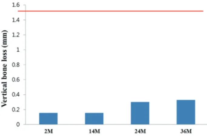
The bone loss values were less than 0.33 ± 0.32 mm after 36 months of implant installation (Fig. 2). These bone loss values are 4.5 times lower than success criteria (≤ 1.5 mm).
There was no significant difference between final prosthesis installation and after 12 months. But, except for the two groups, there was a significant difference among all groups.
DISCUSSION
Studies regarding various surface treatments to enhance osseointegration have led to an increased success rate of implants.2,3,5,6 The SLA treated surface has excellent biocom- patibility and bone affinity.7-10 The bone-implant contact of SLA surfaces achieves a high level of osteoblast differentia- tion occurs actively. It is possible that these properties of the SLA surface influence its osteoconductive ability.11 This virtue could reduce the loading time and increase the possi- bility of applying early loading.11
Fig. 1. Mean and SD of ISQ values measured from implant fixture installation to 14 months after surgery. FI:
at implant installation, 1M: 1 month later, 2M: 2 months later (final prosthesis placement), 3M: 3 months later, 5M: 5 months later, 8M: 8 months later, 14M: 14 months later. Success criterion was a value greater than or equal to 70 (red-line).
J Adv Prosthodont 2018;10:163-6
The Journal of Advanced Prosthodontics 165 Laser treatment of the implant surface rapidly increases
the temperature of titanium and melts the structure, conse- quently increasing the thickness of the oxygen layer.12 After laser treatment, morphological changes and roughness in the titanium surface appear due to the changes in oxygen layer thickness.13 On the surface of laser-treated implant, pre-osteoblast attachment is promoted, pre-osteoblastic dif- ferentiation occurs actively, and bioactivity is increased.14 Altered surface roughness helps the adaptation of fibrin and migration of osteoblasts, consequently leading to the deposition of new bone.
In this study, implant prostheses were installed based on insertion torque value; 10 implants were loaded convention- ally and 13 implants were loaded immediately. At 6 and 12 months after implant prosthesis installation, 23 implants had an ISQ value greater than 70, which was the success cri- teria defined in this clinical trial study. Additionally, the aver- age ISQ values, measured five or six times per patient after surgery, were higher than 70 in each of those 23 implants.
As another success criterion, the analysis of average value of vertical alveolar bone loss was observed to be less than 1.5 mm, consistent with the goal of this study.
Because the laser treated surface of the implants had significantly superior results compared to SLA surface implants in a previous animal study,13 we performed a clini- cal trial by early loading, which confirmed the previous results. By measuring the insertion torque when the implants were placed, the immediate loading application to implant was determined. In the immediate loading case, the ISQ values were also higher than 70. This suggests that when higher initial fixation is obtained, results are likely to be successful in either early or immediate loading. Excellent ISQ results over 70 were observed after 6 and 12 months
post-insertion, which supports the prediction that a high success rate would be maintained through continuous fol- low-up. In this study, the average ISQ value for laser treated implants was 81.4 at 24 weeks after implant placement, sim- ilar to the results of SLA and SLActive surface of implants.
The average bone resorption of 15 patients was 0.33 mm after 36 months, despite different observation periods and research methods from the previous study.
CONCLUSION
Within the limitations of this study, it can be concluded that immediate and early implant loading is possible for laser treated implants.
ORCID
Richard Leesungbok https://orcid.org/0000-0002-8381-723X Sung-Am Cho https://orcid.org/0000-0002-8315-7833 REFERENCES
1. Wismeijer D, Casentini P, Gallucci G, Chiapasco M. ITI treat- ment guide Volume 4, Loading protocol in implant dentistry:
Quintessence; 2010:6-8.
2. Rupp F, Scheideler L, Olshanska N, de Wild M, Wieland M, Geis-Gerstorfer J. Enhancing surface free energy and hydro- philicity through chemical modification of microstructured ti- tanium implant surfaces. J Biomed Mater Res A 2006;76:323- 34.
3. Hofmann AA, Bloebaum RD, Bachus KN. Progression of human bone ingrowth into porous-coated implants. Rate of bone ingrowth in humans. Acta Orthop Scand 1997;68:161-6.
4. Zhao G, Schwartz Z, Wieland M, Rupp F, Geis-Gerstorfer J, Cochran DL, Boyan BD. High surface energy enhances cell response to titanium substrate microstructure. J Biomed Mater Res A 2005;74:49-58.
5. Wennerberg A, Galli S, Albrektsson T. Current knowledge about the hydrophilic and nanostructured SLActive surface.
Clin Cosmet Investig Dent 2011;3:59-67.
6. Hall J, Miranda-Burgos P, Sennerby L. Stimulation of directed bone growth at oxidized titanium implants by macroscopic grooves: an in vivo study. Clin Implant Dent Relat Res 2005;
7:S76-82.
7. Mangano C, Perrotti V, Iezzi G, Scarano A, Mangano F, Piattelli A. Bone response to modified titanium surface implants in nonhuman primates (Papio ursinus) and humans: histological evaluation. J Oral Implantol 2008;34:17-24.
8. Schwartz Z, Martin JY, Dean DD, Simpson J, Cochran DL, Boyan BD. Effect of titanium surface roughness on chondro- cyte proliferation, matrix production, and differentiation de- pends on the state of cell maturation. J Biomed Mater Res 1996;30:145-55.
9. Roccuzzo M, Bunino M, Prioglio F, Bianchi SD. Early loading of sandblasted and acid-etched (SLA) implants: a prospective split-mouth comparative study. Clin Oral Implants Res 2001;
12:572-8.
Fig. 2. Mean and SD of marginal bone loss measured from implant fixture installation to 36 months after surgery. 2M: 2 months later (final prosthesis placement), 14M: 14 months later, 24M: 24 months later, 36M: 36 months later. Cut-off value was 1.5 mm (red-line).
Clinical outcome of immediately and early loaded implants with laser treated surface: a 3-year retrospective study
166
10. Cochran DL, Buser D, ten Bruggenkate CM, Weingart D, Taylor TM, Bernard JP, Peters F, Simpson JP. The use of re- duced healing times on ITI implants with a sandblasted and acid-etched (SLA) surface: early results from clinical trials on ITI SLA implants. Clin Oral Implants Res 2002;13:144-53.
11. Cochran D, Oates T, Morton D, Jones A, Buser D, Peters F.
Clinical field trial examining an implant with a sand-blasted, acid-etched surface. J Periodontol 2007;78:974-82.
12. Hallgren C, Reimers H, Chakarov D, Gold J, Wennerberg A.
An in vivo study of bone response to implants topographical- ly modified by laser micromachining. Biomaterials 2003;24:
701-10.
13. Kang NS, Li LJ, Cho SA. Comparison of removal torques be- tween laser-treated and SLA-treated implant surfaces in rabbit tibiae. J Adv Prosthodont 2014;6:302-8.
14. Gaggl A, Schultes G, Müller WD, Kärcher H. Scanning elec- tron microscopical analysis of laser-treated titanium implant surfaces-a comparative study. Biomaterials 2000;21:1067-73.
15. Cho SA, Jung SK. A removal torque of the laser-treated tita- nium implants in rabbit tibia. Biomaterials 2003;24:4859-63.
J Adv Prosthodont 2018;10:163-6