Introduction
Reconstruction of fully or partially edentulous ar- eas with osseointegrated implants is one of the most reliable prosthetic treatments, with survival rates above 90% in long-term studies.
1,2Despite these high survival rates, there are still complications and fail- ures in implant treatment. Previous studies have re- ported early failures in 0.7% to 7.4% of cases and late failures in 2.1% to 11.3% of cases.
1Therefore, the identification of risk factors associated with implant
failure is essential for treatment planning.
Risk factors for adverse outcomes range from im- plant design to coexisting systemic disease.
3In several studies, diabetes, steroid therapy, osteoporosis, che- motherapy, and head and neck irradiation have been considered to be contraindications for the placement of implants.
4Conversely, in other studies, individual medical problems did not correlate with an increase in implant failures. Rather, implant success was affect- ed by bone quantity and quality and by surgical tech- niques.
5A variety of studies have reported that to-
*Correspondence to: Beom-Seok Chang
Professor, Department of Periodontology, Gangneung-Wonju National University College of Dentistry, 7 Jukheon-gil, Gangneung, 210-702, Republic of Korea Tel: +82-33-640-3188, Fax: +82-33-640-3113, E-mail: [email protected] Received: December 17, 2014/Last Revision: March 3, 2015/Accepted: March 2, 2015
factors affecting the survival of implants: a long-term retrospective study
Susanna Song, Jae-Kwan Lee, Heung-Sik Um, Beom-Seok Chang*
Department of Periodontology and Research Institute of Oral Sciences, Gangneung-Wonju National University College of Dentistry, Gangneung, Republic of Korea
Purpose: The aim of the present study was to evaluate the long-term survival of implants retrospectively and determine the risk factors associated with implant failure. Materials and Methods: Of all implants that were placed at the Department of Periodontology of the Dental Hospital of Gangneung-Wonju National University from January 1998 to December 2012, 2265 implants that were followed up until June 2013 were included in this study. Data were collected from clinical and radiographic examinations from previous visits. The information gathered included gender, age, smoking status, implant diameter, implant length, surface of implant, location of implant within the dental arch, surgical techniques and existence of complications. Results:
The survival rate before loading was 98.9%. The cumulative survival rate after 5 years of loading was 97.2%, and after 15 years of loading was 95.2%. In a simple logistic regression analysis, gender (P = 0.016), smoking status (P = 0.001), location of implant (P
= 0.020) and existence of complications (P = 0.002) were statistically associated with implant failure and included in the multiple regression analysis. As a result of multiple logistic regression analysis, the variables statistically associated with implant failure (P <
0.05) were smoking status (P = 0.049) and existence of complications (P < 0.001). Conclusion: The cumulative survival rate of dental implants after 15 years of loading was 95.2% and that the variables statistically associated with implant failure were smoking status and existence of complications. (J Dent Rehabil Appl Sci 2015;31(1):10-9)
Key words: dental implants; survival rate; retrospective study; risk factors
Copyright© 2015 The Korean Academy of Stomatognathic Function and Occlusion.
It is identical to Creative Commons Non-Commercial License.
cc
ISSN 2233-4084
bacco use has a negative effect on implant survival.
6,7However, no obvious evidence has been presented concerning the influence of tobacco use on the sur- vival rates of implants.
2,7The effect of a history of periodontal disease on implant failure is also contro- versial. Several authors reported that there may be an increased risk for implant failure in periodontally compromised patients,
8,9while other authors insisted that the presence of periodontal pathogens at peri- implant sites does not necessarily indicate future im- plant failure.
10,11Because the results obtained from numerous stud- ies are contradictory, the questions raised by patients cannot be answered. In spite of the wide range of available studies, consistent conclusions cannot yet be drawn on the relationship between these factors and the long-term outcomes of implants. There is a clear need to obtain further scientific evidence in this area. Therefore, the aim of the present study was to evaluate the long-term survival of implants retro- spectively and determine the risk factors associated with implant failure.
Materials and Methods
Data collection
A retrospective chart review was conducted for all implants that were placed at the Department of Peri- odontology of the Dental Hospital of Gangneung- Wonju National University from January 1998 to December 2012 (IRB 2013-13). Of all implants that were included the chart review, only implants that were followed up until June 2013 were included in this study. Data were collected from clinical and ra- diographic examinations from previous visits. The information gathered included the following:
• Gender
• Age (classified into three categories: < 50 years, 50 - 59 years and ≧ 60 years)
• Smoking status (people smoking 1 cigarette or more per day at the time of implant placement were categorized as a smokers)
• Implant diameter (classified into three categories:
< 3.75 mm, 3.75 - 4.5 mm, ≧ 5 mm)
• Implant length (classified into two categories: <
10 mm, ≧ 10 mm, the classification criteria of 10 mm is based on the definition of a short im- plant
12)
• Surface of implant (classified into three catego- ries: blasted, sand-blasted and acid etched, and anodized surface)
• Location of implant within the dental arch (clas- sified into four categories: maxillary anterior, maxillary posterior, mandibular anterior and man- dibular posterior regions)
• Surgical techniques (specific procedures that were performed to place the implants: guided bone re- generation, sinus elevation with a crestal approach and sinus elevation with a lateral approach)
• Complications (biological and mechanical com- plications: peri-implant mucositis, peri-implantitis, implant periapical lesion and fracture of fixture, screw or prosthesis)
• Dates of implant placement, prosthesis place- ment, implant removal and the last follow-up visit An implant failure was defined as the removal of the implant for any reason.
13Implants were regarded as surviving when they were present in the oral cavity at follow-up visits. Total survival time was defined as the period from the date of implant placement to the date of implant removal or the last follow-up visit.
13Implant failure was classified as early failure if it oc- curred before functional loading, or late failure if it occurred after functional loading.
14Statistical analyses
After the data collection, statistical analyses were
performed. The cumulative survival rates of im-
plants were calculated by the life table method at
annual intervals and a Kaplan-Meier survival curve
was obtained. To determine the potential risk factors
associated with implant failure, a simple logistic re-
gression analysis was performed. Implant failure was
the dependent variable and all independent variables
were entered separately as categorical variables. Fol-
lowing the simple regression analysis, independent
variables with P < 0.1 were selected and included in
a multiple regression analysis to eliminate possible
confounding variables. A multiple logistic regression analysis was performed for implant failure as the dependent variable and independent variables with P < 0.05 were considered statistically associated with implant failure. In all types of analyses, each implant was regarded as the analysis unit. Thus multiple im- plants from the same patient had the same patient- related covariates. All analyses were conducted using statistical software (SPSS
TM20, IBM Inc., Chicago, IL, USA).
Results
During the period from January 1998 to December 2012, 5019 implants were placed in 1921 patients. Of these patients, 1146 were excluded from this study because of a lack of follow-up visits. Thus the study group was composed of 2265 implants from 775 pa- tients.
Demographic characteristics
The study group consisted of 296 females and 479 males. The mean age at the last visit was 55.5 years (range: 19 to 81 years). Of 2265 implants, 854 implants (37.7%) were placed in females, and 1411 implants (62.3%) were placed in males. Six hundred thirty-four implants (28.0%) were placed in smok- ers, and 1631 implants (72.0%) were placed in non- smokers (Table 1).
Implant characteristics
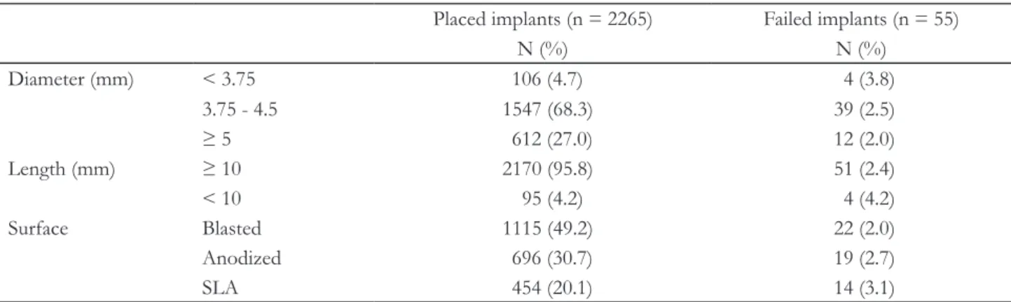
The majority of the implants were of a diameter
≧ 3.75 mm (95.3%), and 4.7% were of a diameter
< 3.75 mm. Most of the implants were ≧ 10 mm (95.8%) in length. The implants had a variety of sur- faces including blasted (49.2%), anodized (30.7%), and SLA surfaces (20.1%) (Table 2).
Table 1. Distribution of implants according to demographic characteristics
Placed implants (n = 2265) Failed implants (n = 55)
N (%) N (%)
Gender Female 854 (37.7) 12 (1.4)
Male 1411 (62.3) 43 (3.0)
Age (yr) < 50 413 (18.2) 13 (3.1)
50 - 59 1083 (47.8) 25 (2.3)
≥ 60 769 (34.0) 17 (2.2)
Smoking No 1631 (72.0) 28 (1.7)
Yes 634 (28.0) 27 (4.3)
Table 2. Distribution of implants according to implant characteristics
Placed implants (n = 2265) Failed implants (n = 55)
N (%) N (%)
Diameter (mm) < 3.75 106 (4.7) 4 (3.8)
3.75 - 4.5 1547 (68.3) 39 (2.5)
≥ 5 612 (27.0) 12 (2.0)
Length (mm) ≥ 10 2170 (95.8) 51 (2.4)
< 10 95 (4.2) 4 (4.2)
Surface Blasted 1115 (49.2) 22 (2.0)
Anodized 696 (30.7) 19 (2.7)
SLA 454 (20.1) 14 (3.1)
Surgical characteristics and the existence of complications
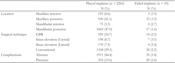
One thousand one hundred and twenty-five im- plants were inserted in the maxilla (195 in the ante- rior and 930 in the posterior) and 1140 in the man- dible (75 in the anterior and 1065 in the posterior).
The guided bone regeneration (GBR) technique was used in 559 implants, and 368 implants were placed after maxillary sinus elevation (198 with the crestal approach and 170 with the lateral approach). The others (1338 implants) were placed without any spe- cific procedure. Postoperative complications includ- ing peri-implant mucositis, peri-implantitis, implant periapical lesions and fractures of the fixture, screw or prosthesis occurred in 354 implants (15.6%) (Table 3).
Implant survival
Of the 2265 inserted implants, 55 (2.4%) failed.
Twenty-five implants (1.1%) failed before loading (early) and 30 (1.3%) failed after loading (late). Ac- cording to the Kaplan-Meier lifetime analysis (Table 4, Fig. 1), the cumulative survival rate after 5 years of loading was 97.2%, and after 15 years of loading was 95.2%.
Table 4. Cumulative survival rate of inserted implants Time (yr)
No. of implants at beginning of
interval
No. of failed implants during interval
Cumulative survival rate
(%)
Place / Load 2265 25 98.9
Load / 1 2082 4 98.7
1 to 2 1699 8 98.2
2 to 3 1398 8 97.6
3 to 4 1093 2 97.4
4 to 5 834 2 97.2
5 to 6 651 1 97.0
6 to 7 478 0 97.0
7 to 8 326 2 96.4
8 to 9 253 3 95.2
9 to 10 141 0 95.2
10 to 11 43 0 95.2
11 to 12 13 0 95.2
12 to 13 2 0 95.2
13 to 14 2 0 95.2
14 to 15 2 0 95.2
Place / Load: placement of implant to time of loading.
Load / 1: time of loading to 1 year.
Table 3. Distribution of the implants according to surgical characteristics and the existence of complications
Placed implants (n = 2265) Failed implants (n = 55)
N (%) N (%)
Location Maxillary anterior 195 (8.6) 3 (1.5)
Maxillary posterior 930 (41.1) 33 (3.5)
Mandibular anterior 75 (3.3) 2 (2.7)
Mandibular posterior 1065 (47.0) 17 (1.6)
Surgical technique GBR 559 (24.7) 14 (2.5)
Sinus elevation (Crestal) 198 (8.7) 7 (3.5)
Sinus elevation (Lateral) 170 (7.5) 4 (2.4)
Conventional 1338 (59.1) 30 (2.2)
Complications Absence 1911 (84.4) 35 (1.8)
Presence 354 (15.6) 20 (5.6)
GBR, guided bone regeneration.
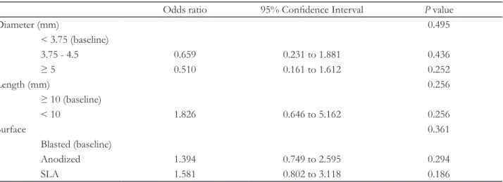
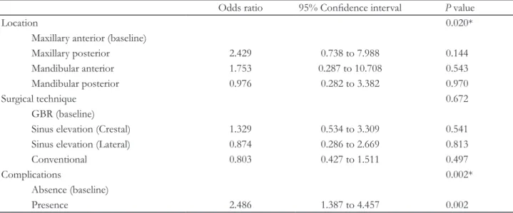
Risk factors associated with implant failure All independent variables including gender, age, smoking status, implant diameter, implant length, surface of implant, location of implant, surgical tech- nique and existence of complications were entered into a simple logistic regression analysis (Table 5 - 7).
Among these variables, gender (P = 0.016), smoking status (P = 0.001), location of implant (P = 0.020) and existence of complications (P = 0.002) were sta- tistically associated with implant failure and included in the multiple regression analysis.
As a result of multiple logistic regression analysis (Table 8), the variables statistically associated with implant failure (P < 0.05) were smoking status (P = 0.049) and existence of complications (P < 0.001).
Table 5. Simple logistic regression analysis for risk factors associated with implant failure (Demographic variables) Odds ratio 95% Confidence Interval
P valueGender 0.016*
Female (baseline)
Male 2.206 1.156 to 4.206 0.016
Age (yr) 0.526
< 50 (baseline)
50 - 59 0.757 0.385 to 1.487 0.419
≥ 60 0.654 0.311 to 1.373 0.262
Smoking 0.001*
No (baseline)
Yes 2.547 1.489 to 4.356 0.001
*Significant association (P < 0.1).
Fig. 1. Kaplan-Meier survival curve. CSR, cumulative survival rate.
100 80 60 40 20 0
CSR (%) Place Place/Load Load/1 yr 1/2 yr 2/3 yr 3/4 yr 4/5 yr 5/6 yr 6/7 yr 7/8 yr 8/9 yr 9/10 yr 10/11 yr 11/12 yr 12/13 yr 13/14 yr 14/15 yr
Time
Table 6. Simple logistic regression analysis for risk factors associated with implant failure (Implant variables) Odds ratio 95% Confidence Interval
P valueDiameter (mm) 0.495
< 3.75 (baseline)
3.75 - 4.5 0.659 0.231 to 1.881 0.436
≥ 5 0.510 0.161 to 1.612 0.252
Length (mm) 0.256
≥ 10 (baseline)
< 10 1.826 0.646 to 5.162 0.256
Surface 0.361
Blasted (baseline)
Anodized 1.394 0.749 to 2.595 0.294
SLA 1.581 0.802 to 3.118 0.186
The implants placed in smokers were 1.839 times more likely to fail than the implants placed in non- smokers. The implants with complications were also at a greater risk of failure compared to the implants without complications, with an odds ratio of 2.977 : 1.
Discussion
The purpose of this retrospective study was to analyze the long-term outcomes of 2265 implants
and determine the risk factors associated with im- plant failure. A retrospective chart review of im- plants placed at the department of periodontology of the dental hospital of Gangneung-Wonju national university from January 1998 to December 2012 was performed.
In the present study, the cumulative survival rate up to 15 years was 95.2%. Previously, Simonis et al.
15reported a long-term cumulative survival rate of 82.94% up to 16 years. Additionally, Roos-Jansåker Table 7. Simple logistic regression analysis for risk factors associated with implant failure (surgical variables and complications)
Odds ratio 95% Confidence interval
P valueLocation 0.020*
Maxillary anterior (baseline)
Maxillary posterior 2.429 0.738 to 7.988 0.144
Mandibular anterior 1.753 0.287 to 10.708 0.543
Mandibular posterior 0.976 0.282 to 3.382 0.970
Surgical technique 0.672
GBR (baseline)
Sinus elevation (Crestal) 1.329 0.534 to 3.309 0.541
Sinus elevation (Lateral) 0.874 0.286 to 2.669 0.813
Conventional 0.803 0.427 to 1.511 0.497
Complications 0.002*
Absence (baseline)
Presence 2.486 1.387 to 4.457 0.002
GBR, guided bone regeneration.
Table 8. Multiple logistic regression analysis for risk factors associated with implant failure
Variables Odds ratio 95% Confidence interval
P valueGender 0.272
Female (baseline)
Male 1.501 0.727 to 3.098 0.272
Smoking 0.049*
No (baseline)
Yes 1.839 1.002 to 3.377 0.049
Location 0.085
Maxillary anterior (baseline)
Maxillary posterior 2.395 0.722 to 7.943 0.153
Mandibular anterior 1.895 0.306 to 11.726 0.492
Mandibular posterior 1.166 0.336 to 4.055 0.809
Complications < 0.001*
Absence (baseline)
Presence 2.977 1.685 to 5.258 < 0.001
*Significant association (P < 0.05).
et al.
2assessed the long-term outcome of implant therapy and reported that the overall survival rate up to 14 years was 95.7%. Thus, the outcome of the present study is consistent with previous studies.
In this study, only 55 implants were removed.
Some of these were concentrated in a small number of individuals. Fifty-five implant removals occurred in 47 patients and 15 of 55 removed implants were in 7 patients. Among them, five were smokers and two were non-smokers. Peri-implantitis occurred in 2 patients and implant periapical lesion occurred in 1 patient.
To identify the risk factors associated with implant failure, statistical analyses were performed. Using multiple logistic regression analysis, a significant relationship was found between implant failure and independent variables including smoking status and existence of complications. Smoking status has pre- viously been regarded as a risk factor for implant fail- ure. Bain and Moy
16reported a failure rate of 11.3%
in smokers and 4.76% in non-smokers. In more recent study, Huynh-Ba et al.
17studied 273 implants placed in the posterior maxilla and reported that smoking markedly increased the risk for implant fail- ure. In terms of existence of complications, Simonis et al.
15referred to peri-implantitis as a complication with a very high risk of implant loss; of 21 cases, only 5 implants were functional after 16 years despite comprehensive treatments including implant debride- ment and administration of systemic antibiotics.
Some variables that were not significantly related to implant failure in multiple regression analysis but reached statistical significance in simple regression analysis included gender and location of implant.
Males (odds ratio = 1.501) and maxillary posterior implants (odds ratio = 2.395) were associated with implant failure. These variables were previously iden- tified as risk factors for implant failure. Zupnik et al.
18reported that gender was the parameter with the strongest correlation with implant failure in a meta- analysis that demonstrated that males had a higher prevalence of destructive periodontal disease.
19Be- cause periodontal disease is widely recognized as a risk factor for peri-implantitis and implant failure,
20,21the higher prevalence of periodontal disease in males
may explain the outcome of the study. Anitua et al.
22reported significantly lower survival for implants placed in the maxilla compared to the mandible. This difference might be the outcome of the less favor- able osseous situation in patients requiring maxillary reconstruction. In addition, Moy et al.
3studied 4680 implants in 1140 patients and reported that implants placed in the maxilla failed at almost twice rate of those placed in the mandible.
The present study had some limitations. First, an inherent limitation of retrospective studies is a risk for bias because only the implants that were followed up were included in the study group. The dropout rate (1146 of 1921 patients, 59.7%) could be consid- ered high and must be considered when interpreting the results. Previous studies have suggested this as a reason for incomplete results.
17,23Second, because the number of failed implants was very low (55 of 2265 implants, 2.4%), definitive conclusions could not be drawn. Alsaadi et al.
24reported that due to the low number of implant failures, only potentially influential factors could be identified. Finally, nu- merous variables that are known to be significantly associated with implant failure were not included in our analyses. For example, diabetes has been signifi- cantly correlated with implant failure.
18Rodrigo et al.
25reported that resonance frequency analysis (RFA) values at restoration placement could significantly predict the outcomes of implants. Quirynen et al.
26reported that a large proportion of failing implants may be explained by the lack of proper supportive periodontal therapy (SPT), and Ong et al.
27proposed that the irregularity of SPT might influence the out- comes and differences between studies. Therefore, well-designed, controlled prospective studies that consider the above limitations are required.
Conclusion
We conclude that the cumulative survival rate of dental implants after 15 years of loading was 95.2%
and that the variables statistically associated with
implant failure were smoking status and existence of
complications.
References
1. Berglundh T, Persson L, Klinge B. A systematic review of the incidence of biological and techni- cal complications in implant dentistry reported in prospective longitudinal studies of at least 5 years. J Clin Periodontol 2002;29:197-212.
2. Roos-Jansåker AM, Lindahl C, Renvert H, Renvert S. Nine- to fourteen-year follow-up of implant treatment. Part I: Implant loss and associations to various factors. J Clin Periodontol 2006;33:283-9.
3. Moy PK, Medina D, Shetty V, Aghaloo TL. Dental implant failure rates and associated risk factors. Int J Oral Maxillofac Implants 2005;20:569-77.
4. Fugazotto PA. Success and failure rates of osseoin- tegrated implants in function in regenerated bone for 6 to 51 months: a preliminary report. Int J Oral Maxillofac Implants 1997;12:17-24.
5. Smith RA, Berger R, Dodson TB. Risk factors as- sociated with dental implants in healthy and medi- cally compromised patients. Int J Oral Maxillofac Implants 1992;7:367-72.
6. Bain CA. Implant installation in the smoking pa- tient. Periodontol 2000 2003;33:185-93.
7. Strietzel FP, Reichart PA, Kale A, Kulkarni M, Wegner B, Küchler I. Smoking interferes with the prognosis of dental implant treatment: a system- atic review and meta-analysis. J Clin Periodontol 2007;34:523-44.
8. Karoussis IK, Salvi GE, Heitz-Mayfield LJ, Brägger U, Hämmerle CH, Lang NP. Long-term implant prognosis in patients with and without a history of chronic periodontitis: a 10-year prospective cohort study of the ITI dental implant system. Clin Oral Implants Res 2003;14:329-39.
9. Evian CI, Emling R, Rosenberg ES, Waasdorp JA, Halpern W, Shah S, Garcia M. Retrospective analy- sis of implant survival and the influence of peri- odontal disease and immediate placement on long- term results. Int J Oral Maxillofac Implants 2004;
19:393-8.
10. Sbordone L, Barone A, Ciaglia RN, Ramaglia L, Iacono VJ. Longitudinal study of dental implants in a periodontally compromised population. J Peri- odontol 1999;70:1322-9.
11. Nevins M. Will implants survive well in patients with a history of inflammatory periodontal disease?
J Periodontol 2001;72:113-7.
12. Morand M, Irinakis T. The challenge of implant therapy in the posterior maxilla: providing a ratio- nale for the use of short implants. J Oral Implantol 2007;33:257-66.
13. Chuang SK, Wei LJ, Douglass CW, Dodson TB.
Risk factors for dental implant failure: a strategy for the analysis of clustered failure-time observations. J Dent Res 2002;81:572-7.
14. Lang NP, Karring T, Meredith N. Group E sum- mary. J Clin Periodontol 2002;29:232-4.
15. Simonis P, Dufour T, Tenenbaum H. Long-term implant survival and success: a 10-16-year follow- up of non-submerged dental implants. Clin Oral Implants Res 2010;21:772-7.
16. Bain CA, Moy PK. The association between the failure of dental implants and cigarette smoking.
Int J Oral Maxillofac Implants 1993;8:609-15.
17. Huynh-Ba G, Friedberg JR, Vogiatzi D, Ioanni- dou E. Implant failure predictors in the posterior maxilla: a retrospective study of 273 consecutive implants. J Periodontol 2008;79:2256-61.
18. Zupnik J, Kim SW, Ravens D, Karimbux N, Guze K. Factors associated with dental implant sur- vival: a 4-year retrospective analysis. J Periodontol 2011;82:1390-5.
19. Shiau HJ, Reynolds MA. Sex differences in destruc- tive periodontal disease: a systematic review. J Peri- odontol 2010;81:1379-89.
20. Baelum V, Ellegaard B. Implant survival in peri- odontally compromised patients. J Periodontol 2004;75:1404-12.
21. Ferreira SD, Silva GL, Cortelli JR, Costa JE, Costa FO. Prevalence and risk variables for peri-implant disease in Brazilian subjects. J Clin Periodontol 2006;33:929-35.
22. Anitua E, Orive G, Aguirre JJ, Ardanza B, Andía I.
5-year clinical experience with BTI dental implants:
risk factors for implant failure. J Clin Periodontol 2008;35:724-32.
23. Scurria MS, Morgan ZV 4th, Guckes AD, Li S,
Koch G. Prognostic variables associated with im-
plant failure: a retrospective effectiveness study. Int
J Oral Maxillofac Implants 1998;13:400-6.
24. Alsaadi G, Quirynen M, Michiles K, Teughels W, Komárek A, van Steenberghe D. Impact of local and systemic factors on the incidence of failures up to abutment connection with modified surface oral implants. J Clin Periodontol 2008;35:51-7.
25. Rodrigo D, Aracil L, Martin C, Sanz M. Diagno- sis of implant stability and its impact on implant survival: a prospective case series study. Clin Oral Implants Res 2010;21:255-61.
26. Quirynen M, Abarca M, Van Assche N, Nevins M, van Steenberghe D. Impact of supportive peri- odontal therapy and implant surface roughness on implant outcome in patients with a history of peri- odontitis. J Clin Periodontol 2007;34:805-15.
27. Ong CT, Ivanovski S, Needleman IG, Retzepi M,
Moles DR, Tonetti MS, Donos N. Systematic re-
view of implant outcomes in treated periodontitis
subjects. J Clin Periodontol 2008;35:438-62.
*교신저자: 장범석
(210-702) 강원도 강릉시 죽헌길 7 강릉원주대학교 치과대학 치주과학교실 Tel: 033-640-3188|Fax: 033-640-3113|E-mail: [email protected] 접수일: 2014년 12월 17일|수정일: 2015년 3월 3일|채택일: 2015년 3월 2일
임플란트의 생존에 영향을 미치는 요인에 대한 장기간의 후향적 연구