Original Article
원고 접수일 2012년 4월 30일, 원고 수정일 2012년 5월 2일, 게재 확정일 2012년 5월 4일
책임저자 한성일
(330-714) 천안시 동남구 단대로 119, 단국대학교 치과대학 치과병원 구강악안면외 과학교실
Tel: 041-550-1991, Fax: 041-551-8988, E-mail: [email protected]
RECEIVED April 30, 2012, REVISED May 2, 2012, ACCEPTED May 4, 2012
Correspondence to Sungil Han
Department of Oral and Maxillofacial Surgery, Dankook University Dental Hospital, Dankook University College of Dentistry
119, Dan-daero, Dongnam-gu, Cheonan 330-714, Korea
Tel: 82-41-550-1991, Fax: 82-41-551-8988, E-mail: [email protected]
CC This is an open access article distributed under the terms of the Creative Commons Attribution Non-Commercial License (http://creativecommons.org/licenses/
by-nc/3.0) which permits unrestricted non-commercial use, distribution, and reproduction in any medium, provided the original work is properly cited.
구치부 단일 임플란트의 생존율에 대한 후향적 연구
한성일ㆍ이재훈
단국대학교 치과대학 구강악안면외과학교실
Abstract
A Retrospective Clinical Study of Survival Rate for a Single Implant in Posterior Teeth
Sungil Han, Jae-Hoon Lee
Department of Oral and Maxillofacial Surgery, Dankook University College of Dentistry
Purpose: Single implants, of which screw loosening has been observed frequently, presents problems such as fixtures fractures, marginal bone loss, and inflammation of the soft tissue around the implant. However, the single implant is more conservative, cost effective, and predictable compared to the 3 unit bridge with respect to the long-term outcome. This study evaluated the survival rate as well as future methods aimed at increasing the survival rate in single implants in posterior teeth.
Methods: Among the implants placed in the Dankook University Dental Hospital department of Oral & Maxillofacial surgery from January 2001 to June 2008, 599 implants placed in the maxillar and mandibular posterior were evaluated retrospectively.
Survival rates were investigated according to implant location, cause of tooth loss, gender, age, general disease, fixture diameter and length, surface texture, implant type and shape, presence of bone graft, surgery stage, surgeons, bone quality and opposite teeth.
Results: Out of 599 single implants in posterior teeth, 580 implants survived and the survival rate was 96.8%. The difference in survival rate was statistically significant according to the implant location. The survival rate was low (84.2%) in implants exhibiting a wide diameter (≥ 5.1 mm) and the surface treated by the acid etching group demonstrated a significantly lower survival rate (91.1%). One stage surgical procedure, which implemented a relatively better bone quality survival rate (100%), was higher than the two stage surgical procedure (96.1%). The survival rate of type IV bone quality (75%) was significantly lower than the other bone quality.
Conclusion: Single posterior teeth implant treatments should use an improved surface finishing fixture as well as careful and safe procedures when performing implant surgery in the maxilla premolar and molar regions since bone quality is poor.
Key words: Single-tooth implants, Missing teeth, Survival rate
서 론
치아우식, 치주질환 혹은 외상 등에 의해서 상실된 치아는 기능 및 심미적인 이유로 보철치료가 필요하다. 가장 일반적인 치아 수복 방법은 인접 자연치를 삭제하고 이를 지대치로 사용하는 브리지(bridge)이다. 그러나 지대치의 치수손상, 부적절한 지대 치 유지형태 및 접착시멘트의 용해에 의한 보철물 탈락, 지대치 파절 및 금관 변연부에 발생하는 이차 치아우식증 등은 고정성 계속가공의치의 문제점이다. 가철성 국소의치 역시 환자의 이물 감 호소, 저작효율의 감소, 잔존골의 흡수, 발음장애, 심미성 부족 등 많은 단점을 가지고 있다. 1960년대 Brånemark 등[1]과 Breine 등[2]은 골 재생에 관한 동물 실험에서 생체와 금속 간의 직접적 결합 현상을 발견하고 골유착(osseointegration)의 원리 를 소개하여 현재의 임플란트 개념을 확립하였다. 즉, 임플란트의 개발로 주위 조직의 손상 없이 기능과 심미성을 회복시킬 수 있게 되었고, 잔존골에 적절한 자극을 줌으로써 더 이상의 골 흡수를 예방할 수 있게 되었다.
임플란트는 완전 무치악 환자에서 의치를 안정화시키기 위해 사용된 이후 장기적 안정성을 가지고 높은 성공률을 보였으며[3], 이후 부분 무치악 환자에서도 사용되어 좋은 결과를 보였다[4].
그리고 단일 치아 결손 증례에서도 Jemt[5]에 의해 처음 시도된 이후로 점차 사용이 확대되고 있다.
일반적으로 임플란트 시술에서 3개 이하의 임플란트 사용은 기계 역학적으로 불리한 것으로 알려져 있으며[6], 실제로 초기의 단일 임플란트 연구에서 교합이 가해진 단일 임플란트 금관의 나사 풀림 현상(screw loosening)이 자주 관찰되었으며, 고정체 파절, 변연골 소실, 깊이 매식된 임플란트 주위의 연조직 염증 및 치관의 심미성 등의 문제점이 발생하였다[7]. 이러한 문제점의 원인으로는 무치악부의 점진적인 골흡수나 상악동의 함기화로 인한 골량 부족 등의 해부학적 한계 요인과, 굴곡 모멘트(bending moment)에 취약한 단일 임플란트의 기계 역학적 요인 등을 들 수 있다[8,9]. 특히, 최후방 구치부에 식립된 단일 임플란트의 경우, 교합면이 근원심으로 커져 원심측으로 캔틸레버가 작용하 게 되어 골 유착이 파괴되거나 파절을 야기할 가능성이 더욱 높다. 하악의 경우 장폭경의 임플란트를 식립하더라도 1개의 임플 란트로는 자연치와 같은 치근면적을 얻기 힘들다[10].
단일치 임플란트는 앞의 문제점들 때문에 교합력이 강하게 작용하는 구치부에서 추천되지 않았으며, 여러 문헌에서 구치부 의 단일치 임플란트 수복은 전치부에 비해 성공률이 낮고 많은 합병증을 유발할 수 있음이 보고되었다[11,12]. 그래서 Rangert 등[6]은 하나의 대구치를 대체하기 위해 굵은 직경의 임플란트나 많은 수의 임플란트가 필요하다고 발표하였고, Lekholm과 Jemt[13], Petorpoulos 등[10]도 직경이 3.75 mm보다 큰 임플란 트를 식립하든가 치근 개수와 같은 2개의 임플란트를 식립하는
것이 나사 풀림이나 파절을 방지할 수 있다고 하였다. 그러나 현재는 하중 분산을 최적화시킨 임플란트 디자인과 표면 처리 개선, 내구성이 강한 지대주 나사 개발 등을 통해 구치부에서 단일치 임플란트의 성공률을 높이고 있다[14].
임플란트의 장기적 성공에 영향을 주는 골유착은 술자의 능력 및 식립부의 골밀도와 골량에 영향을 받을 뿐 아니라 임플란트의 재료, 임플란트의 모양, 수술 방법, 임플란트에 힘이 가해지는 방법 등에 의해 영향을 받는 것으로 알려져 있다[15]. 이러한 요소들이 임플란트의 생존율에 어떻게 영향을 미치는지에 대하여 구치부 단일 임플란트에 관한 연구는 많지는 않지만 점차 증가하 고 있다.
본 연구는 구치부에 식립된 단일 임플란트의 생존율을 알아보 고, 이에 영향을 미치는 요소들을 분석하여, 향후 구치부 단일 임플란트 치료 시 생존율을 높이고자 시행되었다.
연구방법
1. 연구 대상
2001년 1월부터 2008년 6월까지 단국대학교 치과병원 구강악 안면외과에서 임플란트 수술을 받은 환자 중 상악 및 하악 구치부 에 식립된 단일 임플란트를 대상으로 하였다. 총 509명의 환자에 서 599개의 임플란트가 상ㆍ하악 소구치 및 대구치 부위에 식립 되었으며, 브리지 형태의 보철로 연결된 임플란트는 연구에서 제외하였다. 모든 환자들에 대해 구강검사 및 방사선검사를 시행 하였고 심혈관계 질환, 당뇨병, 혈액 질환, 정신질환 등에 대한 전신병력검사를 시행하여 조절되지 않는 절대적인 금기증의 환자 들은 임플란트 수술을 시행하지 않았다. 보철이 끝나고 최소 24개 월부터 84개월까지 다시 내원하였으며, 평균 내원한 기간은 52.2 개월이었다. 총 509명의 환자 중 남자는 303명, 여자는 206명이 었으며 연령은 17세부터 74세까지 범위로 평균 연령은 45.3세였 다.
2. 연구방법 1) 생존율 분석
환자의 진료기록부 및 방사선 사진을 이용하여 임플란트 식립 위치, 치아 상실 원인, 성별, 연령, 전신질환, 임플란트의 직경 및 길이, 표면처리 방법, 경부 유형, 매식체 형태, 골이식 시행 여부, 임플란트 수술 단계, 시술자, 식립 부위의 골질 및 대합치의 종류에 따른 생존율을 조사하였다. 임플란트의 생존율은 Ahlqvist 등[16]이 제시한 아래와 같은 내용들을 평가하였다.
(1) 임상적으로 안정된 보철물로 기능을 하고 있어야 하고,
(2) 임플란트에 동통이 없어야 하고, (3) 주위 연조직이 임상적으
로 건강하거나 약간의 염증만 있어야 하며, (4) 방사선 투과상이
Table 2. Survival rate of implant according to cause of tooth loss
Cause oftooth loss
Upper 1st premolar Upper 2nd premolar Upper 1st molar Upper 2nd molar n Failed Survival (%) n Failed Survival (%) n Failed Survival (%) n Failed Survival (%) Perioproblem
Caries Endodontic
problem Trauma Congenital
missing Unknown Total
21 19 10 3 2 8
1 1
0.740
95.2 100 90.0
100 100 100
19 24 5 2 1 6
1
0.844
94.7 100 100 100 100 100
30 25 7 3 1 9
3 2 1
1 0.982
90.0 92.0 85.7 100 100 88.9
27 19 4 1
7 3 2 1 0
0.760
88.9 89.5 75.0 100
100
Cause of tooth loss
Lower 1st premolar Lower 2nd premolar Lower 1st molar Lower 2nd molar Total n Failed Survival (%) n Failed Survival (%) n Failed Survival (%) n Failed Survival (%) n Failed Survival (%) Perioproblem
Caries Endodontic
problem Trauma Congenital
missing Unknown Total
25 27 8 5
8
100 100 100 100
100 21 27 5 2 2 6
100 100 100 100 100 100
47 37 15 2 1 15
1 1
0.974
97.9 97.3 100 100 100 100
37 24 18 3
11 1
0.821
97.3 100 100 100
100 227 202 72 21 7 70
10 5 3 0 0 1 0.656
95.6 97.5 95.8 100 100 98.6
Table 1. Survival rate of implant according to implant location
Location n Failed Survival (%)
P value
Upper 1st premolarUpper 2nd premolar Upper 1st molar Upper 2nd molar Lower 1st premolar Lower 2nd premolar Lower 1st molar Lower 2nd molar Total
63 57 75 58 73 63 117 93 599
2 1 7 6 0 0 2 1 19
96.8 98.2 90.7 89.7 100 100 98.3 98.9 96.8
0.001
없거나 임플란트 주위로 다른 병적인 소견이 없어야 한다.
2) 통계학적 분석
진료 기록부로부터 얻어진 자료를 바탕으로 연구방법에서 언급 한 요인들을 파악하여 컴퓨터에 엑셀 자료로 저장하였다. 이들 자료를 토대로 하여 IBM SPSS 19.0 프로그램(IBM, New York, NY, USA)을 이용해, 먼저 각각의 요인별로 카이제곱검정(개체수 가 10 이하인 경우 Fisher 검정)을 시행하여 요인에 따른 통계학적 유의성을 검증하였다( P <0.05).
결 과
이번 연구에서 임플란트 식립 이후 총 7년간의 관찰 기간을 가졌으며 평균 재내원 기간은 52.2개월이었다. 임플란트를 식립 한 후 최종 보철물을 완성하기까지 평균 기간은 상악이 9.5개월, 하악은 7.6개월의 시간이 소요되었다. 전체 임플란트에 대한 생존 율은 96.8% (580/599)였으며, 임플란트 식립 시 조건에 따른 생존율은 아래와 같이 조사되었다.
1. 임플란트 식립 위치에 따른 생존율
구치부 단일 임플란트의 식립 위치에 따른 생존율을 보면 상악 제1, 2 소구치가 96.8%, 98.2%, 상악 제1, 2 대구치가 90.7%, 89.7%를 보여 소구치 부위가 대구치 부위보다 높은 생존율을 보였다. 하악 제1, 2 소구치는 모두 100%, 하악 제1, 2 대구치는
98.3%, 98.9% 생존율을 보여 역시 소구치 부위의 생존율이 높았 고, 상악보다는 하악 치아에서 높은 생존율을 보였다. 식립 위치에 있어서 생존율은 위치별로 유의한 차이를 보였다(Table 1).
2. 구치부 단일 치아 상실의 원인에 따른 생존율
임플란트 식립 전 구치부 단일 치아 상실의 원인으로는 치주
질환으로 인한 경우가 가장 많았으며 치아 우식, 치수염, 외상
등의 순으로 치아를 상실한 것으로 나타났다. 전체적으로 치주
질환으로 발치한 경우 임플란트 생존율은 95.6%로 가장 낮았고,
외상의 경우 생존율이 100%로 가장 높았지만 통계적으로 유의성
은 없었다. 각각의 식립 부위에서는 상악 제2 대구치부에서 치수
염으로 인한 발치의 경우 생존율이 75%, 외상으로 인한 경우
100%로 생존율의 차이는 있었지만 통계학적으로는 유의성이 없
었다(Table 2).
Table 3. Survival rate of implant according to gender
Gender Upper 1st premolar Upper 2nd premolar Upper 1st molar Upper 2nd molar
n Failed Survival (%) n Failed Survival (%) n Failed Survival (%) n Failed Survival (%) Male
Female Total
38 25
1 1 0.762
97.4 96.0
31 26
1 0.356
96.8 100.0
41 34
5 2 0.349
87.8 94.1
37 21
4 2 0.877
89.2 90.5
Gender Lower 1st premolar Lower 2nd premolar Lower 1st molar Lower 2nd molar Total n Failed Survival (%) n Failed Survival (%) n Failed Survival (%) n Failed Survival (%) n Failed Survival (%) Male
Female Total
41 32
100.0 100.0
37 26
100.0 100.0
66 51
1 1 0.854
98.5 98.0
54 39
1 0.393
98.1 100.0
345 254
13 6 0.325
96.2 97.6
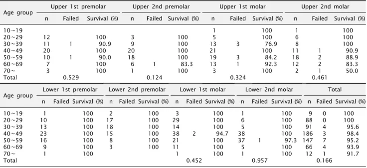
Table 4. Survival rate of implant according to age
Age group Upper 1st premolar Upper 2nd premolar Upper 1st molar Upper 2nd molar n Failed Survival (%) n Failed Survival (%) n Failed Survival (%) n Failed Survival (%) 10∼19
20∼29 30∼39 40∼49 50∼59 60∼69 70∼
Total
12 11 20 10 7 3
1 1 0.529
100 90.9
100 90.0
100 100
3 9 20 18 6 1
1 0.124
100 100 100 100 83.3
100
1 5 13 21 19 13 3
3 3 1 0.324
100 100 76.9
100 84.2 92.3 100
1 6 8 11 18 12 2
1 2 2 1 0.461
100 100 100 90.9 88.9 83.3 50.0
Age group Lower 1st premolar Lower 2nd premolar Lower 1st molar Lower 2nd molar Total n Failed Survival (%) n Failed Survival (%) n Failed Survival (%) n Failed Survival (%) n Failed Survival (%) 10∼19
20∼29 30∼39 40∼49 50∼59 60∼69 70∼
Total
1 10 13 23 16 9 1
100 100 100 100 100 100 100
2 17 18 15 8 3
100 100 100 100 100 100
3 29 14 38 21 11 1
2 0.452
100 100 100 94.7
100 100 100
1 6 5 38 37 5 1
1 0.957
100 100 100 100 97.3
100 100
9 88 91 186 147 66 12
0 0 4 3 7 4 1 0.166
100 100 95.6 98.4 95.2 93.9 91.7
3. 성별 및 연령, 전신 질환에 따른 생존율 1) 성별
총 509명 환자 중 303명의 남자 환자의 구치부에 344개를 식립하여 331개가 생존하여 96.2%의 생존율을 보였고, 206명의 여자 환자의 구치부에 255개를 식립하여 249개가 생존하여 97.6%의 생존율을 보였으나 통계학적으로는 유의한 차이가 없었 다(Table 3).
2) 연령
환자의 연령에 따른 생존율은 70대에서 생존율이 91.7%로 가장 낮게 나타났고, 10대, 20대에서는 100%로 높게 나타났으나, 통계학적으로 유의한 차이가 없었다. 식립 부위에 따른 연령별 생존율에서도 유의성 있는 차이는 나타나지 않았다(Table 4).
3) 전신 질환
구치부 단일 임플란트 중 전신 질환을 가지고 있는 환자에게 식립된 임플란트는 238개로 전체의 40%였고, 전신 질환이 없는 사람에게 식립된 임플란트는 361개로 전체의 60%였다. 전신 질 환의 분포는 고혈압이 가장 많았고, 그 다음으로는 당뇨, B형 간염의 순이었다. 전신 질환에 따른 임플란트 생존율은 정상인과 큰 차이가 없었으나, 골다공증의 경우 80%로 낮게 나타났다.
그러나 통계학적으로 유의하지는 않았다(Table 5).
4. 임플란트 직경과 길이, 표면 처리 방법, 경부 유형 및 매식체 형태에 따른 생존율
1) 임플란트 직경
식립된 임플란트의 직경은 제1, 2 소구치 부위에서는 상, 하악
모두 3.6∼4.0 mm의 임플란트가 가장 많았고, 그 다음이 4.1∼
Table 5. Survival rate of implant according to general disease
Generaldisease
Upper 1st premolar Upper 2nd premolar Upper 1st molar Upper 2nd molar n Failed Survival (%) n Failed Survival (%) n Failed Survival (%) n Failed Survival (%) Normal
HTN DM Hepa B Osteo Others Total
38 10 7 5 3
2
0.851
94.7 100 100 100 100
33 9 7 3 1 4
1
0.981
97.0 100 100 100 100 100
39 13 9 7 1 6
3 1 1 1 1 0.051
92.3 92.3 88.9
1000.0 83.3
25 12 5 8 1 7
3 1 1 1
0.902
88.0 91.7 80.0 87.5 100 100
General disease
Lower 1st premolar Lower 2nd premolar Lower 1st molar Lower 2nd molar Total n Failed Survival (%) n Failed Survival (%) n Failed Survival (%) n Failed Survival (%) n Failed Survival (%) Normal
HTN DM Hepa B Osteo Others Total
54 7 4 4 4
100 100 100 100 100
39 9 5 6 1 3
100 100 100 100 100 100
75 14 10 9 1 8
1 1
0.698
98.7 92.9 100 100 100 100
58 12 8 8 7
1
0.962
98.3 100 100 100 100
361 86 55 50 5 42
11 3 2 1 1 1 0.415
97.0 96.5 96.4 98.0 80.0 97.6
HTN, hypertension; DM, diabetes mellitus; Hepa B, hepatitis B; Osteo, osteoporosis.
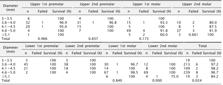
Table 6. Survival rate of implant according to fixture diameter
Diameter(mm)
Upper 1st premolar Upper 2nd premolar Upper 1st molar Upper 2nd molar n Failed Survival (%) n Failed Survival (%) n Failed Survival (%) n Failed Survival (%) 3∼3.5
3.6∼4.0 4.1∼4.5 4.6∼5.0
≥5.1 Total
6 32 20 4 1
1 1
0.966
100 96.9 95.0 100 100
4 31 15 7
1
0.837
100 96.8
100 100
1 15 5 49 5
1 4 2 0.173
100 93.3
100 91.8 60.0
10 8 37 3
2 1 3 0.661
80.0 87.5 91.9 100
Diameter (mm)
Lower 1st premolar Lower 2nd premolar Lower 1st molar Lower 2nd molar Total n Failed Survival (%) n Failed Survival (%) n Failed Survival (%) n Failed Survival (%) n Failed Survival (%) 3∼3.5
3.6∼4.0 4.1∼4.5 4.6∼5.0
≥5.1 Total
5 45 21 2
100
100 100 100
3 38 18 4
100
100 100 100
30 14 67 6
1 1 0.840
96.7
100 98.5
100 12 8 69 4
1 0.000
100 100 100 75.0
19 213 109 239 19
6 2 8 3 0.024
100 97.2 98.2 96.7 84.2
4.5 mm 직경의 임플란트였다. 제1, 2 대구치 부위에서는 상, 하악 모두 4.6∼5.0 mm의 임플란트가 가장 많았고, 3.6∼4.0 mm, 4.1∼4.5 mm 직경의 임플란트 순서였다. 전체적으로 생존 율은 직경에 따라 유의한 차이를 보였다. 5.1 mm 이상에서 84.2%로 가장 낮은 생존율을 보였고, 3∼3.5 mm와 4.1∼4.5 mm에서 100%, 98.2%로 높은 생존율을 보였다. 각각의 위치에 따라 살펴보면 상악 제1 대구치와 하악 제2 대구치에서는 직경 5.1 mm 이상일 때 60%, 75%로 생존율이 두드러지게 감소하였다 (Table 6).
2) 임플란트 길이
식립된 임플란트 길이는 11∼12 mm가 268개로 가장 많았고,
10 mm에서 163개, 13 mm 이상에서 131개 및 8∼9 mm에서 37개 순이었다.
길이에 따른 생존율은 8∼9 mm에서 97.3%, 10 mm에서 95.7%, 11∼13 mm에서 96.6%, 13 mm 이상에서 98.5%로 큰 차이가 없었고 통계학적 유의성도 없었다. 또한 식립 부위에 따른 생존율은 상악 제1 대구치 부위 10 mm에서 83.3%, 13 mm 이상 길이에서는 93.8%, 상악 제2 대구치 부위 10 mm에서 83.3%, 11∼12 mm에서는 93.8% 등으로 다소 차이가 있었지만 통계학적 유의성은 없었다(Table 7).
3) 임플란트 표면 처리 방법
임플란트 표면 처리 방법은 resorbable blast media 기법이
Table 8. Survival rate of implant according to implant surface texture
Surfacetexture
Upper 1st premolar Upper 2nd premolar Upper 1st molar Upper 2nd molar n Failed Survival (%) n Failed Survival (%) n Failed Survival (%) n Failed Survival (%) AE
Grit blasting SLA RBM Anodizing Total
4 8 2 17 32
1
1 0.092
75.0 100 100 94.1
100
3 6 11 17
20 1
0.757
100 100 100 100 95.0
10 13 9 27 16
3
3 1 0.108
70.0 100 100 88.9 93.8
4 6 6 31 11
4 2 0.593
100 100 100 87.1 81.8
Surface texture
Lower 1st premolar Lower 2nd premolar Lower 1st molar Lower 2nd molar Total n Failed Survival (%) n Failed Survival (%) n Failed Survival (%) n Failed Survival (%) n Failed Survival (%) AE
Grit blasting SLA RBM Anodizing Total
9 8 17 18 21
100 100 100 100 100
3 9 10 23 18
100 100 100 100 100
6 20 12 37 42
1
1 0.670
100 95.0
100 100 97.6
6 11 8 33
35 1
0.766 100 100 100 97.0
100 45 81 75 203 195
4 1 0 9 5 0.049
91.1 98.8 100 95.6 97.4
AE, acid etching; SLA, sand blast with large grit and acid etching; RBM, resorbable blast media.
Table 7. Survival rate of implant according to fixture length
Bone quality Upper 1st premolar Upper 2nd premolar Upper 1st molar Upper 2nd molar n Failed Survival (%) n Failed Survival (%) n Failed Survival (%) n Failed Survival (%) 8∼9
10 11∼12
≥13 Total
2 13 27 21
1 1 0.650
100 92.3 96.3 100
4 15 29 9
1 0.805
100 100 96.6
100
2 18 39 16
3 3 1 0.647
100 83.3 92.3 93.8
18 32 8
3 2 1 0.498
83.3 93.8 87.5
Bone quality Lower 1st premolar Lower 2nd premolar Lower 1st molar Lower 2nd molar Total n Failed Survival (%) n Failed Survival (%) n Failed Survival (%) n Failed Survival (%) n Failed Survival (%) 8∼9
10 11∼12
≥13 Total
6 16 39 12
100 100 100 100
2 14 36 11
100 100 100 100
8 24 45 40
2 0.354
100 100 95.6
100 13 45 21 14
1 0.101
92.3 100 100 100
37 163 268 131
1 7 9 2 0.598
97.3 95.7 96.6 98.5
Table 9. Survival rate of implant according to implant type
Implant type Upper 1st premolar Upper 2nd premolar Upper 1st molar Upper 2nd molar n Failed Survival (%) n Failed Survival (%) n Failed Survival (%) n Failed Survival (%) Internal
External Total
52 11
1 1 0.218
98.1 90.9
48 9
1 0.662
97.9 100
57 18
7 0.118
87.7 100
43 15
3 3 0.154
93.0 80.0
Implant type Lower 1st premolar Lower 2nd premolar Lower 1st molar Lower 2nd molar Total n Failed Survival (%) n Failed Survival (%) n Failed Survival (%) n Failed Survival (%) n Failed Survival (%) Internal
External Total
62 11
100 100
47 16
100 100
97 20
1 1 0.212
99.0 95.0
79 14
1 0.672
98.7 100.0
485 114
14 5 0.381
97.1 95.6
Table 10. Survival rate of implant according to implant shape
Implantshape
Upper 1st premolar Upper 2nd premolar Upper 1st molar Upper 2nd molar n Failed Survival (%) n Failed Survival (%) n Failed Survival (%) n Failed Survival (%) Straight
Tapered Total
26 37
1 1 0.799
96.2 97.3
24
33 1
0.390
100 97.0
27 48
3 4 0.691
88.9 91.7
24 34
2 4 0.673
91.7 88.2
Implant shape
Lower 1st premolar Lower 2nd premolar Lower 1st molar Lower 2nd molar Total n Failed Survival (%) n Failed Survival (%) n Failed Survival (%) n Failed Survival (%) n Failed Survival (%) Straight
Tapered Total
30 43
100 100
22 41
100 100
47 70
1 1 0.775
97.9 98.6
45 48
1 0.299
97.8 100
245 354
8 11 0.914
96.7 96.9
Table 11. Survival rate of implant according to bone graft
Bone graft Upper 1st premolar Upper 2nd premolar Upper 1st molar Upper 2nd molar n Failed Survival (%) n Failed Survival (%) n Failed Survival (%) n Failed Survival (%) GBR only
GBR with membrane Sinus No Total
3 14 6 40
1
1 0.784
100 92.9
100 97.5
3 9 6 39
1
0.000
66.7 100 100 100
1 6 44 24
7 0.142
100 100 84.1
100 2 20 36
5 1 0.029
100 75.0 97.2
Bone graft Lower 1st premolar Lower 2nd premolar Lower 1st molar Lower 2nd molar Total n Failed Survival (%) n Failed Survival (%) n Failed Survival (%) n Failed Survival (%) n Failed Survival (%) GBR only
GBR with membrane Sinus No Total
5 24
44
100 100
100 2 17
44
100 100
100 9 23
85 1
1 0.534
100 95.7
98.8 1 8
84
1
0.005
100 87.5
100 24 103 76 396
1 3 12 3 0.000
95.8 97.1 84.2 99.2
GBR, guided bone regeneration.
203개로 가장 많았고, Anodizing, Grit Blasting, sand blast with large grit and acid etching (SLA) 및 acid etching (AE) 순이었다.
표면 처리 방법에 따른 생존율을 살펴보면 SLA군에서 100%, Grit Blasting군에서 98.8% 등으로 대부분 처리 방법들 사이에는 큰 차이를 보이지 않았지만, AE군에서는 91.1%로 가장 낮은 생존율을 보였고 통계학적으로도 유의한 차이가 있었다. 또한 식립 부위별로 살펴보면 상악 제1 소구치 및 제1 대구치 부위에서 는 AE군의 생존율이 각각 75%, 70%로 특히 낮게 나타났다 (Table 8).
4) 임플란트 경부 유형
구치부 단일 임플란트 식립에 사용된 임플란트는 내부연결형이 485개, 외부연결형이 114개로 내부연결형이 많았다. 생존율을 살펴보면 내부연결형이 97.1%, 외부연결형이 95.6%로 비슷한 생존율을 보여주었다(Table 9).
5) 임플란트 매식체 형태
임플란트 매식체의 형태는 직선 형태가 245개(41%), 경사 형 태가 354개(59%)였다. 생존율을 살펴봤을 때 직선 형태가 96.7%, 경사 형태가 96.9%의 생존율을 보여 비슷하였다(Table 10).
5. 식립 시술 유형 1) 골이식
구치부 단일 임플란트 식립 시 골유도 재생술을 시행한 경우는 127개였고 그 중 차폐막을 사용한 경우가 103개였다. 상악동 거상술을 동반한 경우는 상악 제1 대구치에서 51개로 가장 많았 고, 상악 제2 대구치는 25개, 상악 제1, 2 소구치에서 각각 6개였 다.
골이식 여부에 따른 생존율을 살펴보면 골유도 재생술만 시행
한 경우 95.8%, 골유도 재생술과 함께 차폐막을 사용한 경우
97.1%, 상악동 거상술을 시행한 경우에는 84.2%의 생존율을
Table 13. Survival rate of implant according to surgeons
Surgeon Upper 1st premolar Upper 2nd premolar Upper 1st molar Upper 2nd molar n Failed Survival (%) n Failed Survival (%) n Failed Survival (%) n Failed Survival (%) Resident
Professor Total
17
46 2
0.382
100 95.7
13
44 1
0.583
100 97.7
28 47
2 5 0.615
92.9 89.4
13 45
1 5 0.721
92.3 88.9
Surgeon Lower 1st premolar Lower 2nd premolar Lower 1st molar Lower 2nd molar Total n Failed Survival (%) n Failed Survival (%) n Failed Survival (%) n Failed Survival (%) n Failed Survival (%) Resident
Professor Total
25 48
100 100
25 38
100 100
38 79
1 1 0.594
97.4 98.7
34 59
1 0.445
100 98.3
193 406
4 15 0.290
97.9 96.3
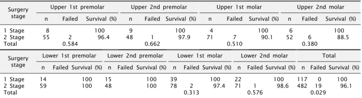
Table 12. Survival rate of implant according to surgery stage
Surgery stage
Upper 1st premolar Upper 2nd premolar Upper 1st molar Upper 2nd molar n Failed Survival (%) n Failed Survival (%) n Failed Survival (%) n Failed Survival (%) 1 Stage
2 Stage Total
8
55 2
0.584
100 96.4
9
48 1
0.662
100 97.9
4
71 7
0.510
100 90.1
6
52 6
0.380
100 88.5
Surgery stage
Lower 1st premolar Lower 2nd premolar Lower 1st molar Lower 2nd molar Total n Failed Survival (%) n Failed Survival (%) n Failed Survival (%) n Failed Survival (%) n Failed Survival (%) 1 Stage
2 Stage Total
14 59
100 100
15 48
100 100
39 78
2 0.313
100 97.4
22 71
1 0.576
100 98.6
117 482
0 19 0.029
100 96.1
보였다. 이는 골이식을 시행하지 않았을 때의 생존율 99.2%와 비교할 때 유의성 있게 낮은 생존율을 보였다(Table 11).
2) 임플란트 수술 단계
1회 임플란트 수술법(1 Stage) 및 2회 임플란트 수술법(2 Stage)에 따른 생존율을 살펴보면 1회법은 100%, 2회법은 96.1%
를 보여 1회법이 조금 높았고 통계적으로 유의성이 있었다. 각 식립 부위별로 살펴보면 전체적으로 2회 수술법이 482개(80%) 로, 1회 수술법 117개(20%)보다 많이 시행되었고 생존율은 2회 수술법이 96.1%로 1회 수술법 100%보다 낮았다. 특히 상악 제1 대구치는 2회 수술법 생존율이 90.1%, 상악 제2 대구치는 2회법 생존율이 88.5%로 다른 부위에 비해서 낮았지만 통계적으로 유의 성은 보이지 않았다(Table 12).
3) 시술자
전체적으로 임플란트는 교수가 406개, 전공의가 193개를 식립 하여 교수가 전공의보다 2배 이상 많이 시행하였다. 하악 제1 대구치를 제외한 모든 부위에서 전공의가 교수보다 높거나 같은 생존율을 보였고, 전체적인 임플란트 생존율은 교수가 식립한 경우 96.3%, 전공의가 식립한 경우 97.9%로 전공의가 임플란트 를 식립한 경우 더 높은 생존율을 보였으나 통계적으로 유의한
차이가 없었다(Table 13).
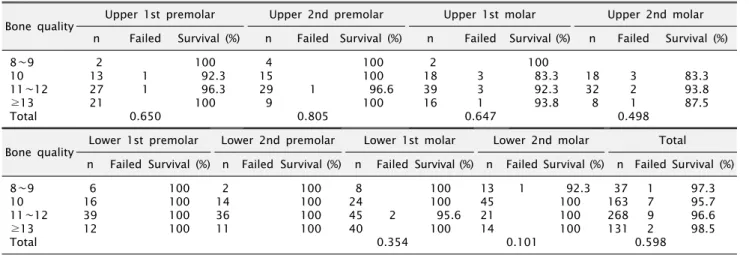
6. 식립 부위의 골질에 따른 생존율
임플란트 식립 부위의 골질에 따른 생존율을 조사한 결과 전체 적으로 Type I골에서 100%, Type II골은 99.4%, Type III골은 95.6%, Type IV골에서는 75% 생존율을 보였고 통계적으로 유의 성이 있었다. 각각의 부위별로 살펴보면 상, 하악 모두 소구치보다 는 대구치 Type IV골에서 90% 이하의 생존율을 보였고, 하악 제1, 2 대구치는 Type IV골에서 0%, 66.7%의 생존율을 보여 특히 낮은 생존율을 나타냈다(Table 14).
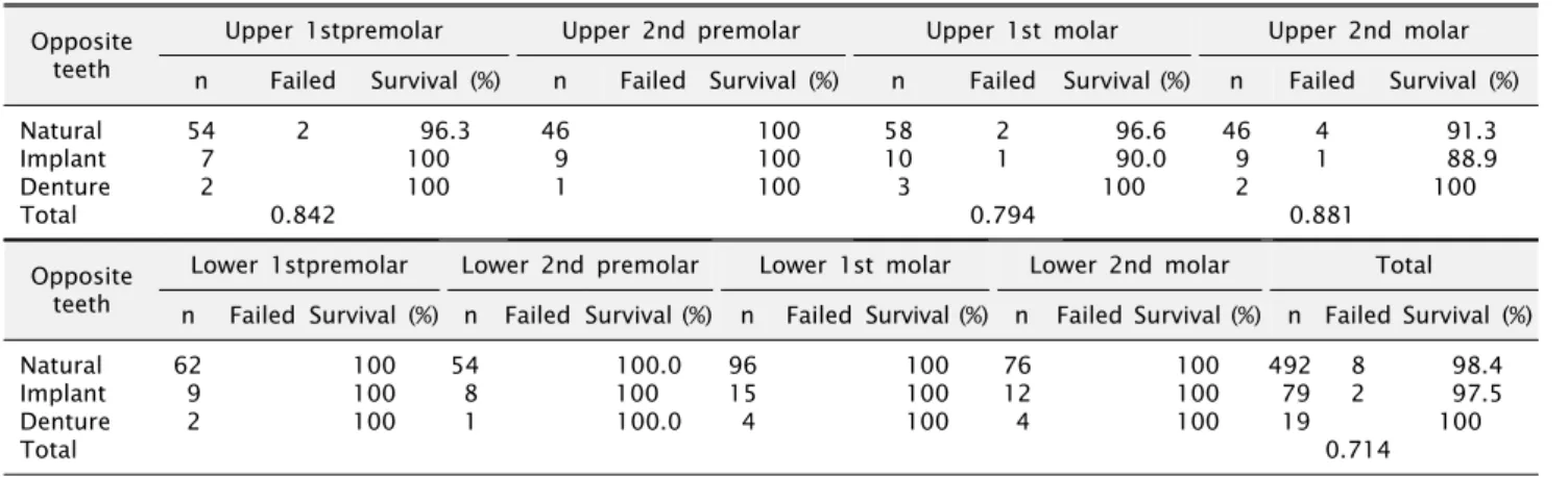
7. 대합치의 종류에 따른 성공률
구치부 단일 치아 임플란트의 대합치는 자연치가 492개로 가장 많았고 임플란트, 의치 순이었다. 생존율을 살펴보면 대합치가 자연치일 경우 98.4%, 임플란트 보철물일 경우 97.5%, 가철성 의치일 경우 100%의 생존율을 보였지만, 통계학적으로 유의성은 없었다(Table 15).
8. 실패의 시기와 유형
이번 연구에서 총 599개의 임플란트 중 19개가 실패하여 3.2%
의 실패율을 보였다. 임플란트의 실패시기를 살펴보면 2차 수술
Table 14. Survival rate of implant according to bone quality
Bone quality Upper 1stpremolar Upper 2nd premolar Upper 1st molar Upper 2nd molar n Failed Survival (%) n Failed Survival (%) n Failed Survival (%) n Failed Survival (%) I
II III IV Total
2 56 5
2 0.879
100 96.4
100
2 48 7
1 0 0.909
100 97.9
100
2 57 16
4 3 0.326
100 93.0 81.3
6 49 3
5 1 0.301
100 89.8 66.7
Bone quality Lower 1stpremolar Lower 2nd premolar Lower 1st molar Lower 2nd molar Total n Failed Survival (%) n Failed Survival (%) n Failed Survival (%) n Failed Survival (%) n Failed Survival (%) I
II III IV Total
49 23 1
100 100 100
46 16 1
100 100 100
3 83 30 1
1 1 0.000
100 98.8
100 0.0
4 68 18 3 1
0.000 100 100 100 66.7
7 258 297 37
0 1 12 6 0.000
100 99.4 95.6 75.0
Table 15. Survival rate of implant according to opposite teeth
Oppositeteeth
Upper 1stpremolar Upper 2nd premolar Upper 1st molar Upper 2nd molar n Failed Survival (%) n Failed Survival (%) n Failed Survival (%) n Failed Survival (%) Natural
Implant Denture Total
54 7 2
2
0.842
96.3 100 100
46 9 1
100 100 100
58 10 3
2 1 0.794
96.6 90.0 100
46 9 2
4 1 0.881
91.3 88.9 100
Opposite teeth
Lower 1stpremolar Lower 2nd premolar Lower 1st molar Lower 2nd molar Total n Failed Survival (%) n Failed Survival (%) n Failed Survival (%) n Failed Survival (%) n Failed Survival (%) Natural
Implant Denture Total
62 9 2
100 100 100
54 8 1
100.0 100 100.0
96 15 4
100 100 100
76 12 4
100 100 100
492 79 19
8 2 0.714
98.4 97.5 100
시행 이전에 실패한 것이 9개로 47.4%, 2차 수술 이후 보철물이 장착되고 기능 부하 6개월 이내에 실패한 것이 8개로 42.1%, 기능 부하 6개월 이후부터 3년 내에 실패한 것이 2개로 10.5%를 나타냈고, 기능 부하 3년 이후에 실패한 임플란트는 없었다.
기능 부하 이전에 실패한 임플란트의 주된 이유는 골유착의 실패로 9개 중 7개에 해당되었다. 그 밖에 일차 수술 이후 임플란 트 주위 골이 급속히 소실되어 2차 수술 시 매식체의 나사선 (thread)이 과도하게 노출된 경우와 임플란트 주변으로 화농성 삼출물과 염증 양상이 지속적으로 반복된 경우가 있었다.
기능 부하 이후에 실패한 임플란트의 경우 골유착이 소실된 경우가 5개였고, 임플란트 주위의 염증이 지속되고 환자가 불편감 을 호소한 경우가 4개였으며, 임플란트 주변골의 점진적인 소실을 보인 경우가 1개였다(Table 16).
고 찰