Ⅰ. 서 론
지난 20여 년 동안 골유착성 임플랜트는 부분 또 는 완전 무치악 환자에게 성공률이 높은 믿을만한 치료방법으로 인정받았다.
1977년 Bra�nemark 등1)이 치근형 골유착성 임플 랜트의 10년 장기 임상관찰 결과를 보고하였고, 1981년 Adell 등2)이 15년 장기 임상관찰 결과보고를 한 이후 많은 치과의사들의 관심을 불러 일으켰으 며, 1982년 Toronto conference3)이후 북미를 비롯 해 전세계적으로 골유착성 임플랜트의 시술이 확대 되었다.
그 이후 전악4,5)과 부분 무치악 환자6-9)들을 대상으 로 한 임플랜트 시술은 호의적인 결과들을 보였다.
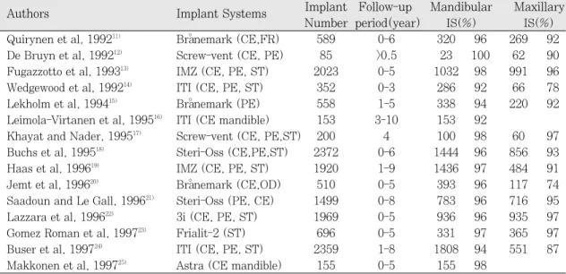
최근에는 가철성 수복물을 피하고, 자연치의 손상을 줄이려는 방향으로 환자의 요구가 변화됨에 따라 임 플랜트의 수요가 점차 증가하고 있으며, 여러 회사 들이 앞다투어 새로운 개념의 임플랜트 시스템을 소 개하고 있다. 하지만 극히 제한된 수의 임플랜트 시 스템만이 장기 임상관찰 결과를 보고하고 있다 (Table I).10-25)
한국에서도 1980년 이래로 임플랜트 시술이 치아 결손부를 치료하는 중요한 치과 술식의 하나가 되었 다. 그러나 한국인에 대한 임플랜트의 장기 임상 관 찰 연구는 아직 부족한 형편이다. 한국인에서의 임 플랜트 생존율은 한국인들의 고유의 악궁 형태, 섭 취하는 식품의 종류나 저작습관 등에 따라 구미인들 과는 약간 다른 결과가 나올 수도 있을 것이다.
Kim 등26)은 평균 3년 동안 follow-up한 임플랜트의 누적생존율이 94%에 이른다고 발표했고 Kim 등27)은 또 다른 연구에서 평균 3년 동안 follow-up한 임플랜 트의 생존율이 96.8%에 이른다고 보고했다. 그러나 이들 연구에는 도중에 탈락된 환자의 수와 시기가 정확히 명시되어 있지 않았으며, 임플랜트 종류에 따른 생존율을 구하지 않았다. 그리고 생존율과 위 험인자에 대한 연구 중 보철적 요인에 대한 부분이 언급되지 않았다.
이에 저자는 고려대학교 의료원 구로병원에서 식 립한 Bra�nemark 임플랜트의 후향적인 연구를 통해 임플랜트의 누적생존율을 구하고 여러 위험요인들 과 임플랜트 생존율과의 상관관계를 규명하고자 본 연구를 시작했다.
Ⅱ. 연구 재료 및 방법
본 연구는 1993년부터 2003년까지 고려대학교의 료원 구로병원 치과의 임플랜트센터에서 임플랜트 치료를 받은 289명의 환자들의 임상기록을 조사하 여, Bra�nemark 임플랜트(Nobel Biocare AB, Gothenburg, Sweden)를 한 개 이상 식립한 83명 환 자의 271개의 임플랜트를 연구대상으로 하였다. 단 보철물에서 다른 회사의 임플랜트와 연결된 경우는 연구대상에서 제외하였다.
101부위에 식립된 총 271개의 임플랜트(3.75 mm: 205개; 4.0 mm: 26개; 5.0 mm Regular Platform: 28개; 5.0 mm Wide Platform: 12개) 중 대한치과보철학회지:Vol. 45, No. 1, 2007
Bra � nemark 임플랜트의 10년 후향적 임상연구
고려대학교의료원 구로병원 보철과, *예방치과
배정윤∙신상완∙조현정∙김영수
*96개의 임플랜트는 상악에, 175개의 임플랜트는 하 악에 식립되었다. 대상 환자들은 16-74세의 남자 54명, 여자 29명 총 83명이었다.
모든 환자들에게 1981년 이후 Adell 등2)이 제안한 외과수술 및 보철술식을 근간으로 하여 시술하였으 며, 다양한 보철수복 방법을 이용하여 수복하였고 이후 follow-up을 위한 내원을 진행하였다. Follow- up을 위한 재내원시 각각의 임플랜트에 대한 임상 검사를 실시하였다. 이를 통해 임플랜트의 생존 또 는 실패에 대한 자료를 모았다. 1997년 Roos28)등이 정의한 기준에 준하여, 사망하거나 drop out된 환자 의 임플랜트의 경우는 설명할 수 없는(unaccounted for) 임플랜트로, 어떤 이유로든 제거한 임플랜트는 실패한(failed) 임플랜트로, 그외의 임플랜트는 생존 한(surviving) 임플랜트로 구분하였다.
본 연구의 관찰기간은 1993년 각 환자의 Stage I surgery (제 1차 수술)일로부터 2003년 12월 이내의 최종 내원일까지로 하였으며, 임플랜트의 예후와 관
들을 검사하기 위하여 노력하였다. 이 요소들은 성 별(male vs. female)/전신적 건강(건강한 사람 vs. 전 신 질환자)/제 1차 수술시의 나이/식립한 날짜/임플 랜트의 길이/임플랜트 직경(3.75 mm vs. 4.0 mm vs. 5.0 mm)/악궁(maxilla vs. mandible)/치아 위 치(incisor vs. canine vs. premolar vs. molar)/대 합치열 상태(natural dentition or fixed prosthesis vs. removable partial denture or overdenture vs. complete denture/Kennedy 부분무치악 분류 (tooth bounded-Class III vs. distal extension-Class I or II)/보철물의 형태(single tooth implant vs. fixed prosthesis vs. removable partial denture vs.
overdenture vs. full fixed bridge vs. hybrid pros- thesis)/보철물의 설계(single implant vs. multiple splinted implants)/abutment의 종류(standard vs. EsthetiCone vs. CeraOne vs. MirusCone vs.
UCLA vs. other abutment)/자연치와의 splint- ing 여부/cantilever 여부 등이다.
Table I. Survival rates of the various implant systems10)
Authors Implant Systems Implant Follow-up Mandibular Maxillary Number period(year) IS(%) IS(%) Quirynen et al, 199211) Bra�nemark (CE,FR) 589 0-6 320 96 269 92 De Bruyn et al, 199212) Screw-vent (CE, PE) 85 >0.5 23 100 62 90 Fugazzotto et al, 199313) IMZ (CE, PE, ST) 2023 0-5 1032 98 991 96 Wedgewood et al, 199214) ITI (CE, PE, ST) 352 0-3 286 92 66 78 Lekholm et al, 199415) Bra�nemark (PE) 558 1-5 338 94 220 92 Leimola-Virtanen et al, 199516) ITI (CE mandible) 153 3-10 153 92
Khayat and Nader, 199517) Screw-vent (CE, PE,ST) 200 4 100 98 60 97 Buchs et al, 199518) Steri-Oss (CE,PE,ST) 2372 0-6 1444 96 856 93
Haas et al, 199619) IMZ (CE, PE, ST) 1920 1-9 1436 97 484 91
Jemt et al, 199620) Bra�nemark (CE,OD) 510 0-5 393 96 117 74 Saadoun and Le Gall, 199621) Steri-Oss (PE, CE) 1499 0-8 783 96 716 95 Lazzara et al, 199622) 3i (CE, PE, ST) 1969 0-5 936 96 935 97 Gomez Roman et al, 199723) Frialit-2 (ST) 696 0-5 331 97 365 97 Buser et al, 199724) ITI (CE, PE, ST) 2359 1-8 1808 94 551 87 Makkonen et al, 199725) Astra (CE mandible) 155 0-5 155 98
The survival rates were similar among the various implant systems. However, the common success criteria was not used by all the authors and there are few studies which reported the reliable results clinically and radiographically with the follow-up periods longer than five years. (CE=complete edentulism; FR=fixed restora- tion; PE=partial edentulism; ST=single tooth implant; OD=overdenture; I=investigated implant; S=sur- viving implant)
임상 데이터를 환자의 치과 의무기록에서 모으고 분석을 위해 SPSS statistical package(SPSS Inc., Chicago, U.S.A.)에 옮겼다. 변수를 코드화하고 임플 랜트의 누적생존율을 life table method에 의해 계산 하였으며 개개의 변수별로 누적생존율에 미치는 영 향을 Cox regression method를 이용하여 통계적으로 분석하였다.
Ⅲ. 연구 결과
1993년과 2003년에 걸쳐 83명의 환자가 271개의 임플랜트 시술을 받았다. 1차 수술시 환자의 나이는 16세에서 74세까지 평균 47.6세였다(Table II).
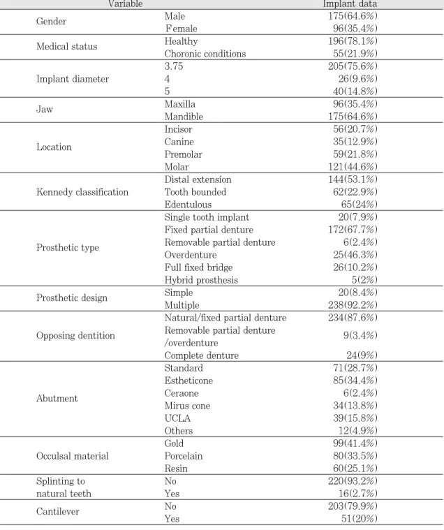
치료받은 환자의 34.9%를 여성이 차지했다. 임플 랜트 데이터를 Table III에 제시하였다. 연구군의 21.9%가 만성 질환을 가지고 있었다. 환자의 64.6%
는 하악에 임플랜트를 식립하였다. 환자의 87.6%에 서 자연치열이나 고정성 보철물에 임플랜트가 대합 하고 있었다. 모두 231개의 regular-diameter 임플랜 트, 40개의 wide-diameter 임플랜트를 식립했다.
통틀어 30개의 임플랜트가 19명의 환자에서 실패 했다(14명의 남자, 5명의 여자). 그리고 1개의 임플 랜트는 최종 보철물의 설계에 사용하지 않은 채 연 조직으로 덮어두었다(sleeper implant). 30개의 임플 랜트 중 13개에서 stage II surgery와 보철물 연결 전 일어나는 early failure가 일어났다. 나머지 17개의 임 플랜트에선 하중을 가한 후 일어나는 late failure가 일어났다.
임플랜트의 길이와 직경에 따른 임플랜트 실패율 이 Table IV에 제시되었다. 상악에서 96개 중 13개
(13.5%), 하악에서 175개 중 17개(9.7%)의 실패가 일어났다. 3.75-mm diameter implant군은 205개 중 18개가 실패해서 8.8%의 실패율을 보였다. 4.0- mm diameter implant군은 11.5%(26개 중 3개), 5.0-mm implant에선 25%(28개 중 7개), 5.0- mm WP implant에선 16.7%(12개 중 2개)의 실패 율을 보였다.
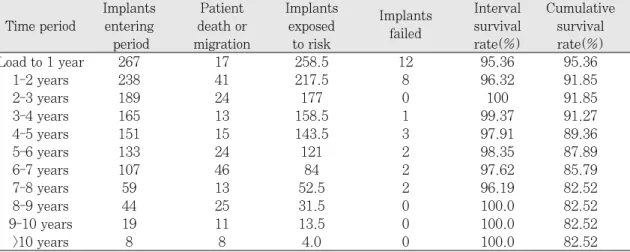
생존 분석을 통해 생존율을 구하였는데 이 때 생존 기간은 임플랜트에 하중을 가한 후를 기준으로 하였 다. 이 결과를 Table V과 Fig 1에 제시하였다. 보철 물의 설계에 포함되지 않은 임플랜트(sleeper)는 분 석에서 제외하였는데 이를 포함시키는 것이 골유착 에 영향을 주는 변수를 평가할 때 도움을 주지 못하 기 때문이다. 임플랜트의 5년째의 89.4%인 누적 생 존율이 10년째에는 82.5%로 감소했다.
임플랜트 생존율에 영향을 미치는 변수를 찾기 위 한 Cox regression analysis에서는 95% 유의수준에 서 임플랜트 길이가 임플랜트 생존율과 상관 관계가 있는 것으로 나타났으며(P=. 011) 그 외 성별, 전신 병력, 임플랜트 직경, 악궁, 구강내 위치, Kennedy 분류, 보철물 형태, 보철물 설계, abutment 종류, 교 합면 재료, splint 여부, cantilever 여부 등은 임플랜 트 생존율과 상관관계가 없었다. 임플랜트 길이에 대한 생존분석에 기초한 그래프를 Fig. 2에 제시했 다. 길이에 따른 누적 생존율(Table VI)을 비교하면 10mm 이하의 임플랜트는 74.5%의 누적 생존율을 보였고 11-15 mm 사이의 임플랜트는 87.7%, 16 mm 이상의 임플랜트는 96.2%의 누적 생존율을 보 였으며 통계적으로 유의한 차이가 있었다.
Table II. Distribution of the subjects’age and sex
Agegroups (years) Males Females Total
10-19 4 1 5
20-29 20 10 30
30-39 21 16 37
40-49 43 29 72
50-59 62 20 82
60-69 15 18 33
70-79 10 2 12
Table III. Numbers of the implants characterized by the independent variables.
Variable Implant data
Gender Male 175(64.6%)
Female 96(35.4%)
Medical status Healthy 196(78.1%)
Choronic conditions 55(21.9%)
3.75 205(75.6%)
Implant diameter 4 26(9.6%)
5 40(14.8%)
Jaw Maxilla 96(35.4%)
Mandible 175(64.6%)
Incisor 56(20.7%)
Location Canine 35(12.9%)
Premolar 59(21.8%)
Molar 121(44.6%)
Distal extension 144(53.1%)
Kennedy classification Tooth bounded 62(22.9%)
Edentulous 65(24%)
Single tooth implant 20(7.9%) Fixed partial denture 172(67.7%) Prosthetic type Removable partial denture 6(2.4%)
Overdenture 25(46.3%)
Full fixed bridge 26(10.2%)
Hybrid prosthesis 5(2%)
Prosthetic design Simple 20(8.4%)
Multiple 238(92.2%)
Natural/fixed partial denture 234(87.6%) Opposing dentition Removable partial denture
9(3.4%) /overdenture
Complete denture 24(9%)
Standard 71(28.7%)
Estheticone 85(34.4%)
Abutment Ceraone 6(2.4%)
Mirus cone 34(13.8%)
UCLA 39(15.8%)
Others 12(4.9%)
Gold 99(41.4%)
Occulsal material Porcelain 80(33.5%)
Resin 60(25.1%)
Splinting to No 220(93.2%)
natural teeth Yes 16(2.7%)
Cantilever No 203(79.9%)
Yes 51(20%)
Table IV. Implant failure rates according to the length and diameter of the fixtures.
Implant size Maxilla Mandible Total
Diameter Length Placed Failed Placed Failed Placed Failed
7 1 1 1 1(100%)
8.5 2 2 0
10 6 1 7 13 1(7.7%)
3.75 mm 11.5 2 2 9 1 11 3(27.3%)
13 25 5 38 3 63 8(15.9%)
15 35 3 29 0 64 3(6.3%)
18 21 0 22 2 43 2(7%)
20 8 8 0
8.5 1 1 0
11.5 5 1 5 1(20%)
4.0 mm 13 12 12 0
15 2 1 3 0
18 5 2 5 2(40%)
8 7 2 7 2(28.6%)
5.0 mm 10 1 1 9 2 10 3(30%)
12 2 1 9 1 11 2(18.2%)
10 5 2 5 2(40%)
5.0 mm(WP) 11.5 4 4 0
13 2 1 3 0
Total 96 13 175 17 271 30
Table V. Cumulative survival rates of Bra�nemark implants depicted as the life table.
Implants Patient Implants Implants Interval Cumulative Time period entering death or exposed failed survival survival
period migration to risk rate(%) rate(%)
Load to 1 year 267 17 258.5 12 95.36 95.36
1-2 years 238 41 217.5 8 96.32 91.85
2-3 years 189 24 177 0 100 91.85
3-4 years 165 13 158.5 1 99.37 91.27
4-5 years 151 15 143.5 3 97.91 89.36
5-6 years 133 24 121 2 98.35 87.89
6-7 years 107 46 84 2 97.62 85.79
7-8 years 59 13 52.5 2 96.19 82.52
8-9 years 44 25 31.5 0 100.0 82.52
9-10 years 19 11 13.5 0 100.0 82.52
>10 years 8 8 4.0 0 100.0 82.52
Table VII. Influence of the implant length on the implant failure reported in the literatures.39) Prosthesis Description of failure
Friberg et al40) IFCD Higher failure with 7 mm implants.
7 mm implants failed more in maxilla(7%) than mandible(3%).
Jemt and Lekholm41) IFCD 72 of 298 7 mm implants placed failed(24%).
Jemt et al42) IOD 7 mm failed more than any other implant length in maxilla.
Van Steenberghe et al43) IFPD 6% failure rate in maxilla with 10 mm implants.
10% failure rate in maxilla with 7 mm implants.
Naert et al44) IFPD 7 and 10 mm were the source of most failures.
Pylant et al45) IFPD 7 of 12 failures were 7 and 10 mm implants.
Jemt and Lekholm46) IFPD 67 of 78 failures were 7 mm implants.
Gunne47) IFPD 12 of 13 failures were 10 mm implants.
Higuchi et al48) IFPD Hihger failure with 7 mm implants in maxilla(18%).
IFCD = implant fixed complete denture : IOD=implant overdenture : IFPD=implant fixed partial denture Table VI. Five year-cumulative survival rates (CSR) according to the
implant length groups.
Implant length CSR
≤10 mm 74.5
11-15 mm 87.7
≥16 mm 96.2
Fig. 1. Cumulative survival rates of the Bra�nemark implant.
Fig. 2. Cumulative survival rates according to the implant length groups.
Ⅳ. 총괄 및 고찰
이번 연구는 고려대학교 의과대학 치과학교실에서 시행된 후향적 연구로 Bra�nemark 임플랜트의 생존 율 및 위험요인과의 상관관계를 보고하는 것이다.
임플랜트의 누적생존율은 5년 follow-up에 89.4%였 고, 10년 follow-up에 82.5%로 나타났다. 이 결과는 Bra�nemark 임플랜트의 5년간 누적생존율이 96.6%
라고 보고한 Ross 등28)의 연구를 비롯하여 다양한 임 플랜트 시스템에서 보고하는 생존율에 비해 상대적 으로 낮은 것이다(Table I).
Adell 등29)은 임플랜트 식립 이후 초기 2년 동안에 가장 높은 구간 실패율을 보이다가 그 이후에 점진 적인 안정화가 진행되어 임플랜트의 실패율이 감소 한다고 하였다. 본 연구에서도 식립 후 초기 2년 동 안에 가장 높은 실패율을 보였다. 임플랜트의 보철 전 초기 실패 원인으로는 골삭제 동안의 골의 과열, 감염, 환자의 건강 상태, 치유기간 동안의 미세한 동 요 등이 제안되었다.30,31)
한편 보철 후 실패의 원인으로는 불량한 구강 위 생, 부적절한 loading 상황, 그리고, framework의 misfit 등이 제안되었다.32,33,34) 최근에 Esposito
M35,36,37)등은 환자의 특성과 더불어 임플랜트 주위염
과 보철물의 과부하가 보철 후 실패의 주요 요인이 라 하였고, 임플랜트의 표면 특성이 실패 양상에 영 향을 줄 수 있다고 하였다. 한편, 본 연구에서는 보 철 후 임플랜트 실패율이 식립 4, 5년 경과 후 갑자 기 높아지는 것으로 나타났는데, 이는 골소실이 심 하게 진행되었으나 보철물이 splinting되어 있어 상 황을 인지하기 어려운 환자가 정기적 검진을 받지 않은 경우 뒤늦게 임플랜트가 제거되었을 가능성을 생각해볼 수 있겠다.
임플랜트 생존율과 상관관계를 보이는 변수를 알 아본 결과 환자의 성별, 전신 병력, 임플랜트 직경, 악궁, 위치, Kennedy 부분 무치악 분류, 보철물 형 태, 보철물 설계, abutment 종류, 교합면 재료, splint 여부, cantilever 여부 등은 임플랜트 생존율과 상관관계가 없는 것으로 나타났고 임플랜트 길이만 이 임플랜트의 생존에 영향을 주는 요인으로 나타났 다(Table VI, Fig. 2).
Wyatt 와 Zarb38)는 7 mm 임플랜트의 성공율은
75%이고, 10 mm 이상 임플랜트의 성공률은 90%
이상이었다고 보고하였다. 그 외에도 많은 연구들에 서 이와 유사한 결과들을 보고하였다(Table VII). 또 한 많은 연구자들이 7 mm 임플랜트를 상악에 식립 했을 때 가장 높은 실패율이 나타났다고 보고하였 다.40,41,43,48)
임플랜트를 지지하는 골의 면적은 임플랜트의 길 이를 연장함으로써 증가시킬 수 있다. 길이 증가의 중요성은 치조정 부위의 골접촉 면에서는 의미가 없 지만 초기 고정과 전체 골접촉 면적의 양을 증가시 키는 측면에서 의미가 있다. 임플랜트의 길이 증가 를 통해 임플랜트의 removal torque나 전단강도를 증 가시킬 수 있는 것으로 보고된 바 있다.49)
그러나 최근의 연구50,51)에 따르면 하악 구치부에 식립된 8 mm 길이의 hydroxyapatite-coated 임플랜 트에서 4%-6%의 낮은 실패율을 보인 것으로 나타 나 임플랜트의 표면 특성을 변화시킴으로써 짧은 임 플랜트에서도 성공률을 증가시킬 수 있음을 알 수 있다. 본 연구에서 사용된 임플랜트는 대부분 machined type의 임플랜트로 앞으로의 연구에서는 임플랜트의 표면 특성 변화에 따른 누적생존율 변화 에 대한 분석이 있어야 할 것으로 사료된다.
또한 최근의 연구에 따르면 임플랜트의 길이를 증 가시켜도 치조정 부위에서의 임플랜트 주위 응력을 감소시키는 데는 효과가 없으며, 일단 초기 고정을 위한 최소한의 길이가 확보되었다면 임플랜트의 폭 이 길이보다 더 중요한 요인인 것으로 보고되었다.49) 이번 연구에서는 임플랜트의 직경과 생존율이 통계 학적으로 상관관계가 없는 것으로 나타났지만, reg- ular diameter 임플랜트의 9.1%가 실패한 반면, wide diameter 임플랜트의 22.5%가 실패한 것으로 나타나 Shin 등52)이 보고한 wide diameter 임플랜트 군이 regular diameter 임플랜트 군보다 실패율이 12.5% 높게 나타났다는 연구 결과와 큰 차이가 없는 것으로 분석되었다. 앞으로의 연구에서는 임플랜트 의 생존율에 영향을 줄 가능성이 높은 변수들만을 사용하여 좀더 특이적인 분석을 시행할 필요가 있다 고 생각된다.
Albrektsson과 Zarb53)는 임플랜트 연구의 신뢰성을 높이기 위해 2개 이상의 기관에서 50명 이상의 환자 를 대상으로, 5년 이상 정기적으로 관찰해야 한다고
보고하였다. 그러나 본 연구에서는 과거의 자료를 진료기록부와 방사선 사진을 통해 취득할 수밖에 없 었기 때문에 자료의 신뢰성을 진료기록부의 충실성 에 의존할 수밖에 없었고, 중도탈락된 환자의 수가 많아 식립된 임플랜트의 완전한 follow-up이 이루어 지지 않았다. 또한 시간소요에 대한 문제점과 현실 적으로 환자의 동의를 구하기 어렵다는 문제점으로 인해 보철물을 제거하고 시행해야 하는 개개 임플랜 트의 동요도 검사를 하지 못했으며 표준화된 기준점 과 조사각을 통해 얻어진 치근단 방사선사진 상에서 임플랜트 인접 치조골의 변연골 수준을 측정하지 못 해 임플랜트의 성공기준에 따른 성공율은 얻지 못하 고 생존율만 구할 수밖에 없었다.
우리나라에는 아직까지 단일 임플랜트 시스템에 대한 장기간의 임상관찰 보고가 거의 없다. 치과의 사는 임플랜트 제조업체로부터 임상적으로 적절한 예후를 가지는 임플랜트를 공급받아 식립할 의무가 있음을 알아야 하고, 환자에게 임플랜트 치료에 동 반되는 이득과 위험에 대해 설명할 의무가 있음을 알아야 할 것이다. 본 연구를 기초로 하여 국내에서 도 앞으로 단일 시스템에 대하여 임플랜트 성공기준 에 근거하여 성공률을 평가하는 전향적인 연구가 시 행되어야 할 것이다.
Ⅴ. 결 론
후향적인 연구를 통해 Bra�nemark 임플랜트의 누 적생존율을 구하고, 임플랜트의 생존율에 영향을 미 치는 인자를 분석하여 다음과 같은 결과를 얻었다.
1. 10년 간의 Bra�nemark 임플랜트의 누적생존율은 82.52%였다.
2. Cox regression analysis에 의해 임플랜트의 길이 는 임플랜트의 누적생존율에 영향을 미치는 것으 로 분석되었다(P<. 05).
3. Bra�nemark implant의 장기적인 임상결과는 받 아들여질만 하였다.
앞으로 임플랜트의 성공에 영향을 미치는 인자들 을 분석하기 위하여 더 많은 수의 환자와 임플랜트 를 대상으로 하는 장기간의 전향적인 연구가 요구 된다.
참고문헌
1. Bra�nemark, PI, Hansson, BO, Adell R, Breine U, Lindstrom J, Hallen O, Ohman A. Osseointegrated implants in the treat- ment of the edentulous jaw. Experience from a 10-year period. Scand J Plast Reconstr Surg 1977:11:Suppl.16, and as a mono- graph from Almqvisit & Wiksell interna- tional, Stockholm 1977.
2. Adell R, Lekholm U, Rockler B, Bra�nemark PI. A 15 year study of osseointegrated im- plants in the treatment of the edentulous jaw. Int J Oral Surg 1981;10:387-416.
3. Zarb GA. Proceedings on the Toronto Conference on osseointegration in clinical dentistry. St. Louis, C. V. Mosby Co, 1983.
4. Cox JF, Zarb GA. The longitudinal clini- cal efficacy of osseointegrated dental im- plants: a 3 year report. Int J Oral Maxillofac Implants 1987;2:91-100.
5. Zarb GA, Schmitt A. The longitudinal clinical effectiveness of osseointegrated dental implants: the Toronto study. Part III: Problems and complications encoun- tered. J Prosthet Dent 1990;64:185-94.
6. Van Steenberghe D, Lekholm U, Bolender C, Folmer T, Henry P, Herrmann I, Higuchi K, Laney W, Linden U, Astrand P. Applicability of osseointegrated oral implants in the rehabilitation of partial edentulism: a prospective multicenter study on 558 fixtures. Int J Oral Maxillofac Implants 1990;5:272-281.
7. Zarb GA, Schmitt A. The longitudinal clinical effectiveness of osseointegrated dental implants in posterior partially edentulous patients. Int J Prosthodont 1993;6:189-196.
clinical effectiveness of osseointegrated dental implants in anterior partially eden- tulous patients. Int J Prosthodont 1993;
6:180-188.
9. Lekholm U, van Steenberghe D, Herrmann I, et al. Osseointegrated implants in the treatment of partially edentulous jaws: A prospective 5 year multicenter study. Int J Oral Maxillofac Implants 1994;9:627-635.
10. Mithridade Davarpanah, Hennry Martinez.
Clinical manual of implant dentistry:
Quintessence Publishing Co, Ltd, 2003.
11. Quirynen M, Naert I, van Steenberghe D, Nys L. A study of 589 consecutive implants supporting complete fixed prostheses.
Part I: Periodontal aspects. J Prosthet Dent 1992;68:655-663.
12. De Bruyn H, Collaert B, Linden U, Flygare L. A comparative study of the clinical efficacy of Screw Vent implants versus Branemark fixtures, installed in a peri- odontal clinic. Clin Oral Implants Res 1992;3:32-41.
13. Fugazzotto PA, Gulbransen HJ, Wheeler SL, Lindsay JA. The use of IMZ osseoin- tegrated implants in partially and completely edentulous patients: success and failure rates of 2,023 implant cylinders up to 60 months in function. Int J Oral Maxillofac Implants 1993;8:617-621.
14. Wedgwood D, Jennings KJ, Critchlow HA, Watkinson AC, Shepherd JP, Frame JW, Laird WR, Quayle AA. Experience with ITI osseointegrated implants at five cen- tres in the UK. Br J Oral Maxillofac Surg 1992;30:377-381.
15. Leimola Virtanen R, Peltola J, Oksala E, Helenius H, Happonen RP. ITI titani- um plasma sprayed screw implants in the treatment of edentulous mandibles: A follow up study of 39 patients. Int J Oral
Maxillofac Implants 1995;10:373-378.
16. Khayat PG, Nader NA. Interet et indica- tions d’un implant visse a hexagone interne:
Le Screw Vent. J Parodontal 1995;14:
31-41.
17. Buchs AU, Hanh J, Vassos DM. The prospective clinical study of 2372 Steri Oss HA coated threaded implants Six year post restoration update results. Presented at the 1995 SteriOss international con- ference. 1995.
18. Haas R, Mensdorff Pouilly N, Mailath G, Watzek G. Survival of 1,920 IMZ im- plants followed for up to 100 months.
Int J Oral Maxillofac Implants 1996;11:
581-588.
19. Jemt T, Chai J, Harnett J, et al. A 5 year prospective multicenter follow up report on overdentures supported by osseointegrat- ed implants. Int J Oral Maxillofac Implants 1996;11:291-298.
20. Saadoun AP, Le Gall MG. An 8 year compilation of clinical results obtained with Steri Oss endosseous implants.
Compend Contin Educ Dent 1996;17:669- 688.
21. Lazzara RJ, Siddiqui AA, Binon P, et al. Retrospective multicenter analysis of 3 endosseous dental implants placed over a five year period. Clini Oral Implants Res 1996;7:73-83.
22. Gomez Roman G, Schulte W, d’Hoedt B, Axman Krcmar D. The Frialit 2 implant system: five year clinical experience in sin- gle tooth and immediately postextraction applications. Int J Oral Maxillofac Implants 1997;12:299-309.
23. Buser D, Mericske Stern R, Bernard JP, Behneke A, Behneke N, Hirt HP, Belser UC, Lang NP. Long term evaluation of non submerged ITI implants. Part 1: 8 year life
table analysis of a prospective multi cen- ter study with 2359 implants. Clin Oral Implants Res 1997;8:161-172.
25. Makkonen TA, Holmberg S, Niemi L, Olsson C, Tammisalo T, Peltola J. A 5 year prospective clinical study of Astra Tech den- tal implants supporting fixed bridges or over- dentures in the edentulous mandible.
Clin Oral Implants Res 1997;8:469-475.
26. Kim YS, Lee DK, Min SK, Lee J, Moon C.
Clinical Study On Success Rate Of Osseointegrated Dental Implants. Korean Assoc Maxillofac Plast Reconstr Surg 2002;24:137-147. Korean.
27. Kim JS, Chang HH, Chang CH, Rhyu SH, Kang JH. Preprosthetic Stage Dental Implant Failure. J Korean Assoc Oral Maxillofac Surg 2001;27:178-183. Korean.
28. Roos J, Sennerby L, Lekholm U, Jemt T, Grondahl K, Albrektsson T. A qualitative and quantitative method for evaluating im- plant success: a 5 year retrospective analysis of the Bra�nemark implant. Int J Oral Maxillofac Implants 1997;12:504-514.
29. Adell R, Eriksson B, Lekholm U, Bra�ne- mark PI, Jemt T. Long term follow up study of osseointegrated implants in the treat- ment of totally edentulous jaws. Int J Oral Maxillofac Implants 1990;5:347- 359.
30. Albrektsson T, Bra�nemark PI, Hansson HA, Lindstrom J. Osseointegrated titanium implants. Requirements for ensuring a long lasting, direct bone to implant an- chorage in man. Acta Orthop Scand 1981;
52:155-170.
31. Bra�nemark PI. Osseointegration and its ex- perimental background. J Prosthet Dent 1983;50:399-410.
32. Jemt T. Implant treatment in resorbed
Res 1993;4:187-194.
33. Skalak R. Biomechanical considerations in osseointegrated prostheses. J Prosthet Dent 1983;49:843-848.
34. Lekholm U, Adell R, Lindhe J, Bra�nemark PI, Eriksson B, Rockler B, et al. Marginal tissue reactions at osseointegrated titanium fixtures. II. A crosssectional retrospec- tive study. Int J Oral Maxillofac Surg 1986;15;53-61.
35. Esposito M, Thomsen P, Ericson LE, Sennerby L, Lekholm U. Histopathologic observations on late oral implant fail- ures. Clin Implant Dent Relat Res 2000;
2:18-32.
36. Esposito M, Hirsch JM, Lekholm U, Thomsen P. Biological factors contributing to failures of osseointegrated oral im- plants. (II). Etiopathogenesis. Eur J Oral Sci 1998;106:721-764.
37. Esposito M, Hirsch JM, Lekholm U, Thomsen P. Biological factors contributing to failures of osseointegrated oral im- plants. (I). Success criteria and epidemi- ology. Eur J Oral Sci 1998;106:527-551.
38. Wyatt CC, Zarb GA. Treatment outcomes of patients with implant supported fixed par- tial prostheses.Int J Oral Maxillofac Implants 1998;13:204-211.
39. Goodacre, Kan, and Rungchrassaeng.
Clinical complications of osseointegrated implants J Prosthet Dent 1999;81;537-552.
40. Friberg B, Jemt T, Lekholm U. Early failures in 4,641 consecutively placed Branemark dental implants: a study from stage 1 surgery to the connection of com- pleted prostheses. Int J Oral Maxillofac Implants 1991;6:142-146.
41. Jemt T, Lekholm U. Implant treatment in edentulous maxillae: a 5 year follow up re-
jaw resorption. Int J Oral Maxillofac Implants 1995;10:303-311.
42. Jemt T, Book K, Linden B, Urde G.
Failures and complications in 92 consec- utively inserted overdentures supported by Bra�nemark implants in severely resorbed edentulous maxillae: a study from prosthetic treatment to first annual check up. Int J Oral Maxillofac Implants 1992;7:162- 167.
43. Van Steenberghe D, Lekholm U, Bolender C, Folmer T, Henry P, Herrmann I, Higuchi K, Laney W, Linden U, Astrand P. Applicability of osseointegrated oral implants in the rehabilitation of partial edentulism: a prospective multicenter study on 558 fixtures. Int J Oral Maxillofac Implants 1990;5:272-281.
44. Naert I, Quirynen M, van Steenberghe D, Darius P. A six year prosthodontic study of 509 consecutively inserted implants for the treatment of partial edentulism. J Prosthet Dent 1992;67:236-245.
45. Pylant T, Triplett RG, Key MC, Brunsvold MA. A retrospective evaluation of en- dosseous titanium implants in the partially edentulous patient. Int J Oral Maxillofac Implants 1992;7:195-202.
46. Jemt T, Lekholm U. Oral implant treatment
in posterior partially edentulous jaws: a 5 year follow up report. Int J Oral Maxillofac Implants 1993;8:635-640.
47. Gunne J, Jemt T, Linden B. Implant treatment in partially edentulous pa- tients: a report on prostheses after 3 years. Int J Prosthodont 1994;7:143- 148.
48. Higuchi KW, Folmer T, Kultje C. Implant survival rates in partially edentulous pa- tients: a 3 year prospective multicenter study. J Oral Maxillofac Surg 1995;53:264.
49. Carl E.Misch. Contemporary implant den- tistry. Mosby Co, 1999
50. Buchs AU, Hahn J, Vassos DM. Efficacy of threaded hydroxyapatite coated im- plants placed in the posterior mandible in support of fixed prostheses. Implant Dent 1996;5:106-110.
51. Teixeira ER, Wadamoto M, Akagawa Y, Kimoto T. Clinical application of short hydroxylapatite coated dental implants to the posterior mandible: a five year survival study. J Prosthet Dent 1997;
78:166-171.
52. Shin SW, Bryant SR, Zarb GA. A retro- spective study on the treatment outcome of wide bodied implants. Int J Prosthodont 2004;17:52-58.
Reprint request to:
Sang-Wan Shin, D.D.S., M.PH., Ph.D., MSc.
Department of Prosthodontics, Graduate School of Clinical Dentistry, Korea University 97, Gurodonggil, Guro-Gu, Seoul, 152-703, Korea
Statement of problems: There are few studies which reported the survival rates of the spe- cific dental implant systems in the Korean population with the follow-up periods longer than 5 years.
Purpose: This retrospective clinical study was aimed to evaluate cumulative survival rate (CSR) of Bra�nemark implants followed for 10 years and to determine risk factors for implant failure.
Material and methods: A total of 271 Bra�nemark implants in 83 patients were investigated with several identified risk factors. Life table analysis was undertaken to examine the CSR. Cox regression method was conducted to assess the association between potential risk factors and over- all CSR.
Results: Thirty implants failed. The 10-year implant CSR was 82.5%. Cox regression analysis demonstrated a significant predictive association between overall CSR and implant length (P<.05).
Conclusion: An acceptable long-term result of Bra�nemark implant was achieved and implant length showed a significant association with the CSR.
A 10-YEAR RETROSPECTIVE CLINICAL STUDY OF BRA �NEMARK IMPLANTS
Jung-Yoon Bae, D.D.S., M.S.D., Sang-Wan Shin, D.D.S., M.Ph., Ph.D., MSc., Hyun-Jung Cho, D.D.S., M.S.D., Young-Soo Kim*, D.D.S., M.S.D., Ph.D.
Department of Prosthodontics and *Preventive Dentistry, Guro Hospital, Korea University Medical Center ABSTRACT