1연세대학교 의과대학 내과학교실, 2진단방사선과학 교실, 3폐질환연구소, 4BK21 의과학 사업단, 5암전이 연구센터 정우영1, 변민광1, 박무석1, 한창훈1, 강신명1, 이도연2, 김영삼1,3, 김세규1,3-5, 김성규1, 장 준1,3
Risk Factors of Recurrent Hemoptysis after Bronchial Artery Embolization
Wou Young Chung1, M.D., Min Kwang Byun1, M.D., Moo Suk Park1, M.D., Chang Hoon Hahn1, M.D., Shin Myung Kang1, M.D., Do Yon Lee2, M.D., Young Sam Kim1,3, M.D., Se Kyu Kim1,3-5, M.D., Sung Kyu Kim1,3, M.D., Joon Chang1,3, M.D.
1Department of Internal Medicine, 2Radiology, 3The Institute of Chest Diseases, 4Brain Korea 21 Project for Medical Sciences, and 5Cancer Metastasis Research Center Yonsei University College of Medicine, Seoul, Korea
서 론 : 대량객혈은 치료가 이루어 지지 않을 경우, 50% 이상의 사망률을 보이는 호흡기 영역의 가장 위급한 응급상황의 하나이며 여러 원인에 의해 발생 될 수 있다. 1973년 레미 등에 의해 처음 보고된 이후로 기관지동맥 색전술은 대량의 재발성 객혈의 치료로 확립되어 그 효과가 입증 되었다.
그러나 기관지동맥 색전술의 재발률은 10~52%로 보고되어 객혈의 재발을 예측 할 수 있는 위험 요소들에 대한 연구가 필요하다.
재료 및 방법 : 2000년 1월부터 2005년 1월까지 세브란스병원에 100 cc 이상의 대량 객혈로 내원하여 기관지동맥 색전술 을 시행 받은 66명 환자들을 대상으로 하여 기관지동맥 색전술 후 재발의 빈도와 재발과 관련이 있는 위험 요인에 대해 분석하였다.
결 과 : 5년 간 기관지동맥 색전술을 시행 받은 환자는 75명이었고, 장기간 추적관찰이 되어 결과 분석이 가능했던 환자 는 66명이었다. 이들의 평균 나이는 54.9 ± 15.9세이었고, 남자가 48명, 여자가 18명이었다. 원인 질환은 결핵 20명, 기관지 확장증 및 기타 양성질환 23명, 악성 종양 7명이었고, 평균 20.4개월 간의 추적 관찰 기간 동안 23명(34.9%)에서 치료가 필요한 대량 객혈이 재발되었다. 환자의 성별과 나이, 이전에 객혈로 시술 받은 과거력, 객혈의 원인 질환, 분포 혈관의 수 등은 대량 객혈의 재발과 유의한 관계가 없었으나, 병변의 양측성, 흉막 비후, 객혈의 양은 유의한 인자로 관찰되었고, 로그 회귀분석 결과에서도 동일하였다.
결 론 : 기관지동맥 색전술을 시행한 후 객혈의 재발을 예측할 수 있는 위험 인자로 흉막 비후, 병변의 양측성, 객혈의 양이 중요하게 작용하며, 이에 대한 대규모 임상 연구가 필요하다.
(Tuberc Respir Dis 2006; 60: 65-71)
Key words : 객혈; 기관지동맥 색전술; 위험 요인
Address for correspondence : Joon Chang, M.D., Department of Internal Medicine, Yonsei University College of Medicine, Severance Hospital, CPO box 8044, Seoul, Korea.
Phone : 82-2-2228-1952 Fax : 82-2-393-6884 E-mail : [email protected]
Received : Oct. 12. 2005 Accepted : Oct. 28. 2005
Introduction
When massive hemoptysis is untreated, it has a mortality rate of over 50 percents. It is considered as one of most dreaded of all respiratory emergen- cies and can have a variety of underlying causes.
Bronchial artery embolization (BAE) was first reported in 1973 by Remy1 and it has become an
established procedure in the management of massi- ve and recurrent hemoptysis. Its efficacy is widely documented thereafter by number of articles1-11. An immediate control of hemoptysis is achieved in 73 to 98%, with a mean follow of less than one mon- th1-4. Immediate success rates have increased re- cently because of the introduction of superselective embolization and the refinement of embolic agents and techniques4. However, the long-term success rate of BAE is known to be unfavorable. Long- term recurrence rates are reported to be 10 to 52%, with a mean follow up period ranging from one to 46 months1-7.
The variety of factors influencing that control failure has been described.
Author No. of patients
Follow up (months)
Recurrence rate (%)
Risk factor for recurrence Other variables
Osaki S4 22 46 50
Bronchiectatic change on CT scan, Pulmonary-bronchial artery shunt
Age, sex, underlying disease, morhphologic change, amount of hemoptysis, angiographic finding
Yeo DS5 146 6 63 Pleural lesion on chest X-ray Embolic material, underlying disease, feeding vessel, shunt, bilaterality
Kim SO6 75 60 54.5 Underlying disease, amount of bleeding, extent of lung lesion
NR†,
Tamura S8 40 20 60 Pleural thickening on chest X-ray NR†,
Kim BC14 47 72 40
None* Age, sex, bilaterality, shunt, feeding artery, neovascularity, underlying disease,
Ko DS15 46 12 41.3
Multiple feeding vessel Incomplete embolization Previous history of hemoptysis
Hypervascularity, underlying disease, age, sex
* No statistically valid risk factor among candidate variables.
† NR = not reported
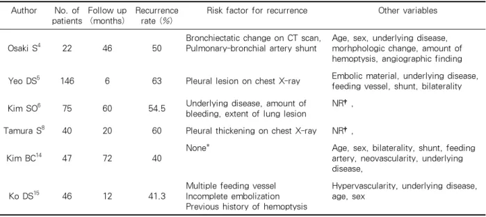
Table 1. Literature reviews of bronchial artery embolization outcomes Bronchiectatic change on high resolution CT
scan (HRCT)4, broncho-pulmonary shunt4, pleural thickening5,7, underlying lung diseases6, the amount of bleeding8, multiple feeding vessels9, incomplete embolization10, and previous hemoptysis history11 are possible risk factors of recurrent bleeding events. But these findings vary from article to article and there is not yet a proven condition to predict the recurrence.
This study is designed to survey previously do- cumented possible risk factors of recurrence in tho- se who underwent BAE in our hospital, during a long period. Furthermore, since all patients had taken HRCT, we focused on radiological findings such as pleural thickening to be possible risk fac- tors of the recurrence.
Materials and methods
Seventy-five patients underwent bronchial artery embolization due to massive hemoptysis more than 100 cc amount of bleeding in Severance Hospital from Jan. 2000 to Jan. 2005. Among them, nine
patients’ data were not available and could not be contacted with. Finally 66 (48 males, 18 females) patients’ medical records were analyzed retrospec- tively with a mean follow up period of 20.4 months (ranging from 1 month to 56 months).
The recurrence of massive hemoptysis after BAE was defined that a gross hemoptysis more than 100 cc amount of blood occurred again which needed to undergo treatments such as BAE or lung resection surgery.
Demographic characteristics such as gender, age, the duration of symptom, the amount of hemopty- sis, previous history of treatment for hemoptysis, the main medical condition causing hemoptysis, bi- laterality of pulmonary lesion, the number of fee- ding vessels, and the presence of pleural thickening were our parameters analyzed.
For comparison of various risk factors between recurrent massive hemoptysis group and controlled hemoptysis group, Pearson’s Chi-square test (for n
> 5) and Fisher’s exact test (for n ≤ 5) were used.
For continuous variables, Student’s T-test was used. Statistical validity is defined if p-value is less
Relapsed group Controlled group p-value
Gender
Male 17 31 -
Female 6 12 NS
Total 23 43 -
Age (yrs) 56.5 ± 16.2 54.3 ± 16.2 NS
Duration of symptom (day) 6.3 ± 12.7 8.7 ± 19.5 NS
Amount of hemoptysis (cc) 217 ± 98 153 ± 94 0.008
Previous intervention
Yes (n=9) 5 (55%) 4(45%)
No (n=57) 18 (32%) 39(68%) NS
NS = not significant
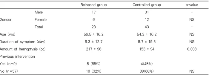
Table 2. Demographic features, duration, amount of hemoptysis and previous intervention history
Relapsed group (n=23) Controlled group (n=43) p-value
Active tuberculosis 5(25%) 15(75%)
NS Malignancies
Primary lung cancer 2(50%) 2(50%)
Metastais to the lung 2(67%) 1(33%)
Other benign diseases
Bronchiectasis 5(26%) 14(74%)
Aspergilloma 8(57%) 6(43%)
Others 1(17%) 5(83%)
* Other diseases include 1 bronchial artery aneurysm, 2 lung abscesses, and 2 bronchitis
NS = not significant
Table 3. Underlying diseases and previous history of bronchial artery embolzation intervention than 0.05.
To verify the compounding factors between the risk factors whose characters were diverse con- founding variables, we did multivariate analysis using logistic regression analysis with 95% confi- dence interval.
Results
Among 66 patients whose data were available, 23 (34.9%) patients had recurred massive hemoptysis during a mean follow up period of 20.4 months (ranging from 1 month to 54 months). Eight out of
23 recurred patients had pneumonectomy or lobec- tomy of lung (3 pneumonectomies and 5 lobecto- mies) in following event, and remaining 12 patients had to undergo another BAE. Four patients died due to uncontrolled hemoptysis (one died after pne- umonectomy).
Concerning the factors influencing relapse, age and sex did not play any role and duration of sym- ptom had not any significance, but the amount of hemoptysis had a statistical significance (p =0.008 by T-test; Table 2).
As to the underlying diseases, we had 20 active tuberculosis, 19 benign diseases including 19 bron-
Relapsed group (n = 23) Controlled group (n = 43) p-value Bilaterality of lesion
Yes 8(62%) 5 0.008
No 15(28%) 38
Pleural thickening
Present 12(75%) 4 0.001
Absent 11(22%) 39
Number of feeders
1 10(30%) 23(70%) NS
> 1 13(39%) 23(61%)
NS = not significant
Table 4. Radiologic findings and the recurred massive hemoptysis
Variables Odds ratio 95% C.I p-value
Gender Male
Female
1
0.59 0.53-4.37 NS
Age <50
≥50
1
0.42 0.34-2.42 NS
Previous intervention Yes No
1
0.51 0.28-2.76 NS
Underlying disease Active tuberculosis 1
Malignancy 1.76 0.77-11.84 NS
Other benign disease 0.62 0.80-4.86 NS
Duration of symptom ≤ 7days
> 7 days
1
1.14 0.16-1.73 NS
Amount of hemoptysis ≤ 200 cc
> 200 cc
1
10.21 10.08-50.23 0.008
Bilaterality of lesion No Yes
1
13.93 1.41-138.32 0.018
Number of feeding vessel 1
>1
1
2.20 0.36-13.39 NS
Pleural thickening No Yes
1
20.84 3.38-128.45 0.001
NS = not significant
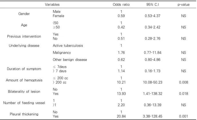
Table 5. Multivariate analysis of different variables
chiectasis, 14 aspergillomas, and 6 others (including 1 bronchial artery aneurysm, 2 lung abscesses, and 2 bronchitis), and 7 malignancies (4 primary lung cancer and 3 metastatic malignancies), we did not find any statistical significance between them (Ta- ble 3). Nine patients had a history of previous BAE
treatments for hemoptysis, but they showed no increased risk of relapse of major hemoptysis (Ta- ble 3). Bilateral lesion on radiographic finding and pleural thickening on HRCT had increased risk of recurred major hemoptysis (p = 0.018 and 0.001, re- spectively, by Chi-square test and Fisher’s exact
test; Table 4). Number of feeding vessels had not any statistical value concerning the relapse.
Univariate analysis showed that reliable risk fac- tors for recurrent hemoptysis after BAE were amo- unt of hemoptysis, bilaterality of lesion, and pleural thickening on HRCT. To verify the compounding factors between the risk factors whose characters were diverse confounding variables, we did multi- variate analysis using logistic regression analysis with 95% confidence interval. Multivariate analysis showed same results as univariate analysis (Table 5).
Discussion
We analyzed the result of BAE in our institution during a long period. In previous literatures, long- term recurrence rate have been reported to be 10 to 52%, with a mean follow up period ranging from one to 46 months1-11.
Remy et al.1 reported that of 49 patients treated for hemoptysis, an immediate arrest was achieved in 41, but 34 patients had experienced re-bleeding in follow up period beyond the 18 months. Ulfacker et al.3 reported that an immediate control of hemop- tysis was achieved in 33 out of 41 patients (80.5%) while hemoptysis recurred in 9 of 33 patients (27.3
%) in the long-term follow up (mean 24.8 months).
In our analysis, BAE effectively controlled 65.1% of life threatening massive hemoptysis (23 re-blee- dings in 66 cases) in a mean follow up period of 20.4 months.
Regarding the factor of recurrence after BAE, Osaki et al.4 concluded that bronchiectatic change on CT scan and pulmonary-bronchial shunt had some statistical significance. Kim et al.6 described the underlying lung disease and amount of bleeding as reliable risk factors for the recurrence, in a study involving 75 patients with a result estimating 54.5
% of re-bleeding rate after 3 years. But in the stu-
dy of Kim et al14 published 5 years earlier, those factors had no significant impact on the recurrence of hemoptysis. The former used Caplan-Mayer survival analysis with each variable and the latter used Chi-square univariate analysis. The diversity of previously proposed risk factors may be expla- ined by variability of their criteria on recurrence, sample size, underlying diseases, follow-up time, and statistic tool used.
In this study, the amount of hemoptysis had so- me statistical relation with the recurrent event.
Though the analysis of underlying disease had no statistical significance, active tuberculosis tended to have more control rate compared to aspergilloma and cancer. The effective anti-tuberculosis drug therapy must have reduced the recurrent hemopty- sis but its relatively modest prevalence in our series (30% compared to 43-52% in other domestic studies)5,6,12,14 lead to overall no statistical signifi- cance.
The fact that bilaterlity of lesion on initial chest X-ray was higher in relapse group can be explai- ned by the extent of the lung disease accounts for more serious pathology as it was commented by Kim et al.6
In 1993, Tamura et al.8 described pleural thicke- ning as a risk factor for recurrent bleeding after BAE. According to them, in the presence of pleural thickening, non-bronchial systemic feeder vessels that originate from various arteries (e.g., intercos- tals artery, branches of the subclavian and axillary arteries, internal mammary artery and inferior ph- renic artery) may develop along the pleural surface and become enlarged as a result of the inflamma- tory process. In our study 16 cases showed pleural thickening on chest radiography and 12 (75%) of them experienced recurrent massive bleeding, whi- ch was significantly higher than 22% of no pleural thickening group.
This study is a retrospective review of medical records, which often should underestimate strength of variable. But the presence of pleural thickening which had the highest odds ratio can be a reliable risk factor for the recurrence of hemoptysis after BAE, and this should be verified in a prospective study with larger number of patients because this study has the limitation caused by small size po- pulation involved with various underlying diseases.
Summary
Background :
Hemoptysis, when massive and untreated, has a mortality rate of over 50 percents, is considered as one of most dreaded of all respiratory emergencies and can have a variety of underlying causes.
Bronchial artery embolization (BAE) has become an established procedure in the management of ma- ssive and recurrent hemoptysis, and its efficacy is widely documented thereafter by number of arti- cles.
However, the long-term success rate of BAE is known to be unfavorable. Risk factors influencing that control failure are inevitably needed.
Materials and methods :
Seventy-five patients underwent bronchial artery embolization due to massive hemoptysis in Seve- rance Hospital from Jan. 2000 to Jan. 2005. Nine patients’ data were not available and could not be contacted with. Finally 66 patients’ (48 males, 18 females) medical records were analyzed retrospec- tively during a mean follow up period of 20.4 mon- ths (ranging from 1 month to 54 months).
Results :
Among 66 patients whose data were available, 23 (34.9%) patients had recurrent major hemoptysis.
Patients’ age, sex, underlying disease, previous in- tervention history, and number of feeding vessels
had no statistical validity as risk factors of recurred major hemoptysis. But bilaterality of lesion, amount of hemoptysis, and pleural thickening were revealed as meaningful factors for predicting relapse (p = 0.008, 0.018, and 0.001, respectively).
Conclusion :
According to our series, patients presenting with larger amount of hemoptysis, pleural thickening of chest radiography and bilateral lesion are associa- ted with increased risk of major hemoptysis in pa- tients treated with BAE.
References
1. Remy J, Arnaud A, Fardou H, Giroud R, Vousin C.
Treatment of hemoptysis by embolization of bronchial arteries. Radiology 1977;122:33-7.
2. Swanson KL, Johnson CM, Prakash UB, McKusick MA, Andrews JC, Stanson AW. Bronchial artery em- bolization: experience with 54 patients. Chest 2002;
121:789-95.
3. Ulfacker R, Kaemmerer A, Neves C, Picon P.
Management of massive hemoptysis by bronchial artery embolization. Radiology 1983;146:627-34.
4. Osaki S, Naknishi Y, Wataya H, Wataya H, Takaya- ma K, Inoue K, et al. Prognosis of bronchial artery embolization in the management of hemoptysis.
Respiration 2000;67:412-6.
5. Yeo DS, Lee SY, Hyun DS, Lee SH, Kim SC, Choi YM, et al. Effect of bronchial artery embolization in management of massive hemoptysis. Tuberc Respir Dis 1999;46:53-64.
6. Kim SO, Oh IJ, Kim KS, Yu YK, Lim SC, Kim YC, et al. Recurrent hemoptysis after bronchial artery embolization. Tuberc Respir Dis 2001;51:364-72.
7. Katoh O, Kishikawa T, Yamada H, Matsumpoto S, Kudo S. Recurrent bleeding after arterial emboliza- tion in patients with hemoptysis. Chest 1990;97:541-6.
8. Tamura S, Kodama T, Otsuka N, Kihara Y, Nisikawa K, Yuki Y, et al. Embolotherapy for persistent hemop- tysis: the significance of pleural thickening. Cardiova- sc Intervent Radiol 1993;16:85-8.
9. White R. Bronchial artery embolotherapy for control of acute hemoptysis: analysis and outcome. Chest 1999;115:912-5.
10. Mal H, Rullon I, Mellot F, Brugiere O, Sleiman C, Menu Y, et al. Immediate and long term results of
bronchial artery embolization for life threatening he- moptysis. Chest 1999;115:996-1001.
11. Lee TW, Wan S, Choy DK, Chan M, Arifi A, Yim AP.
Management of massive hemoptysis: a single institu- tion experience. Ann Thorac Cardiovas Surg 2000;6:
232-5.
12. Yoon W, Kim JK, Kim YH, Chung TW, Kang HK.
Bronchial and non bronchial systemic artery emboli- zation for life threatening hemoptysis: a comprehen- sive review. Radiographics 2002;22:1395-409.
13. Yu-Tang Goh P, Lim M, Teo N, En Shen Wong D.
Embolization for hemoptysis: a six year review. Cadi- ovasc Intervent Radiol 2002;25:17-25.
14. Kim BC, Kim JM, Kim YS, Kim SM, Choi WY, Lee KS, et al. Effect of bronchial artery embolization in the management of massive hemoptysis: factors in- fluencing rebleeding. Tuberc Respir Dis 1996;43:
590-9.
15. Ko DS, Kwon SY, Lee CT, Han SK, Shim YS, Lee JH.
Effectiveness of bronchial artery embolization in he- moptysis patients and risk factors of recurrence. Tu- berc Respir Dis 2004;57(Suppl):128.