• 검색 결과가 없습니다.

A Case of Catamenial Hemoptysis Treated by Bronchial Artery Embolization

N/A
N/A
Protected

Academic year: 2021

Share "A Case of Catamenial Hemoptysis Treated by Bronchial Artery Embolization"

Copied!
4
0
0

로드 중.... (전체 텍스트 보기)

전체 글

(1)

A Case of Catamenial Hemoptysis Treated by Bronchial Artery Embolization

Suk Pyo Shin, M.D.

1

, Chi Young Park, M.D.

1

, Ji Hyun Song, M.D.

1

, Hong Min Kim, M.D.

1

, Daniel Min, M.D.

1

, Sang Hwan Lee, M.D.

1

, San Ha Kang, M.D.

1

, Gyeong Sik Jeon, M.D.

2

and Ji-Hyun Lee, M.D.

1

Departments of

1

Internal Medicine and

2

Radiology, CHA Bundang Medical Center, CHA University, Seongnam, Korea

Catamenial hemoptysis is a rare condition, characterized by recurrent hemoptysis associated with the presence of intrapulmonary or endobronchial endometrial tissue. Therapeutic strategies proposed for intrapulmonary endometriosis with catamenial hemoptysis consist of medical treatments and surgery. Bronchial artery embolization is a well-established modality in the management of massive or recurrent hemoptysis, but has seldom been used for the treatment of catamenial hemoptysis. We report a case of catamenial hemoptysis associated with pulmonary parenchymal endometriosis, which was successfully treated by a bronchial artery embolization.

Keywords: Endometriosis; Hemoptysis; Embolization, Therapeutic

nary parenchyma or in the airway

3

.

Various treatment modalities such as hormonal therapy, surgery or medical conservative treatment have been at- tempted, but controversies exist about optimal management of catamenial hemoptysis. Bronchial artery embolization (BAE) is a well established minimally invasive treatment modality for hemoptysis and few have been reported for the management of catamenial hemoptysis. Here, we describe a case of catamenial hemoptysis caused by pulmonary paren- chymal endometriosis successfully treated with BAE.

Case Report

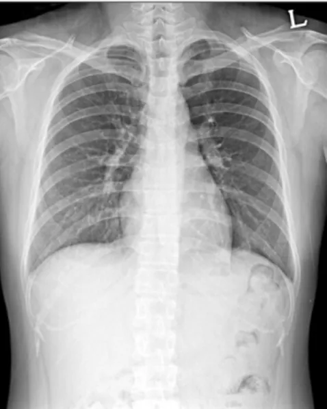
A 34-year-old married woman was admitted to pulmonary department with a 4-day history of hemoptysis. Hemoptysis occurred from the 3rd day of menstruation till 6th day and the total amount of hemoptysis was about 150 mL. She had no history of previous hemoptysis event. She had medical history of an appendectomy 20 years ago and pelvic inflammatory disease 2 years ago. She gave birth by normal spontaneous vaginal delivery (gravida 1, para 1) 10 years ago and she had not had a past history of obstetric or gynecological procedures before developing hemoptysis. Her medical history was oth- erwise unremarkable and she did not have a significant family history. She denied using smoking, excessive alcohol and illicit Copyright © 2014

The Korean Academy of Tuberculosis and Respiratory Diseases.

All rights reserved.

Introduction

Thoracic endometriosis is a rare disorder characterized by a presence of functional endometrial tissue within the pleura, the lung parenchyma or the airway

1

. The tissue is responsive to circulating sex hormones and clinical manifestations are re- lated to the menstrual cycle. Clinically, thoracic endometriosis includes four well-recognized entities, namely, catamenial pneumothorax, catamenial hemothorax, catamenial hemop- tysis, and lung nodules

2

. In catamenial hemoptysis, the source of bleeding is an endometrial implant located in the pulmo-

CASE REPORT

http://dx.doi.org/10.4046/trd.2014.76.5.233

ISSN: 1738-3536(Print)/2005-6184(Online) • Tuberc Respir Dis 2014;76:233-236

233

Address for correspondence: Ji-Hyun Lee, M.D.

Division of Respiratory and Critical Care Medicine, Department of Internal Medicine, CHA Bundang Medical Center, CHA University, 59 Yatap-ro, Bundang-gu, Seongnam 463-712, Korea

Phone: 82-31-780-6140, Fax: 82-31-780-6143 E-mail: [email protected]

Received: Oct. 14, 2013 Revised: Nov. 5, 2013 Accepted: Nov. 21, 2013

cc

It is identical to the Creative Commons Attribution Non-Commercial

License (http://creativecommons.org/licenses/by-nc/3.0/).

(2)

SP Shin et al.

234 Tuberc Respir Dis 2014;76:233-236 www.e-trd.org

drugs. She had no complaints of weight loss, fever, dyspnea, palpitations, gastrointestinal symptoms or a history of bleed- ing. Her physical examination was within normal limits. Chest X-ray (Figure 1) had no abnormal findings. Chest computed tomography (CT) scan was performed on the 2nd hospital day, which was fourth day of menstruation and the CT scan showed a focal consolidation with adjacent ground glass opacity (GGO) in the posterior basal segment of the left lower lobe (Figure 2A), but there’s no evidence of enlarged bronchial artery or vascular abnormality. Fiberoptic bronchoscopy showed a small amount of blood clot in the bronchi of left lower lobe. No endobronchial lesion was detected during the investigation. Bronchial washing fluid showed no acid fast bacilli, bacteria or abnormal cells. Hemoptysis was spontane- ously resolved with the cessation of menstruation and did not

recur during the rest of admission period. We assumed the illness as catamenial hemoptysis due to pulmonary endome- triosis, and she was discharged. After discharge from the first admission, follow-up chest CT scan was performed for follow- ing up the lung lesion at 26th day of menstrual cycle. Previ- ously noted focal consolidation with adjacent GGO lesion was almost disappeared and only a noncalcified 5-mm-sized nod- ule was left (Figure 2B). Four days passed and at the first day of the next menstrual cycle, hemoptysis recurred. The amount of hemoptysis ranged 100 to 150 mL and she was admitted again. Chest CT scan was performed and reappeared larger nodule with focal GGO was seen at the same location (Figure 2C). To control the bleeding, we decided to perform bronchial arteriography embolization. Digital subtraction technique with a digital subtraction angiography unit was used. Femoral artery was punctured, then thoracic aortography and followed selective left bronchial arteriography were done with a 5-Fr catheter. Diagnostic angiography showed a small nodular staining on left lower lung zone (Figure 3A) that corresponds with the lesion of previous chest CT scan. After determination of the pathologic vascularity, embolization was performed with 355 to 500 μm Contour polyvinyl alcohol particles. After embolization, control angiography showed occlusion of the artery feeding the lesion (Figure 3B). There was no more he- moptysis after the embolization. After discharge, she has been followed up for 5 months without hemoptysis. We diagnosed this case as catamenial hemoptysis because the hemoptysis events and size change of the lesion on chest CT scans were synchronized with the menstruation cycle, and both the CT scan and the bronchoscopy excluded other possible causes of hemoptysis.

Discussion

Catamenial hemoptysis is a cyclic pulmonary hemorrhage Figure 1. Initial posteroanterior chest radiograph shows no specific

abnormality on the lung field.

Figure 2. (A) Chest computed tomography (CT) scan performed on 4th day of menstruation shows focal consolidation with surrounding ground

glass opacity in the posterior basal segment of the left lower lobe. (B) In the CT scan on the 26th day of menstruation, previously noted focal con-

solidation with surrounding ground glass opacity in the left lower lobe has been resolved. Residual small noncalcified nodule is noted. (C) In the

CT scan on the 1st day of the next menstruation, focal ground glass opacity and nodule is noted at the same location as previous one.

(3)

Bronchial artery embolization in catamenial hemoptysis

http://dx.doi.org/10.4046/trd.2014.76.5.233 235

www.e-trd.org

that is synchronized with female menstruation, which is asso- ciated with the presence of intrapulmonary or endobronchial ectopic endometrial tissue. The mechanism regarding tho- racic endometriosis is not fully understood and three theories have been proposed to explain the presence of intrathoracic endometrial implants: coelomic metaplasia, retrograde men- struation with subsequent transperitoneal-transdiaphragmat- ic migration of endometrial tissue and lymphatic or hematog- enous embolization from the uterus or pelvis

4

. None of these theories can explain all the clinical manifestations, and the disease probably has a multifocal etiology. The theory of coe- lomic metaplasia is based on the concept that both endome- trium and pleural mesothelium share the same embryologic origin. Pathologic stimuli induce precursor cells of the pleura into endometrial cells. The second theory is based on the con- cept that movement of fluids in the peritoneal cavity follows predictable patterns: namely “peritoneal fluid circulation.” It implies a preferable flow direction from the pelvis to the right subdiaphragmatic area through the right paracolic gutter

5

. During the process of the flow, endometrial tissue could enter into the thorax through either congenital or acquired dia- phragmatic defects

5,6

. However, these theories cannot explain the occurrence of intrapulmonary endometriosis. The last theory of transplantation of endometrium through lymphatic or vascular embolization can explain intrapulmonary endo- metriosis. Trauma or manipulation of uterine tissue would be a factor predisposing to microembolization. For example, the nationwide Korean report of 19 patients with catamenial hemoptysis showed that 16 (84%) patients had a history of obstetric or gynecological procedures before development of hemoptysis

7

. Also, in a study that followed 4 patients with catamenial hemoptysis, all patients had history of one or two dilatations and curettages before diagnosis of catamenial

hemoptysis

8

. However, this patient had not had a past history of obstetric or gynecological procedures before developing hemoptysis.

The diagnosis of pulmonary parenchymal endometriosis is usually assumed on the basis of the clinical history and the exclusion of other causes of recurrent hemoptysis. Chest roentgenogram may reveal pulmonary opacities or nodular infiltrates, but findings could be normal even in a patient with current bleeding

4

. The diagnostic use of bronchoscopy is lim- ited, because most cases of pulmonary endometriosis involve the distal pulmonary parenchyma rather than the mucosa of large bronchi and also the bleeding site may only be apparent during menstruation. Chest CT scan is useful for detection of the lesion and exclusion of other causes for hemoptysis. CT signs of pulmonary endometriosis include ill-defined or well- defined nodules, thin-walled cavities, bullous formations and ground glass opacities

2

. These lesions, which are expressions of the endometrial implants and/or secondary hemorrhage, may change in size during the menstrual cycle

4,8

. The lesion on CT scans in this patient also showed the characteristic change according to her menstrual cycle.

There is no guideline for the treatment of catamenial he- moptysis. Hormonal therapy has been considered as the first choice in patients with thoracic endometriosis. It includes oral contraceptives, progestational drugs, danazol, and gonado- tropin-releasing hormone agonists which suppress the endo- metrial tissue. It has been proved to be effective in controlling symptoms, but controlled trials on the efficacy of these drugs are lacking. Also, heavy side effects of the hormonal therapy have been observed and symptoms often recur after discon- tinuation

7

. Moreover patients who consider pregnancy cannot take these drugs.

Medical conservative management could be another op-

Figure 3. (A) Left bronchial angiography

shows a small nodular staining in left

lower lung field (arrow). (B) Left bron-

chial angiography after embolization

shows no distal flow and disappearance

of nodular staining.

(4)

SP Shin et al.

236 Tuberc Respir Dis 2014;76:233-236 www.e-trd.org

tion, because most of the hemoptysis events associated with pulmonary emdometriosis are not lethal and most patients from case series are almost women of childbearing ages. In a study reported in Korea, 4 patients taken only conservative management had been followed approximately for five years and hemoptysis spontaneously disappeared after several epi- sodes of minor bleeding

8

.

Surgical treatment has been advocated if medical treatment fails, intolerable drug-related side effects occur, or symptoms recur after the cessation of hormonal therapy

3

. Wedge resec- tion or lobectomy can be applied to these cases according to the extent and location of the lesion. Video-assisted thoraco- scopic surgery, endoscopic laser treatment

9

or open surgery can be done

7

.

In our case we had observed the patient after the first epi- sode, but the hemoptysis recurred in the next menstruation cycle. Since the amount of bleeding was more than that of the previous episode, we decided to try BAE prior to surgery.

In general, BAE is a well-known alternative to surgery in the management of hemoptysis. Clinically, BAE has been widely applied for the treatment of hemoptysis caused by bron- chiectasis, tuberculosis, aspergillosis, lung cancer or chest trauma. Possible rare complications of BAE are spinal cord injury, esophageal ulceration, stroke, bronchial infarction and transient chest pain. BAE may be more lifesaving and provide better long term control of recurrent bleeding and give better quality of life than medical conservative management alone does in massive or even if not massive but socially or physi- cally recurrent troublesome hemoptysis

10,11

.

Blood supply of lung parenchymal endometriosis has not been well described. In some case reports or case series, pathologic findings of the removed pulmonary endometrial tissues revealed the presence of expanded bronchovascular bundles

12

or thin-walled large capillaries or bronchial arter- ies

13

.

Despite these findings, BAE has not been frequently used to control catamenial hemoptysis. We found only one report from Kervancioglu et al.

14

, in which hemoptysis with multiple pulmonary endometriosis was successfully treated by BAE without recurrence for 3 months of follow-up. On the other hand, Katoh et al.

15

found no abnormalities on bronchial and pulmonary angiograms in their clinically suspected pulmo- nary endometriosis patients. In this case, we found a small nodular staining in bronchial angiogram matched to the lesion on the chest CT scan. After BAE, the patient has not shown hemoptysis during the follow up period of 5 months with regular menses. In conclusion, we reported a patient of catamenial hemoptysis treated with BAE. This case suggests the possibility that BAE might be an alternative therapeutic strategy for the patient with catamenial hemoptysis of intra- pulmonary endometriosis.

Conflicts of Interest

No potential conflict of interest relevant to this article was reported.

References

1. Honore GM. Extrapelvic endometriosis. Clin Obstet Gynecol 1999;42:699-711.

2. Volkart JR. CT findings in pulmonary endometriosis. J Com- put Assist Tomogr 1995;19:156-7.

3. Terada Y, Chen F, Shoji T, Itoh H, Wada H, Hitomi S. A case of endobronchial endometriosis treated by subsegmentectomy.

Chest 1999;115:1475-8.

4. Alifano M, Trisolini R, Cancellieri A, Regnard JF. Tho- racic endometriosis: current knowledge. Ann Thorac Surg 2006;81:761-9.

5. Kirschner PA. Porous diaphragm syndromes. Chest Surg Clin N Am 1998;8:449-72.

6. Korom S, Canyurt H, Missbach A, Schneiter D, Kurrer MO, Haller U, et al. Catamenial pneumothorax revisited: clinical approach and systematic review of the literature. J Thorac Cardiovasc Surg 2004;128:502-8.

7. Kim CJ, Nam HS, Lee CY, Yum HK, Yang SH, Seo KH, et al.

Catamenial hemoptysis: a nationwide analysis in Korea. Res- piration 2010;79:296-301.

8. Ryu JS, Song ES, Lee KH, Cho JH, Kwak SM, Lee HL. Natural history and therapeutic implications of patients with catame- nial hemoptysis. Respir Med 2007;101:1032-6.

9. Puma F, Carloni A, Casucci G, Puligheddu C, Urbani M, Por- caro G. Successful endoscopic Nd-YAG laser treatment of endobronchial endometriosis. Chest 2003;124:1168-70.

10. Mal H, Rullon I, Mellot F, Brugiere O, Sleiman C, Menu Y, et al.

Immediate and long-term results of bronchial artery emboliza- tion for life-threatening hemoptysis. Chest 1999;115:996-1001.

11. Kim YG, Yoon HK, Ko GY, Lim CM, Kim WD, Koh Y. Long- term effect of bronchial artery embolization in Korean pa- tients with haemoptysis. Respirology 2006;11:776-81.

12. Flieder DB, Moran CA, Travis WD, Koss MN, Mark EJ. Pleuro- pulmonary endometriosis and pulmonary ectopic decidu- osis: a clinicopathologic and immunohistochemical study of 10 cases with emphasis on diagnostic pitfalls. Hum Pathol 1998;29:1495-503.

13. Jelihovsky T, Grant AF. Endometriosis of the lung. Thorax 1968;23:434-7.

14. Kervancioglu S, Andic C, Bayram N, Telli C, Sarica A, Sirikci A.

Bronchial artery embolization in the management of pulmo- nary parenchymal endometriosis with hemoptysis. Cardio- vasc Intervent Radiol 2008;31:824-7.

15. Katoh O, Yamada H, Aoki Y, Matsumoto S, Kudo S. Utility of

angiograms in patients with catamenial hemoptysis. Chest

1990;98:1296-7.

수치

Figure 2. (A) Chest computed tomography (CT) scan performed on 4th day of menstruation shows focal consolidation with surrounding ground  glass opacity in the posterior basal segment of the left lower lobe

참조

관련 문서

Time series of vertical cross section of potential vorticity and wind vector calculated by Case 1 along the A-A' line indicated at Fig.. Same

3) A comparison of the stoichiometric equation with the experimental kinetic expression can suggest whether or not we are dealing with an elementary reaction. 4) If one

Указание ложной информации может привести к аннулированию визы и разрешения на пребывание, а также может повлечь за собой уголовное

벨라루스에서는 특히 간장의 경우 상당 소비량이 소위 초밥과 같은 싸지 않은 식품들과 함께 소비가 되는 편인데, 이에 사람들의 낮아진 가처 분 소득도 간장의 판매량

자료원 : 벨라루스 통계청, 벨라루스 관세청, https://connect.ihs.com(Global Trade Atlas), www.belta.ru, www.news.tut.by,

참고로 대한민국은 2014년 9월에 Hi-Tech Park 내에 ‘한-벨 정보접근센터’를 개소하여, 센터 내 한국문화체험라운지, 인터넷 라운지, ICT랩, 세미나룸을 갖추고

Hairdressing Appliances And Hand Dryers, Space-Heating And Soil-Heating Apparatus, Water Heaters, Immersion Heaters, Smoothing Irons, Microwave Ovens, Ovens,

- 실제로 KOTRA 민스크 무역관에서 온라인 주문을 시도해 본 결과 평소에는 익일 배송예약시간의 경우 예약을 하는데 큰 어려움이 없었으나 현재 배송예약을