http://dx.doi.org/10.5468/ogs.2016.59.4.286 pISSN 2287-8572 · eISSN 2287-8580
Introduction
Endometriosis is a common gynecological disorder caused by ectopic implantation of endometrial glandular and stromal cells outside the uterine cavity [1-3]. It is classified according to three pathological categories; peritoneal lesions, deep infil- trative lesions, and ovarian endometriotic lesions [4]. Among these, ovarian endometrioma is the only subtype which can be suspected preoperatively using pelvic ultrasonography.
European Society of Human Reproduction and Embryology
Preoperative risk factors in recurrent endometrioma after primary conservative surgery
Seung Joo Chon, Seung Hyeong Lee, Joo Hyun Choi, Ji Sung Lee
Department of Obstetrics and Gynecology, Gil Hospital, Gachon University College of Medicine, Incheon, Korea
Objective
Endometriosis is a common gynecological disorder caused by ectopic implantation of endometrial glandular and stromal cells outside the uterine cavity. Among several types of endometriosis, endometrioma is the only subtype that could be determined preoperatively using pelvic ultrasonography, and guidelines recommend pathologic confirmation of endometrioma greater than 3 cm in diameter. However, although surgery is performed in cases of endometrioma, endometrioma has a high cumulative rate of recurrence. Therefore, because determining the possibility of recurrence before performance of initial surgery is important, we examined preoperative factors associated with recurrent endometrioma.
Methods
This was a retrospective, comparative study including 236 patients who visited the outpatient clinic between January 2009 and December 2011. Patients who were pathologically diagnosed with endometrioma were included in this study. They were followed up postoperatively and were divided into two groups according to presence of recurrent endometrioma.
Results
We examined associations between baseline factors and recurrent endometrioma. In multivariate analysis, dysmenorrhea and cyst septation were statistically significant after adjusting with age, parity, surgical staging and postoperative management. We examined cumulative recurrence free survival within cases of recurrent endometriosis, based on the presence of inner cyst septation. The cumulative recurrence free survival was lower in cases with septation.
Conclusion
Our study found that recurrent endometrioma is more likely in patients with inner cyst septation and the recurrence occurred within a shorter duration of time than in patients without inner cyst septation on preoperative ultrasonography. Therefore intensive caution and postoperative long term medical therapy would be appropriate in patients with inner cyst septation on preoperative ultrasonography before undergoing primary surgery for endometrioma.
Keywords: Endometriosis; Preoperative; Recurrence; Septation; Ultrasonography
Articles published in Obstet Gynecol Sci are open-access, distributed under the terms of the Creative Commons Attribution Non-Commercial License (http://creativecommons.
org/licenses/by-nc/3.0/) which permits unrestricted non-commercial use, distribution, and reproduction in any medium, provided the original work is properly cited.
Copyright © 2016 Korean Society of Obstetrics and Gynecology
Received: 2015.10.22. Revised: 2015.12.21. Accepted: 2015.12.23.
Corresponding author: Ji Sung Lee
Department of Obstetrics and Gynecology, Gachon University College of Medicine, 21 Namdong-daero 774beon-gil, Namdong-gu, Incheon 21565, Korea
Tel: +82-32-460-3254 Fax: +82-32-460-3290
E-mail: [email protected]
http://orcid.org/0000-0002-2076-7634
guidelines recommend pathologic confirmation of endome- trioma greater than 3 cm in diameter to exclude malignan- cies [3].
Although pathological and final diagnosis could only be made after surgery, suspected diagnosis could be made based on clinical symptoms along with ultrasonographic findings, based on typical sonographic findings and improvements in ultrasonographic technologies. Endometrioma can be easily detected, and the recurrence rate is also considerably high.
The cumulative rate of endometrioma recurrence is 12%
to 30% after 2 to 5 years of postoperative follow up [5-7].
Therefore, it is important to determine the possibility of recur- rence before performance of the initial surgery.
Many studies have reported on risk factors associated with recurrence of endometrioma after laparoscopic excisions. As- sociations of age at surgery [8,9], the revised American Society for Reproductive Medicine (r-ASRM) score [5,8-10], previous medical treatment [6,9], size of the largest cyst [6], previous operative history due to endometriosis [5], and the r-ASRM adnexal adhesion scores, particularly ovarian adhesion score [11] with high recurrence of the disease have been reported.
However, few studies on prognostic factors associated with recurrence of endometrioma on preoperative detailed findings have been reported.
Factors related to operative field and postoperative manage- ment have been reported. However discovery of more pre- operative factors would be helpful in counseling patients on their future prognosis. Therefore, in this retrospective study, we examined preoperatvie factors associated with recurrent endometrioma.
Materials and methods
1. Study population
This was a retrospective, comparative study, including 236 pa- tients who visited the outpatient clinic in Gil Hospital, Gachon Medical School between January 2009 and December 2011.
Patients initially suspected of endometrioma using ultraso- nography and pathologically diagnosed with endometrioma were included. After the operation, patients were followed up serially and divided into two groups according to presence of recurrent endometrioma (group 1, n=37) or not (group 2, n=199). Patients suspected of endometrioma on peoperative ultrasonography but not diagnosed with pathologically, and
who only had endometriotic spots in the pelvic cavity without endometrioma were excluded. Patients who previously under- went surgery due to endometrioma in other hospitals were also excluded because initial information regarding before and during the primary operation was incomplete.
The study was conducted in accordance with the ethical standards of the Helsinki Declaration, and was approved by the Gachon Medical School, Gil Hospital, institutional review board (GCIRB 2015-293).
2. Variable measurements and surgery
Data on age, height and weight were collected at the time of the primary operation due to endometrioma. Body mass index was calculated by weight (kg) divided by square meters of height (m
2). Serum blood test was performed for measure- ment of CA 125, and ultrasonography was done to examine uterus and both adnexa. Two experienced sonographers per- formed sonography of all the enrolled patients using Accuvix- XQ (Samsung Medison, Seoul, Korea), and Voluson E8 (GE Healthcare Korea, Seoul, Korea). Diameter and symmetricity of anterior and posterior uterine walls were measured and the shape was described. A patient was suspected to have adeno- myosis when uterine enlargement with multiple, fine, linear areas of attenuation throughout the lesion and poor definition of the endo-myometrial junction were noted. The maximum diameter of endometrioma was measured in centimeters by measuring the largest diameter of the ovarian cyst with dif- fuse, homogeneous, low-level internal echogenecity. Not only size, but also laterality and features of endometrioma were examined, and described according to the presence of septa- tion and nodularity.
Laparoscopic excision of ovarian endometrioma was per-
formed as follows. Upon entering the pelvic cavity, uterus and
both adnexa were examined. If pelvic adhesion was observed,
dissection and adhesiolysis were performed initially using a
sharp monopolar. The endometriotic ovary was then freed
from adjacent adhesion bands and a sharp incision was made
superficially on the ovarian cortex so that the cyst wall could
be identified. Once the ovarian cyst wall was exposed, the
wall was held with tooth forceps and contralateral force was
applied in order to strip off the cystic lesion only. To prevent
massive active bleeding during ovarian cystectomy, bipolar for-
ceps were used to cause focal coagulation of actively bleeding
lesions. Because the incision made on the ovarian cortex was
not large, the ovary was not sutured, but it was put together
with hemostatic glue (Greenplast kit, Green Cross, Yongin, Korea). Fulguration was performed if endometriotic spots were found in the pelvic cavity.
Operation type was divided according to six categories; right ovarian cystectomy, left ovarian cystectomy, bilateral ovarian cystectomies, right salpingo-oophorectomy, left salpingo-oo- phorectomy, and unilateral ovarian cystectomy with unilateral salpingo-oophorectomy. Because this study was conducted in order to monitor occurrence of recurrent endometrioma, cases of bilateral salpingo-oophorectomy were excluded. The opera- tive field was examined and data on pelvic adhesion, posterior cul de sac obliteration, laterality of involved ovary, and r-ASRM stage were reviewed. After the operation, serum CA 125 was
measured once more 3 months later, and options for adjuvant treatment were classified according to three categories; none, gonadotropin-releasing hormone agonist only, and gonado- tropin-releasing hormone agonist with hormone therapy.
Data on patients with recurrent endometrioma were also collected. Associated symptoms, duration of disease free survival, serum CA 125, laterality and maximum diameter of recurrent endometrioma, options and duration of treatment of recurred cases were reviewed retrospectively.
3. Criteria and definitions
Endometrioma was suspected if a homogeneous hypoecho- genic ovarian cyst with a thick wall was visible on ultrasonog-
Table 1. Baseline characteristics of patients according to presence of recurrent endometrioma before primary operation
Baseline characteristics Non-recurrent (n=199) Recurrent (n=37) P-value
Age at 1st operation (yr) 31.470±7.682 30.810±7.359 0.583
BMI at 1st operation (m/kg
2) 21.017±3.563 22.181±37.177 0.017
Parity 0.758
0 134 (67.3) 26 (70.3)
1 30 (15.1) 5 (13.5)
2 32 (16.1) 5 (13.5)
3 3 (1.5) 1 (2.7)
Symptoms
None 31 (15.6) 3 (8.1) 0.236
Dysmenorrhea 138 (69.3) 32 (86.5) 0.033
Dyspareunia 4 (2.0) 1 (2.7) 0.789
Irregular bleeding 38 (19.1) 6 (16.2) 0.680
Lower abdominal discomfort 61 (30.7) 14 (37.8) 0.390
Palpable mass 4 (2.0) 1 (2.7) 0.789
Preoperative CA 125 (U/mL) 67.643±85.917 68.138±75.675 0.348
Ultrasonography
AP diameter of uterus (cm) 5.331±1.043 5.468±1.116 0.417
Presence of globular uterus 37 (18.6) 7 (18.9) 0.963
Maximum diameter of EMS (cm) 5.693±2.569 6.611±2.830 0.041
Laterality of EMS 0.042
Bilateral 54 (27.1) 13 (35.1)
Unilateral 145 (72.9) 24 (64.9)
Right only 56 (28.1) 13 (35.1)
Left only 89 (44.8) 11 (29.8)
Cyst septation 82 (41.2) 22 (62.9) 0.018
Cyst nodularity 119 (59.8) 27 (77.1) 0.051
Follow-up duration (mo) 25.090±15.653 60.270±32.778 <0.001
BMI, body mass index; AP, anteroposterior; EMS, endometrioma.
raphy. Variations including hyperechogenic intracystic nodules or septations were also examined and recorded. Recurrent endometrioma was defined as an ovarian endometrioma equal to or larger than 3 cm detected on ultrasonography.
The last follow up date was considered the last date of visit to the outpatient clinic of the obstetrics and gynecologic depart- ment in our hospital, and follow-up duration was calculated in months, from the day of the first visit to the day of the last follow-up.
4. Statistical analysis
Continuous data were expressed as mean±standard deviation, and categorical data were expressed as numbers of cases (per- centages). Because the number of enrolled patients was small, the Mann-Whitney U-test was performed in univariate analy- sis to compare differences between non-recurrent group and recurrent group. One-sample t-test was used for analysis of data only on recurrent endometrioma. Univariate analysis was performed to determine possible risk factors for recurrence and factors with P-value <0.05 were included in multivariate
Table 2. Intraoperative findings in primary operation according to presence of recurrent endometrioma
Intraoperative findings Non-recurrent (n=199) Recurrent (n=37) P-value
Operation name 0.918
ROC 44 (22.1) 11 (29.7)
LOC 69 (4.7) 9 (24.3)
BOC 53 (26.6) 13 (35.1)
RSO 6 (3.0) 1 (2.7)
LSO 19 (9.5) 1 (2.7)
UOC+USO 8 (4.0) 2 (5.4)
Intraoperative findings
Pelvic adhesion 153 (76.9) 34 (91.9) 0.039
PCDS obliteration 0.004
None 47 (23.6) 1 (2.9)
Partial 87 (43.7) 16 (45.7)
Complete 65 (32.7) 18 (51.4)
Laterality of involved ovary 0.169
Bilateral 53 (26.6) 15 (42.9)
Unilateral 146 (73.3) 20 (57.1)
Right only 54 (27.1) 11 (31.4)
Left only 92 (46.2) 9 (25.7)
r-ASRM stage 0.074
I 1 (0.5) 0 (0.0)
II 12 (6.0) 1 (2.7)
III 81 (40.7) 11 (29.7)
IV 105 (52.8) 25 (67.6)
Postoperative CA 125 (U/mL) 19.429±24.601 14.922±9.371 0.767
Adjuvant treatment 0.099
None 45 (22.6) 6 (16.2)
GnRHa only 133 (66.8) 23 (62.2)
GnRHa+HT 21 (10.6) 8 (21.6)
ROC, right ovarian cystectomy; LOC, left ovarian cystectomy; BOC, bilateral ovarian cystectomy; RSO, right salpingo-oophorectomy; LSO, left
salpingo-oophorectomy; UOC, unilateral ovarian cystectomy; USO, unilateral salpingo-oophorectomy; PCDS, posterior cul de sac; r-ASRM,
revised-American Society of Reproductive Medicine; GnRHa, gonadotropin releasing hormone agonist; HT, hormone therapy.
analysis using a step-wise variable selection method. In multi- variate analysis, logistic regression analysis was performed for identification of significant variables which contribute to recur- rent endometrioma. Kaplan-Meier analysis was performed to determine cumulative recurrence free survival depending on presence of septation or nodularity only among patients with recurrent endometrioma, and the log-rank test was performed for comparison of significant differences between the curves obtained in each graph. PASW ver. 18.0 (SPSS Inc., Chicago, IL, USA) was used for data analysis, and a P-value less than 0.05 was considered statistically significant.
Results
Preoperative baseline characteristics of patients with and with- out recurrent endometrioma are listed and analyzed (Table 1).
Statistically significant difference in body mass index, presence of dysmenorrhea, maximum diameter, bilaterality and side of endometrioma, presence of cyst septation, and follow up duration was observed between the two groups. Patients with
recurrent endometrioma were more obese (body mass index, 21.017±3.563 vs. 22.181±37.177 m/kg
2; P=0.017), and had dysmenorrhea (69.3% vs. 86.5%, P=0.033). They also had greater maximum diameter of endometrioma (5.693±2.569 vs. 6.611±2.830 cm, P=0.041), higher proportions of bilater- ality (27.1% vs. 42.9%, P=0.042) and cyst septation (41.2%
vs. 62.9%, P=0.018) as well as longer follow-up period (25.090±15.653 vs. 60.270±32.778 months, P<0.001) than those without recurrent endometrioma.
Intraoperative findings and postoperative management were analyzed according to presence of recurrent endometrioma (Table 2). In sub-classification analysis of operation type, later- ality of involved ovary, r-ASRM stage, and adjuvant treatment, no differences were observed between the two groups. How- ever, regarding pelvic adhesion along with posterior cul de sac obliteration, patients with recurrent endometrioma showed higher prevalence than those without recurrent endometrio- ma (pelvic adhesion, 76.9% vs. 91.9%, P=0.039; posterior cul de sac obliteration, 76.4% vs. 97.1%, P=0.004).
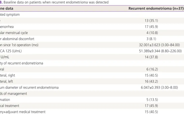
In evaluation of baseline data on patients with recurrent endometrioma, 35.1% of cases with recurrent endometrioma
Table 3. Baseline data on patients when recurrent endometrioma was detected
Baseline data Recurrent endometrioma (n=37)
Associated symptom
None 13 (35.1)
Dysmenorrhea 17 (45.9)
Irregular menstrual cycle 4 (10.8)
Lower abdominal discomfort 3 (8.1)
Duration since 1st operation (mo) 32.001±3.623 (3.00–84.00)
Serum CA 125 (U/mL) 51.389±9.344 (8.80–226.00)
≥35 U/mL 14 (37.8)
Laterality of recurrent endometrioma
Bilateral 6 (16.2)
Unilateral, right 15 (40.5)
Unilateral, left 16 (43.2)
Maximum diameter of recurrent endometrioma 6.047±0.393 (3.00–8.00)
Methods of management
Observation 5 (13.5)
Medical treatment 17 (45.9)
Surgery+adjuvant medical treatment 15 (40.5)
Duration of medical treatment
Medical treatment (mo) 20.647±5.479 (3.00–72.00)
Adjuvant medical treatment (mo) 11.333±2.890 (1.00–37.00)
showed no associated clinical symptoms, while the most common symptom was dysmenorrhea (45.9%) (Table 3).
The mean duration since the first operation to the point of recurrence was 32.001±3.623 (range, 3 to 84) months and the mean value of serum CA 125 was 51.389±9.344 (range, 8.80 to 226.0) U/mL, including values ≥35 U/mL in 37.8% of recurred cases. 83.7% of the cases had unilaterally recurred endometrioma, and the maximum diameter of recurred endo- metrioma was 6.047±0.393 (range, 3.00 to 8.00) cm in size.
When recurrent endometrioma was detected, 45.9% of cases underwent medical treatment, 40.5% underwent both surgi- cal and medical treatment, whereas the rest (13.5%) under- went no treatment at all.
Associations between baseline factors and recurrent endo- metrioma were examined (Table 4). Multivariate analysis was performed using dysmenorrhea, bilaterality of endometrioma, and cyst septation, which were statistically significant in uni- variate analysis. Among these three factors, dysmenorrhea (odds ratio, 3.436; 95% confidence interval, 1.150 to 10.290;
P=0.026), and cyst septation (odds ratio, 2.293; 95% con- fidence interval, 1.066 to 4.937; P=0.033) were statistically
significant after adjusting with age, parity, surgical staging, postoperative management, and follow-up duration.
Cyst septation and nodularity were the two ultrasonographi- cally detailed descriptive factors in recurrent ovarian endome- trioma in this study. Cumulative recurrence free survivals with- in recurrent endometriotic cases were examined based on the presence of these two factors each (Figs. 1, 2). The cumulative recurrence free survival was lower in cases with septation (log rank, P=0.003), but it was similar in cases with and without nodularity (log rank, P=0.726).
Discussion
Our study concluded that the presence of dysmenorrhea and cyst septation on preoperative ultrasonography may be as- sociated with recurrent endometrioma. Among patients with recurrent endometrioma, cumulative recurrence free survivals were significantly different between groups with and without septation. Patients with inner cyst septation showed shorter cumulative recurrence free survival than those in the other Table 4. Risks of recurrent EMS according to various factors
R isk factors of recurrent EMS Univariate analysis Multivariate analysis Exp(B) (95% CI) P-value Exp(B) (95% CI) P-value Associated symptom
Dysmenorrhea 2.830 (1.055–7.615) 0.038 3.436 (1.150–10.290) 0.026
Dyspareunia 1.356 (0.150–12.470) 0.789 - -
Irregular bleeding 0.827 (0.323–2.111) 0.680 - -
Lower abdominal discomfort 1.387 (0.671–2.861) 0.388 - -
Palpable mass 1.362 (0.152–12.471) 0.787 - -
Preoperative CA 125
≥35 U/mL 1.007 (0.997–1.010) 0.973 - -
Ultrasonography
AP diameter of uterus 1.130 (0.821–1.561) 0.462 - -
Presence of globular uterus 1.030 (0.421–2.511) 0.962 - -
Maximum diameter of EMS 1.128 (0.998–1.271) 0.053 - -
Bilaterality of EMS 2.102 (1.021–4.331) 0.042 1.630 (0.755–3.530) 0.213
Cyst septation 2.422 (1.152–5.071) 0.018 2.293 (1.066–4.937) 0.033
Cyst nodularity 2.270 (0.983–5.249) 0.054 - -
Adjusted variables: age, parity, surgical staging, postoperative management, follow-up duration.
CI, confidence interval; AP, anteroposterior; EMS, endometrioma.
group while no statistically significant difference in cumulative recurrence free survival was observed between groups, divided according to presence of nodularities.
Pelvic pain and dyspareunia are the main symptoms related to endometrioma. Pathogenesis of pain related to these symptoms is not generally categorized according to these in- dividualized symptoms, but according to degrees of infiltrated endometriosis; ovarian endometrioma, superficial peritoneal endometriosis and deep infiltrating endometriosis [12]. Deep infiltrating endometriosis, the most severe form may be ex- plained by the significantly decreased apoptosis and increased proliferation activity [12]. However, in cases of recurrent en- dometriosis, associations between types of endometriosis and various symptoms related to pain have not been elucidated.
Our study showed association of the presence of dysmen- orrhea before initial surgery with recurrent endometrioma.
Diverse symptoms related to endometrioma are difficult to individualize because they are mostly from homogenous background. Mechanisms of dysmenorrhea have not been uniformly determined, however direct and indirect effects of focal bleeding from endometriotic implants, actions of inflam- matory cytokines in the peritoneal cavity, and irritation or
direct infiltration of nerves in the pelvic floor are causes [13].
In other words, presence of dysmenorrhea would mean that the disease itself has progressed based on the mechanisms mentioned above. In addition, operating on an endometriotic lesion would only decrease the severity of symptoms and re- move visualized cystic lesions, whereas it would not clear basic mechanisms that have occurred and are occurring in the pelvic cavity. This would naturally lead to recurrence of endometrio- sis in various forms after a certain period of time.
Prevalence of primary endometrioma is known to be higher in the left ovary in patients with unilateral endometrioma [14- 18]. Difference in occurrence of laterality due to variations in anatomy and peritoneal fluid flow in a pelvic cavity has been reported [19]. However, results regarding laterality of recurrent endometrioma have been inconclusive. Our study concluded that recurrent endometrioma does not depend on laterality of primary endometriotic lesions, but patients who initially had bilateral endometrioma would have higher recurrence rates.
Like our study, one retrospective study reported that frequency and laterality of unilateral recurrent endometriomas were not different between the two sides [20]. On the other hand, a cross-sectional study suggested that recurrence of endome-
Duration since 1st operation torecurrence (mo) 0 20 40 60 80 100
Log rank, P=0.003
Cumulative recurrent free survival
1.0
0.8
0.6
0.4
0.2
0.0
Fig. 1. Cumulative recurrent free survival according to presence of septation within endometrioma in cases with recurrent endome- trioma. People having inner cystic septation was associated with recurrence within shorter period of time (log rank, P=0.003) than people without septation.
People having inner cystic septation People not having inner cystic septation
Duration since 1st operation torecurrence (mo) 0 20 40 60 80 100
Log rank, P=0.726
Cumulative recurrent free survival
1.0
0.8
0.6
0.4
0.2
0.0
Fig. 2. Cumulative recurrent free survival according to presence of nodularity within endometrioma in cases with recurrent endo- metrioma. People with and without inner cystic nodularity had no difference in duration of recurrence since 1st operation (log rank, P=0.726).
People having inner cystic nodularity People not having inner cystic nodularity