Follow up after bronchial artery
embolization in patients with hemoptysis
Wou Young Chung
Department of Medicine
Follow up after bronchial artery
embolization in patients with hemoptysis
Wou Young Chung
Department of Medicine
Follow up after bronchial artery
embolization in patients with hemoptysis
Directed by Professor Joon Chang
The Master’s Thesis submitted to the Department of Medicine,
the Graduate School of Yonsei University
In partial fulfillment of the requirements for the degree of Master
of Medicine
Wou Young Chung
This certifies that the Master’s Thesis
of Wou Young Chung is approved.
---
〔Thesis Supervisor : Joon Chang〕
---
〔 Se Kyu Kim : Thesis Committee Member #1〕
---
〔 Do Yun Lee : Thesis Committee Member #2〕
The Graduate School
Yonsei University
Acknowledgements
I sincerely address my acknowledgement to my thesis supervisor, Professor Joon Chang, who provided every support for this series.
Other thesis committee members, Professor Se Kyu Kim and Do Yun Lee paid so much time and attention for the achievement of this article. Finally I am always sorry for my beloved wife Hee Jin to whom I acknowledge how much I owe.
T
T
T
TABLE OF CONTENTS
ABLE OF CONTENTS
ABLE OF CONTENTS
ABLE OF CONTENTS
page
page
page
page
Ⅰ. INTRODUCTION ---10
Ⅱ. MATERIALS AND METHODS ---12
Ⅲ. RESULTS ---13
Ⅳ. DISCUSSION --- 16
Ⅴ. CONCLUSION ---18
7
LIST OF TABLES
Table 1:
Previous studies about bronchial artery embolization outcomes--11
Table 2:
Demographic features, duration and symptoms ---14
Table 3: Underlying diseases and previous history of bronchial
artery embolization intervention ---14
Table 4: Radiologic findings and the recurred major hemoptysis ---15
ABSTRACTS
Follow up after bronchial artery embolization in patients with
hemoptysis
Wou Young Chung
Department of Medicine
The Graduate School, Yonsei University
( Directed by Professor Joon Chang )
Bac Bac Bac
Backgroundkgroundkgroundkground: Hemoptysis, when massive and untreated, has a mortality rate of
over 50 percents, is considered one of most dreaded of all respiratory emergencies and can have a variety of underlying causes.
Bronchial artery embolization (BAE) has become an established procedure in the management of massive and recurrent hemoptysis, since it was first reported in 1973 by Remy. And its efficacy is widely documented thereafter by number of articles.
An immediate control of hemoptysis is achieved in 73 to 98%, with a mean follow of less than one month. Immediate success rates have increased recently because of the introduction of superselective embolization and the refinement of embolic agents and techniques.
However, the long term success rate of BAE is known to be unfavorable. Long term recurrence rate have been reported to be 10 to 52%, with a mean follow up period ranging from one to 46 months. Variety of factors influencing that control failure has been described by number of authors.
Materials and methods: Materials and methods: Materials and methods:
Materials and methods: Seventy five patients underwent bronchial artery embolization due to massive hemoptysis in Severance Hospital between January 2000 and January 2005. Nine patients’ data were not available and could not be contacted with. Finally 66 patients’ (48 males, 18 females) medical records were analyzed retrospectively during a mean follow up period of 2.74 years (ranging from 4 months to 75 months).
Results Results Results
9
recurrent major hemoptysis during a mean period of 2.74 years (ranging from 4months to 75 months).
Patients’ demographic characteristics, hemoptysis etiology, previous
intervention history and number of feeding vessels had no statistical validity as risk factors of recurred major hemoptysis. But bilaterality of lesion, amount of hemoptysis and pleural thickening were revealed as meaningful factors for
predicting relapse (P < 0.05).
C C C
Conclusion: onclusion: onclusion: onclusion: According to our series, patients presenting with larger amount of
hemoptysis, pleural thickening of chest radiography and bilateral lesion are associated with increased risk of major hemoptysis in patients treated with bronchial artery embolization.
Key words: Key words: Key words:
Follow up after bronchial artery embolization in patients with
hemoptysis
Wou Young Chung
Department of Medicine
The Graduate School, Yonsei University
( Directed by Professor Joon Chang )
Ⅰ. Introduction Ⅰ. Introduction Ⅰ. Introduction Ⅰ. Introduction
Hemoptysis, when massive and untreated, has a mortality rate of over 50 percents, is considered one of most dreaded of all respiratory emergencies and can have a variety of underlying causes.
Bronchial artery embolization (BAE) has become an established procedure in the management of massive and recurrent hemptysis, since it was first reported
in 1973 by Remy1. And its efficacy is widely documented thereafter by number
of articles1-11.
An immediate control of hemoptysis is achieved in 73 to 98%, with a mean
follow of less than one month1-4. Immediate success rates have increased
recently because of the introduction of superselective embolization and the
refinement of embolic agents and techniques4. However, the long term success
rate of BAE is known to be unfavorable. Long term recurrence rate have been reported to be 10 to 52%, with a mean follow up period ranging from one to
46months1-7. Variety of factors influencing that control failure has been
described by number of authors.
Bronchiectatic change on high resolution CT scan (HRCT)4,
broncho-pulmonary shunt4, pleural thickening5,7, underlying lung diseases6, the amount of
bleeding8, multiple feeding vessels9, incomplete embolization10, and previous
hemoptysis history11are possible risk factors of recurrent bleeding events. But
these findings vary from article to article and there is not yet a proven condition to predict the recurrence.
This study is designed to survey previously documented possible risk factors of recurrence in those who underwent BAE in our hospital, during
11 relatively a long period.
Since nearly all patients had taken HRCT, we focused on radiological
findings such aspleural thickening to be possible risk factors of the recurrence.
Ta Ta Ta
Table1ble1ble1.... Pble1 PPPrevious studies about revious studies about revious studies about revious studies about bbbbronchiaronchial artery embolizationronchiaronchial artery embolizationl artery embolizationl artery embolization outcomes outcomes outcomes outcomes 4444
Study Recurrence,% Follow up period
Months max. median Patients Remy et al. 1977 Immediate Within 18 months 16 69 30 - 49 Uflacker et al. 1985 Immediate Long-term 19.5 27.3 47 24 41 33 Cremashi et al. 1993 Immediate Within 12 months Within 1-14 years 4 16a 14 168 - 209 Osaki et al. 2000 Immediate Long-term hemoptysis Minor hemosputa 0 27 23 88 47 22
a Recurrent severe hemoptysis
Ⅱ Ⅱ Ⅱ
Ⅱ. . . . Materials and methodsMaterials and methodsMaterials and methodsMaterials and methods
Seventy five patients underwent bronchial artery embolization due to massive hemoptysis in Severance Hospital between January 2000 and January 2005. Nine patients’ data were not available and could not be contacted with. Finally 66 (48 males, 18 females) patients’ medical records were analysed retrospectively during a mean follow up period of 2.74 years (ranging from 4 months to 75 months).
Recurrence of major hemoptysis is defined if one of following conditions is fulfilled; if another gross hemoptysis (more than 100cc/day) happens or the patient had to 1) undergo another bronchial artery embolization, or 2) lung resection surgery due to hemoptysis, or 3) was advised one of these procedures or 4) died because of hemoptysis.
Minor hemoptysis including blood tinged sputum was also recorded but that was not comprised in recurred major hemoptysis group if further serious event did not occur.
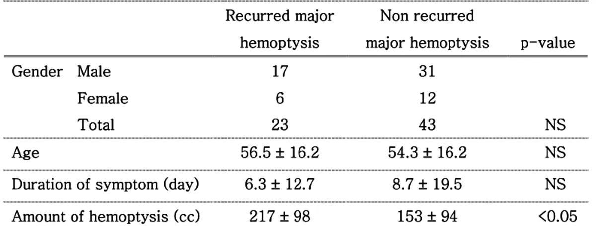
Demographic characteristics such as gender and age, duration of symptom, amount of hemoptysis, previous history of treatment for hemoptysis, the main medical condition causing hemoptysis, bilaterality of pulmonary lesion, number of feeding vessels and presence of pleural thickening were our parameters analysed.
For comparison of various risk factors between recurrent major hemoptysis group and controlled hemoptysis group, Pearson’s Chi-square test and Student’s T-test were used. Statistical validity is defined p-value less than 0.05.
13 Ⅲ
Ⅲ Ⅲ
Ⅲ. . . . ResultsResultsResultsResults
Among 66 patients whose data were available, 23 (34.9%) patients had
recurred major hemoptysis during a mean follow up period of 2.74 years (ranging from 4 months to 75 months). Eight of these 23 recurred patients had pneumonectomy or lobectomy of lung (3 pneumonectomies and 5 lobectomies) in following event, and 4 died with uncontrolled hemoptysis (one died after pneumonectomy). Out of the remaining 15 relapsed patients, 12 had to undergo another bronchial artery embolization.
Concerning the factors influencing relapse, demographic findings did not play any role and duration of symptom had not any significance, but the amount of
hemoptysis had a statistical significance (P < 0.05; Table 2).
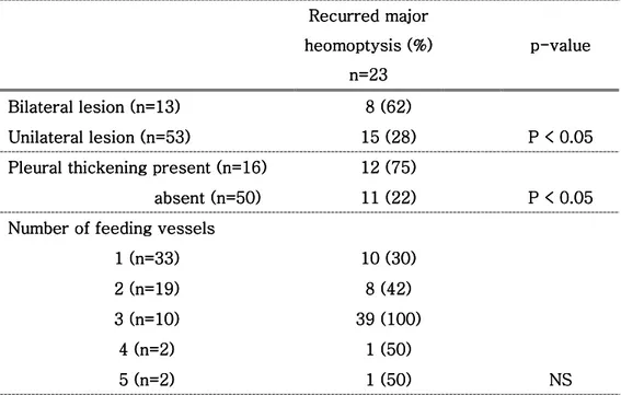
As to the underlying diseases, we had 19 bronchiectasis, 7 malignancies (4 primary lung cancer and 3 metastatic malignancies), 20 tuberculosis, 14 aspergillomas and 6 others (including 1 bronchial artery aneurysm, 2 lung abscesses, and 2 unknown causes) and we did not find any statistical significance between them (Table 3). Nine patients had a history of previous BAE treatments for hemoptysis, but they showed no increased risk of relapse of major hemoptysis (table 3). Bilateral lesion on radiographic finding or pleural
thickening on HRCT had increased risk of recurred major hemoptysis (P < 0.05;
Table 4). Number of feeding vessels had not any statistical value concerning the relapse.
In present study, reliable risk factors for recurrent hemptysis after BAE were: amount of hemoptysis, bilaterality of lesion and pleural thickening on HRCT.
Minor hemoptysis including blood tinged sputum occurred in 30 patients who were classified in non recurred group for major hemoptysis. So the fraction of patients who experienced any bleeding after the procedure was 80.3% (53 out of 66). We also analysed the risk factors for all patients who had at least one minor bleeding event. But there was no reliable risk factor that can predict bleeding events comprising minor hemoptysis.
Table Table Table
Table 222.... D2 DDDemographic featuresemographic features, duration emographic featuresemographic features, duration , duration and , duration and and and amount of hemoptysisamount of hemoptysisamount of hemoptysis amount of hemoptysis
RRRRecurredecurredecurredecurred major major major major hemoptys hemoptyshemoptys hemoptysisisisis Non recurred Non recurred Non recurred Non recurred m m m
major hemoptysisajor hemoptysisajor hemoptysis ajor hemoptysis pppp---value-valuevaluevalue G
G G
Genderenderenderender Male Male Male Male Female Female Female Female Total Total Total Total 17 17 17 17 6 6 6 6 23 23 23 23 31 31 31 31 12 12 12 12 43 43 43 43 NS NS NS NS Age Age Age Age 56.5 56.5 56.5 56.5 ±±±± 16.2 16.2 16.2 16.2 54.3 54.3 54.3 54.3 ±±±± 16.2 16.2 16.2 16.2 NS NS NS NS Duration of symptom Duration of symptom Duration of symptom
Duration of symptom ( ( (day) (day)day)day) 6.3 6.3 6.3 6.3 ±±±± 12. 12.7 12. 12.7 77 8.7 8.7 8.7 8.7 ±±±± 19.5 19. 19. 19.555 NS NS NS NS Amount of hemoptysis Amount of hemoptysis Amount of hemoptysis Amount of hemoptysis (cc)(cc)(cc) (cc) 217 217 217 217 ±±±± 98 98 98 98 153 153 153 153 ±±±± 9 9 9 9444 4 <0.05 <0.05 <0.05 <0.05 Table Table Table
Table 333.... U3 UUnderlying diseasUnderlying diseasnderlying diseasnderlying diseases es es es andand previousandand previous previous previous historyhistoryhistoryhistory of of of of bronchial artery bronchial artery bronchial artery bronchial artery embolzation
embolzation embolzation
embolzation interventioninterventioninterventionintervention
RecurredRecurredRecurredRecurred major major major major heomoptysis heomoptysis heomoptysis heomoptysis (%)(%)(%)(%) n nn n=23=23=23=23 p pp
p----valuevaluevaluevalue
Underlying diseases Underlying diseases Underlying diseases Underlying diseases Bronchiectasis Bronchiectasis Bronchiectasis Bronchiectasis (n=19)(n=19)(n=19)(n=19) 5555 (26)(26)(26) (26) Malignancy Malignancy Malignancy Malignancy (n=7) (n=7) (n=7) (n=7) Primary lung ca Primary lung caPrimary lung ca
Primary lung cancerncerncerncer (n= (n= (n=4 (n=44)))) 4
4 4 4 4 (57)(57)(57) (57) 2 (50) 2 (50) 2 (50) 2 (50) Metasta MetastaMetasta
Metastasis to the lungsis to the lungsis to the lung (n=sis to the lung (n= (n= (n=3333)))) 2 (67)2 (67)2 (67)2 (67) Tuberculosis Tuberculosis Tuberculosis Tuberculosis (n=20) (n=20) (n=20) (n=20) 5555 (25)(25)(25) (25) Aspergilloma Aspergilloma Aspergilloma Aspergilloma (n=14)(n=14)(n=14)(n=14) 8888 (57)(57)(57) (57) Other* Other* Other* Other* (n=6)(n=6)(n=6) (n=6) 1111 (17)(17)(17) (17) NSNSNSNS Previous intervention Previous intervention Previous intervention Previous intervention Yes Yes Yes Yes (n=9) (n=9) (n=9) (n=9) 5555 (55)(55)(55) (55) No No No No (n=57) (n=57) (n=57) (n=57) 1818 (1818 (32) ( (32)32) 32) NSNSNSNS * **
* Other diseases include 1 bronchial artery aneurysm, 2 lun Other diseases include 1 bronchial artery aneurysm, 2 lung Other diseases include 1 bronchial artery aneurysm, 2 lun Other diseases include 1 bronchial artery aneurysm, 2 lunggg a a a abscesses, bscesses, bscesses, bscesses,
and and and
15 Table 4
Table 4 Table 4
Table 4.... Radiologic findings and the recurre Radiologic findings and the recurre Radiologic findings and the recurre Radiologic findings and the recurred majd majd majd major hemoptysisor hemoptysisor hemoptysisor hemoptysis
RecurredRecurredRecurredRecurred major major major major heomoptysis heomoptysis heomoptysis heomoptysis (%)(%)(%)(%) n=23 n=23 n=23 n=23 p pp
p----valuevaluevaluevalue
Bilateral lesion Bilateral lesionBilateral lesion
Bilateral lesion (n=13)(n=13)(n=13) (n=13) 8888 (62)(62)(62) (62) Unilateral lesion
Unilateral lesionUnilateral lesion
Unilateral lesion (n=53)(n=53)(n=53)(n=53) 15151515 (28)(28)(28)(28) P < 0.05P < 0.05 P < 0.05P < 0.05 Pleural thickening prese
Pleural thickening presePleural thickening prese
Pleural thickening presentntntnt (n=16)(n=16)(n=16) (n=16) 12121212 (75)(75)(75)(75) absent
absent absent
absent (n=50)(n=50)(n=50)(n=50) 11111111 (22)(22)(22)(22) P < 0.05P < 0.05 P < 0.05P < 0.05 Number of feeding vessels
Number of feeding vesselsNumber of feeding vessels
Number of feeding vessels 1 1 1 1 (n=33)(n=33)(n=33)(n=33) 10101010 (30)(30)(30)(30) 2 2 2 2 (n=19)(n=19)(n=19)(n=19) 8888 (42)(42)(42) (42) 3 3 3 3 (n=10)(n=10)(n=10)(n=10) 33339999 (100)(100)(100) (100) 4 44 4 (n=2)(n=2)(n=2)(n=2) 1111 (50)(50)(50) (50) 5 55 5 (n=2)(n=2)(n=2)(n=2) 1111 (50)(50)(50) (50) NSNSNSNS
Ⅳ Ⅳ Ⅳ
Ⅳ. . . . DiscussionDiscussionDiscussionDiscussion
We analyzed the result of bronchial artery embolization in our institution during a relatively long period. In previous literatures, long term recurrence rate have been reported to be 10 to 52%, with a mean follow up period ranging
from one to 46 months1-11.
Remy et al.1 reported that of 49 patients treated during hemoptysis, an
immediate arrest was achieved in 41 but 6 of these patients suffered a relapse in 2-7 months after BAE, but there was no recurrent bleeding in the remaining 35 patients. However, only 7 of 35 (20%) patients had not experienced re-bleeding in follow up period beyond the 18 months. Ulfacker et al. reported that an immediate control of hemoptysis was achieved in 33 of 41 patients (80.5%) while hemoptysis recurred in 9 of 33 patients (27.3%) in the long term follow up (mean 24.8months). In our analysis, bronchial artery embolization effectively controlled 65.1% of life threatening massive hemoptysis (23 recurs in 66 cases) in a mean follow up period of 31.9 months.
Regarding the factor of recurrence after BAE, Osaki et al.4concluded that
bronchiectatic change on CT scan and pulmonary bronchial shunt had some
statistical significance. Kim et al.6 described the underlying lung disease and
amount of bleeding as reliable risk factors for the recurrence, in a study involving 75 patients with a result estimating 54.5% of re-bleeding rate after 3 years.
The diversity of previously proposed risk factors may be explained by variability of their criteria on recurrence, sample size, underlying diseases, follow-up time and embolization technique.
In this study, the amount of hemoptysis had some statistical relation with the recurrent event. Though the analysis of underlying disease had no statistical validity, tuberculosis tended to have more control rate compared to aspergilloma and cancer. The effective anti-tuberculosis drug therapy must have reduced the recurrent hemoptysis but its relatively modest prevalence in
our series (30% compared to 43-52% in other domestic studies)5,6,12,14 lead to
overall no statistical significance.
In 1993, Tamura et al. described pleural thickening as a risk factor for
recurrent bleeding after bronchial artery embolization8. According to them, in
the presence of pleural thickening, non bronchial systemic feeder vessels that originate from various arteries (e.g., intercostals artery, branches of the subclavian and axillary arteries, internal mammary artery and inferior phrenic
17
artery) may develop along the pleural surface and become enlarged as a result of the inflammatory process. In our study 16 cases showed pleural thickening on chest radiography and 12 (75%) of them experienced recurrent major bleeding, which is statistically valid compared with 22% of no pleural thickening group.
This study is a retrospective review of medical records, which often should underestimate strength of variable. But any hemoptysis including minor hemoptysis and blood tinged sputum occurred in 53 patients (80.3%). Perhaps because this portion is too high, no reliable risk factor that can predict minor bleeding was found. However minor bleeding event especially blood tinged sputum is considered as a natural course that can easily happen after embolization and is nowhere the target point of the procedure.
Ⅴ Ⅴ Ⅴ
Ⅴ. Conclusion. Conclusion. Conclusion. Conclusion
The amount of hemoptysis, bilaterality of lesion, and the presence of pleural thickening were revealed as reliable risk factors for relapse of gross hemoptysis after initial bronchial artery embolization.
In a well designed prospective study based on this series, one should take account of the extent of destroyed lung, the baseline pulmonary function and the selection of the same interventional radiology expert.
19 References References References References
1. Remy J, Arnaud A, Fardou H, Giroud R, Vousin C. Treatment of hemoptysis by embolization of bronchial arteries. Radiology 1977;122:33-37.
2. Karen L, Swanson KL, Prakash UB: Bronchial artery embolization. Experience with 54 patients. Chest 2002;121:749-759.
3. Ulfacker R, Kaemmerer A, Neves C, Picon P, Rizzon CF, Oliveira C. Management of massive hemoptysis by bronchial artery embolization. Radiology 1983;146:627-634.
4. Osaki S, Naknishi Y, Wataya H, Wataya H, Takayama K. Prognosis of bronchial artery embolization in the management of hemoptysis. Respiration 2000;67:412-416.
5. Yeo DS, Lee SY, Hyun DS, Lee SH, Kim SC, Choi YM et al. Effect of bronchial artery embolization in management of massive hemoptysis. Tuberc Respir Dis 1999;46:53-64.
6. Kim SO, Oh IJ, Kim KS, Kim KS, Yu YK, Lim SC. Recurrent Hemoptysis after bronchial artery embolization. Tuberc Respir Dis 2001;51:364-372.
7. Katoh O, Kishikawa T. Recurrent bleeding after arterial embolization in patients with hemoptysis. Chest 1990;97:541-546.
8. Tamura S, Kodama T, Otsuka N, Kihara Y, Nisikawa K, Yuki Y et al. Embolotherapy for persistent hemoptysis. The significance of pleural thickening. Cardiovasc Intervent Radiol 1993;16:85-88.
9. White R. Bronchial artery embolotherapy for control of acute hemoptysis. Analysis and outcome. Chest 1999;115:912-915.
10. Mal H, Rullon I, Mellot F, Brugiere O, Sleiman C, Menu Y, Fournier M. Immediate and long term results of bronchial artery embolization for life threatening hemoptysis. Chest 1999; 115:996-1001.
11. Lee TW. Management of massive hemoptysis. A single institution experience. Ann Thorac Cardiovas Surg 2000.;6:232-235
12. Yoon W, Kim JK, Kim YH, Chung TW, Kang HK. Bronchial and non bronchial systemic artery embolization for life threatening hemoptysis: a comprehensive review. Radiographics 2002; 22:1395-1409.
13. Yu-Tang Goh P, Lim M, Teo N, En Shen Won Wong D. Embolization for hemoptysis, a six year review. Cadiovasc Intervent Radiol 2002;25:17-25. Epub 2001 Nov23.
the management of massive hemoptysis: factors influencing rebleeding. Tuberc Respir Dis 1996;4:590-599.
21 Abstract (in Korean)