Radiological Findings and Outcomes of Bronchial Artery Embolization in Cryptogenic Hemoptysis
Management of cryptogenic massive hemoptysis is difficult, and conservative treatment may be inadequate to stop the hemorrhage. Surgery is not a reasonable option because there is no underlying identifiable pathology. This study aimed to investigate the radiologic findings and bronchial artery embolization outcomes in cryptogenic hemoptysis, and to compare the results with non-cryptogenic hemoptysis. We evaluated 26 patients with cryptogenic hemoptysis and 152 patients with non-cryptogenic hemoptysis. A comparison of the bronchial artery abnormalities between the cryptogenic and non-cryptogenic hemoptysis groups showed that only extravasation was more statistically significant in the cryptogenic hemoptysis group than in the non-cryptogenic hemoptysis group, while the other bronchial artery abnormalities, such as bronchial artery dilatation, hypervascularity, and bronchial-to-pulmonary shunting, showed no significant difference between groups.
Involvement of the non-bronchial systemic artery was significantly greater in the non- cryptogenic hemoptysis group than in the cryptogenic hemoptysis group. While 69.2% of patients with cryptogenic hemoptysis also had hypervascularity in the contralateral bronchial arteries and/or ipsilateral bronchial artery branches other than the bleeding lobar branches, this finding was not detected in non-cryptogenic hemoptysis. Embolization was performed on all patients using polyvinyl alcohol particles of 355-500 µm. Hemoptysis ceased in all patients immediately after embolization. While recurrence of hemoptysis showed no statistically significant difference between the cryptogenic and non-cryptogenic hemoptysis groups, it was mild in cryptogenic hemoptysis in contrast to mostly severe in non-cryptogenic hemoptysis. Transarterial embolization is a safe and effective technique to manage cryptogenic hemoptysis.
Keywords: Cryptogenic Hemoptysis; Computed Tomography; Angiography; Embolization Selim Kervancioglu,1 Nazan Bayram,2
Feyza Gelebek Yilmaz,1 Maruf Sanli,3 and Akif Sirikci1
Departments of 1Radiology, 2Pulmonology, and
3Thoracic Surgery, Faculty of Medicine, Gaziantep University, Gaziantep, Turkey
Received: 13 September 2014 Accepted: 6 January 2015 Address for Correspondence:
Selim Kervancioglu, MD
Department of Radiology, Gaziantep University, Faculty of Medicine, University Avenue, 27310, Gaziantep, Turkey Tel: +90.532-475-7528, Fax: +90.342-360-3928 E-mail: [email protected]
http://dx.doi.org/10.3346/jkms.2015.30.5.591 • J Korean Med Sci 2015; 30: 591-597
INTRODUCTION
Hemoptysis is bleeding originating from the lower airways; it has a wide clinical spectrum that can reach life-threatening di- mensions. The most important causes of hemoptysis are bron- chiectasis, chronic bronchitis, tuberculosis, and malignancy (1).
Etiological causes are usually investigated with chest radiogra- phy, computed tomography (CT), and bronchoscopy. Etiologi- cal causes cannot be identified in 7%‒22% of cases, and the con- dition is defined as cryptogenic hemoptysis (2). Angiography and bronchial artery embolization (BAE) are important modal- ities allowing for rapid diagnosis and treatment in hemoptysis (3). Management of cryptogenic massive hemoptysis is difficult and surgery is not a reasonable option, as there is no underly- ing, identifiable pathology. The current study was performed to evaluate CT and angiographic findings and BAE outcomes in cryptogenic hemoptysis.
MATERIALS AND METHODS
In this retrospective study, we evaluated patients who had un- dergone BAE from January 2005 to January 2013 due to crypto- genic hemoptysis. Additionally, the angiography findings and the BAE results in patients with cryptogenic hemoptysis were compared with those in patients with non-cryptogenic hemop- tysis in the same period. Diagnosis of cryptogenic hemoptysis was made in those without any specific disease history (e.g., bron- chiectasis, tuberculosis, malignancy), when no endobronchial and parenchymal abnormalities (except for hemorrhagic find- ings) were observed after the patients were examined by bron- choscopy and CT, and when the laboratory findings were nor- mal. In our hospital, hemoptysis is classifying as mild (< 30 mL/
24 hr), moderate (30-100 mL/24 hr), and severe (> 100 mL/24 hr), and angiography and BAE is performing in severe hemop- tysis. All patients were hospitalized, and standard treatment was administered to correct hypoxemia and hemodynamic in-
stability with fluid replacement and blood products. All patients underwent chest radiography, thorax CT, and fiber-optic bron- choscopy to identify the cause of hemoptysis and localize the side with bleeding.
Angiography was performed on all patients using the right femoral approach with a Philips Allura XPER FD DSA machine.
First, non-selective angiography was performed with a 5F pig- tail catheter to visualize the aortic arch and thoracic aorta and to investigate the bronchial artery anatomy and the presence of systemic collateral vessels. Then, a 4F or a 5F Cobra or Simmons catheter were used to catheterize the bronchial artery on the bleeding side and visualize it with a nonionic contrast medium.
Ipsilateral intercostals arteriograms and, when necessary, sub- clavian arteriograms were obtained by selective catheterizations.
Bronchial arteriography criteria that can lead to a diagnosis of hemoptysis are bronchial artery enlargement, hypervascular- ization, bronchial-to-pulmonary shunting, and extravasation of the contrast material into the bronchial lumen. Following se- lective catheterization of the bleeding bronchial artery or other systemic artery, embolization was performed under fluoroscop- ic guidance by using 355-500 µm polyvinyl alcohol (PVA) parti- cles (Contour; Boston Scientific, Natick, MA, USA).
Statistical analysis
To compare the angiographic findings and the outcomes of em- bolization in the cryptogenic and non-cryptogenic hemoptysis groups, Fisher’s exact test was used. All analyses were perform- ed using SPSS for Windows version 22.0. A two-sided P value less than 0.05 was considered statistically significant.
Ethics statement
The study was approved by the institutional review board of the Medical School of Gaziantep University (Approval No. 2013-131).
Informed consent was waived by the board.
RESULTS
Cryptogenic hemoptysis
This study evaluated 26 patients (19 men, 7 women) with a mean age of 40.4 yr (range, 20-60 yr) who received endovascular treat- ment after being diagnosed with cryptogenic hemoptysis. Table 1 lists clinical characteristics of patients.
All patients had chest radiography. In 13 patients (50%), these were normal, and in the other 13 patients (50%), they were ab- normal, showing alveolar infiltrates or consolidation. CT exam- ination was performed on all patients. Upon CT imaging, 20 pa- tients (76.9%) had ground-glass or scattered nodular infiltrates, while 6 patients (23.1%) had hazy consolidation. In addition to
Table 1. Clinical characteristics of patients
Characteristics No. (%) of patients
Male gender 19 (73.1)
Smoker 14 (53.8)
Chronic obstructive pulmonary disease 2 (7.7)
Diabetes mellitus 2 (7.7)
Systemic arterial hypertension 3 (11.5)
Mitral valve insufficiency 2 (7.7)
Tricuspid valve insufficiency 1 (3.8)
Hepatitis B 1 (3.8)
Hepatitis C 1 (3.8)
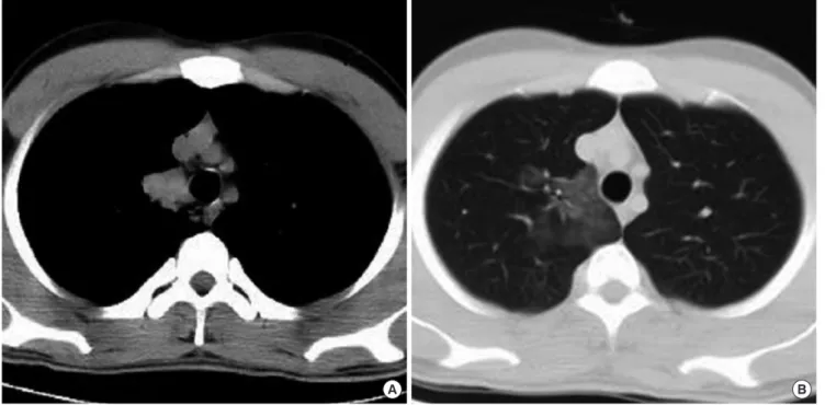
A B
Fig. 1. Axial CT scan (A) Before BAE, demonstrates mass image at paramediastinal area of right upper lobe. (B) 4 days after BAE, demonstrates mass image disappearance.
infiltrates, 1 patient (3.8%) also had liquefied material in seg- mental and lobar bronchi, and 1 patient (3.8%) had a mass im- age that disappeared on the fourth day after the BAE (Fig. 1). In all cases, bronchoscopy was performed before angiography to identify endobronchial lesions that could cause hemoptysis.
Bronchoscopy identified active bleeding and the blood clot that showed the bleeding side. The side of bleeding identified at bron- choscopy was compatible with CT findings, in all patients. Nine
patients (34.6%) had only left lung hemorrhage, while 16 patients (61.5%) had only right lung hemorrhage. One patient (3.8%) had hemorrhage in both lungs. Hemorrhage was located to up- per lobe in 14 patients (53.8%), lower lobe in 7 patients (26.9%), and right middle lobe in 2 patients (7.7%). Three patients (11.5%) had hemorrhage in multiple lobes.
Diagnostic angiography revealed abnormalities in all patients.
While all patients had bronchial artery abnormalities, 4 patients
C D
A B
Fig. 2. Images of a patient by CT scan and angiography. (A) Axial CT scan obtained with parenchymal lung window demonstrates hazy consolidation at left upper lobe. (B) Left second intercostal arteriography demonstrates dilatation and hypervascularity. (C) Bronchial arteriography shows combined right and left bronchial trunk with hypervascularity, more prominent in the left upper lobe. (D) CT scan obtained 10 months after embolization demonstrates normal lung parenchyma.
(15.4%) also had systemic artery abnormalities, including 3 pa- tients (11.5%) who had intercostal artery abnormalities and 1 patient (3.8%) who had abnormalities of 1 lateral thoracic artery and 2 intercostal arteries (Fig. 2). The most commonly detected abnormalities of bronchial artery were dilatation in 18 patients (69.2%) and hypervascularity in 25 patients (96.2%) (Fig. 3). Four patients (15.4%) had bronchial-to-pulmonary shunting while 4 patients (15.4%) had extravasation into the bronchial lumen. In addition to bleeding bronchial artery branches, 18 patients (69.2%) exhibited hypervascularities in contralateral bronchial arteries and/or ipsilateral bronchial artery branches other than bleed- ing lobar branches (Fig. 3, 4). Intercostal and lateral thoracic ar- teries abnormalities were also dilatation and hypervascularity.
Bronchial arteries were investigated bilaterally in 10 patients.
Of those, 7 patients had bronchial artery abnormalities on both sides (Fig. 4). In 3 of 10 patients, the bronchial artery on the blee- ding side was thinner than the bronchial artery on the opposite side (Fig. 4). Interestingly, 71.4% of the patients (5 patients) with bilateral bronchial artery abnormalities were smokers.
Hemoptysis ceased in all patients after embolization. Embo- lization was repeated in 3 patients (11.5%) after 1, 6, and 7 mon- ths, respectively, as there was mild recurrence of hemoptysis. In the remaining 23 patients (88.5%), hemoptysis was not observ- ed during long-term follow up. After the last embolization, no hemoptysis was reported during a mean follow-up period of 29.2 ± 15.6 months (6-61 months). There were no major com- plications related to the procedure.
Non-cryptogenic hemoptysis
A total of 152 patients (97 men, 55 women) with a mean age of 47.5 yr (range, 25-85 yr) who received endovascular treatment after being diagnosed with non-cryptogenic hemoptysis were evaluated. Of those, 22 patients (14.5%) were smokers.
CT examination revealed bronchiectasis in 95 patients (62.5%), cavitary lesion in 26 patients (17.1%), pleuroparenchymal fibro- tic changes due to tuberculosis in 16 patients (10.5%), lung can- cer in 11 patients (7.2%), and metastasis in 4 patients (2.6%). All of the patients also had infiltrates and/or consolidation in the CT images. Of the patients with non-cryptogenic hemoptysis, 87 (57.2%) had unilateral lung abnormality, while 65 (42.8%) had abnormality in both lungs; moreover, 134 patients (88.2%) had abnormality in more than one lobe.
Diagnostic angiography revealed abnormalities in all of the patients. While all of the patients had bronchial artery abnor- malities, 54 (35.5%) also had nonbronchial systemic artery ab- normalities, including abnormalities in the intercostal artery, thyrocervical trunk, internal mammary artery, and lateral tho- racic artery. The most commonly detected abnormalities of the bronchial artery were dilatation in 80 patients (52.6%) and hy- pervascularity in all patients (100%). Thirty-nine patients (25.7%) had bronchial-to-pulmonary shunting while 2 (1.3%) had ex- travasation into the bronchial lumen. A comparison of the bron- chial artery abnormalities between the cryptogenic and non- cryptogenic hemoptysis groups showed that only extravasation was statistically significant in the cryptogenic hemoptysis group
Fig. 3. In a patient with right upper lobe hemorrhage, arteriography of right intercos- tal bronchial trunk demonstrates dilatation of right bronchial artery with hypervascu- larity in all, more prominent upper lobe.
Fig. 4. In a patient with left lower lobe hemorrhage, arteriography of combined right and left bronchial trunk demonstrates mild dilatation of right bronchial artery with hy- pervascularity in all, more prominent upper lobe, and thin left bronchial artery with extravasation of the contrast material into the lower lobe bronchial lumen (arrow).
than the non-cryptogenic hemoptysis group (P = 0.004), while the other bronchial artery abnormalities had no significant dif- ference between groups (for all, P > 0.05). Conversely, the in- volvement of the nonbronchial systemic artery was significantly greater in the non-cryptogenic hemoptysis group than the cryp- togenic hemoptysis group (P = 0.044).
Of the 65 patients with bilateral lung abnormalities in CT ex- aminations, 55 had bilateral bronchial artery abnormalities in angiography. Of the 87 patients with unilateral lung abnormali- ties, 38 were investigated for bilateral bronchial arteries, and no abnormality was identified in the contralateral bronchial artery.
Only 5 patients with unilateral lung abnormality who were in- vestigated for bronchial artery bilaterally (as both bronchial ar- teries originated from a trunk) were smokers.
Because most of the patients had diffuse parenchymal disor- ders in the lung(s), they also had diffuse bronchial artery abnor- malities. Only 18 patients (11.8%) had abnormality in one lobe of the lung in the CT images, and abnormalities in the ipsilater- al bronchial artery branches, other than bleeding lobar branch- es, were not identified in any of the patients. Of those patients, only 3 were smokers.
Hemoptysis ceased in all patients after embolization. Recur- rence of hemoptysis appeared 14 days to 21 months after the embolization in 38 patients (25%). Of those, 20 (13.2%) were re- embolized 1 to 6 times, as the hemoptysis was severe. Other pa- tients were treated medically. Recurrence of hemoptysis had no statistically significant difference between the cryptogenic and non-cryptogenic hemoptysis groups (P = 0.205).
DISCUSSION
Diagnostic radiological examinations used for hemoptysis are chest radiography and thorax CT. The role of CT is identifica- tion of the cause of bleeding and pulmonary lobe with bleed- ing. In cryptogenic hemoptysis, the cause of bleeding cannot be identified; CT findings show infiltration and/or consolidation on the bleeding side parenchyma and liquefied material in the bronchial system. In addition to these findings, the current study found a mass image that resolved after BAE in one patient. The mass image arose as a result of hemoptysis; it was not the cause of hemoptysis. This state shows that diagnosis of a tumor can- not be done until a control CT examination reveals an unresolv- ed mass image after the BAE. To the best of our knowledge, this resolving CT finding in cryptogenic hemoptysis has not been reported previously in the literature.
The efficacy and safety of BAE in treating hemoptysis caused by various etiologies has been analyzed in both the short term and long term. In 90% of cases, embolization stops the bleeding immediately, and 70% of patients do not have hemoptysis re- currences during one year of follow up (4, 5). In the current study, hemoptysis was ceased immediately after the embolization in
all patients with non-cryptogenic hemoptysis, while 75% of those patients had no recurrence of hemoptysis in the long fol- low-up period. In contrast to hemoptysis with known etiolo- gies, in cryptogenic hemoptysis, information about the role of BAE is limited. A limited number of studies evaluating the role of BAE in cryptogenic hemoptysis have reported that bleeding stopped immediately after embolization in 85%‒95% of cases (2, 6, 7). In one of these studies, 15% of the cases had recurrences in three months (7). For cryptogenic hemoptysis cases that have undergone BAE, long-term follow-up results are insufficient. In the current study, BAE was efficient at immediately controlling hemoptysis in all cases. Mild recurrence of hemoptysis occurred in three cases (11.5%), and re-embolization was performed.
While the rate of recurrence of hemoptysis was lower in the cryptogenic hemoptysis group than the non-cryptogenic he- moptysis group, there was no statistically significant difference between the groups. Otherwise, the recurrence of hemoptysis was mild in the cryptogenic hemoptysis group; in contrast, the most severe hemoptysis recurrence occurred in the non-cryp- togenic hemoptysis group. Furthermore, only one re-emboliza- tion was sufficient in the recurrence of cryptogenic hemoptysis, while multiple re-embolizations were needed in the recurrence of non-cryptogenic hemoptysis. In all cases with cryptogenic hemoptysis, after the last embolization, no hemoptysis occurred during a mean follow-up period of 29.2 ± 15.6 months.
The angiography findings in cryptogenic hemoptysis cases cited in the literature are bronchial artery enlargement, hyper- vascularization, retrograde bronchial-to-pulmonary shunting, and contrast extravasation into the bronchial lumen (2, 3, 6, 7).
The current study found that dilatation of the bronchial artery and hypervascularity were the most commonly detected ab- normalities, consistent with previous reports. In contrast, in this study, extravasation into the bronchial lumen occurred slightly more often than in previous reports. Comparison of the angio- graphic findings for the cryptogenic hemoptysis patients and the non-cryptogenic hemoptysis patients showed no significant difference in bronchial artery enlargement, hypervasculariza- tion, and bronchial-to-pulmonary shunting, while extravasa- tion into the bronchial lumen was significantly greater in the cryptogenic hemoptysis patients than in the non-cryptogenic hemoptysis patients.
Bronchial artery dilatation is reported as the most common angiographic finding. In our study, in 30% of the cryptogenic hemoptysis patients who had bilateral bronchial arteriography, the bronchial artery on the bleeding side was thinner than the contralateral bronchial artery. This finding made us conclude that other angiography findings, such as hypervascularization, bronchial-to-pulmonary shunting, and contrast extravasation into the bronchial lumen, were more valuable and significant as diagnostic findings.
An important finding of the current study was that 69.2% of
patients with cryptogenic hemoptysis had, in addition to bleed- ing bronchial artery branches, hypervascularities in contralat- eral bronchial arteries and/or ipsilateral bronchial artery bran- ches other than bleeding lobar branches. Menchini et al. (7) re- ported bilateral bronchial artery abnormality in 76% of the pa- tients who underwent bronchial arteriography bilaterally. Al- though the current study found similar results; Hypervasculari- ty in ipsilateral bronchial artery branches other than bleeding lobar branches was also found. To the best of our knowledge, this angiography finding has not been reported previously in the literature. In contrast to the cryptogenic hemoptysis cases, in the non-cryptogenic hemoptysis cases the contralateral bron- chial artery and the ipsilateral bronchial artery branches, other than the bleeding lobar branches, had no abnormalities.
Another important finding of the current study was the ab- normality of nonbronchial systemic arteries in 4 patients (15.4%).
In hemoptysis, nonbronchial systemic artery abnormalities are observed during inflammatory conditions with the involvement of pleura (8). Our cases were regarded as cryptogenic hemopty- sis, as they did not have any parenchymal or pleural abnormali- ties. To the best of our knowledge, abnormality of the nonbron- chial systemic artery in cryptogenic hemoptysis has not been reported previously in the literature. In the current study, as ex- pected, abnormalities of the nonbronchial systemic arteries were significantly greater in the non-cryptogenic hemoptysis group than in the cryptogenic hemoptysis group.
Angiographic findings show us that there are conditions af- fecting one or both lungs widely in the pathophysiology of cryp- togenic hemoptysis. The fact that 53.8% of our patients smoked supports this hypothesis, because smoking has chronic inflam- matory effect. In recent years, the literature has mentioned the relationship between smoking and cryptogenic hemoptysis (2, 7). Smoking induced inflammatory changes are seen mostly on the upper zones of the lungs (7, 9, 10). Supporting this, a study reported bleeding in the upper lobes in 71% of the cryptogenic hemoptysis cases in smokers (7). Similarly, the most effected side of lung is the upper lobe in our study.
The goal of BAE is to stop systemic arterial inflow in fragile vessels in the pathological region of the lungs, thereby decreas- ing perfusion pressure and preventing bleeding (11). The objec- tive in endovascular treatment of hemoptysis is occlusion of the bleeding distal bed, and the embolic material used should be particles. Absorbable gelatin sponge can be used in emboliza- tion; however, as it resorbs over time and recanalizes, it is not a preferred embolic material for hemoptysis. The most commonly used embolization material in BAE is PVA particles. PVA is bio- compatible and nonbiodegradable, providing for permanent embolization (12). We used PVA when performing BAE on our patients and did not encounter any significant complications, either during or after the procedure. Trisacrylgelatin microsph- eres also are being used now as embolization material in BAE (3).
The complications of BAE are quite limited and can be relat- ed to the femoral arterial approach, selective catheterization or embolization. The main complications are arterial occlusion, perforation, dissection, pseudoaneurysm, arteriovenous fistula, hemorrhage, non-target embolization and postembolization syndrome. The major risk of the BAE is the paraplegia caused by occlusion of the spinal artery. In the present study, no treat- ment complications occurred.
A limitation of the current study is that it focused only on pa- tients with cryptogenic hemoptysis who underwent transarte- rial embolization and excluded patients who received other me- dical or surgical treatments. The study sought to evaluate arteri- al abnormalities and embolization results in cryptogenic hemo- ptysis.
In conclusion, bronchial artery dilatation is one of the most commonly observed angiographic findings, yet hypervascular- ization, bronchial-to-pulmonary shunting, and contrast extrav- asation are considered more valuable and significant in crypto- genic hemoptysis. Angiographic findings show that whatever the underlying cause for cryptogenic hemoptysis, it involves the lung(s) in a diffuse manner. Nonbronchial systemic arteries can also be affected in cryptogenic hemoptysis. BAE is an effective and reliable treatment option for cryptogenic hemoptysis, with excellent short-term and long-term results.
DISCLOSURE
The authors have no conflicts of interest to disclose.
AUTHOR CONTRIBUTION
Conception and coordination of the study: Kervancioglu S, Yil- maz FG, Sirikci A. Acquisition of radiological data: Kervanciog- lu S, Yilmaz FG, Sirikci A. Acquisition of clinical data: Bayram N, Sanli M. Analysis and interpretation of data: Kervancioglu S, Yilmaz FG, Sirikci A. Drafting the article: Kervancioglu S. Revis- ing article critically for important intellectual content: all au- thors. Final approval of the version to be published: all authors.
ORCID
Selim Kervancioglu http://orcid.org/0000-0003-4678-8959 Nazan Bayram http://orcid.org/0000-0002-4692-2639 Feyza Gelebek Yilmaz http://orcid.org/0000-0002-9900-5981 Maruf Sanli http://orcid.org/0000-0002-6548-1664
Akif Sirikci http://orcid.org/0000-0002-0432-7831
REFERENCES
1. Bruzzi JF, Rémy-Jardin M, Delhaye D, Teisseire A, Khalil C, Rémy J. Multi- detector row CT of hemoptysis. Radiographics 2006; 26: 3-22.
2. Delage A, Tillie-Leblond I, Cavestri B, Wallaert B, Marquette CH. Cryp- togenic hemoptysis in chronic obstructive pulmonary disease: character- istics and outcome. Respiration 2010; 80: 387-92.
3. Samara KD, Tsetis D, Antoniou KM, Protopapadakis C, Maltezakis G, Siafakas NM. Bronchial artery embolization for management of massive cryptogenic hemoptysis: a case series. J Med Case Rep 2011; 5: 58.
4. Swanson KL, Johnson CM, Prakash UB, McKusick MA, Andrews JC, Stanson AW. Bronchial artery embolization : experience with 54 patients.
Chest 2002; 121: 789-95.
5. Shigemura N, Wan IY, Yu SC, Wong RH, Hsin MK, Thung HK, Lee TW, Wan S, Underwood MJ, Yim AP. Multidisciplinary management of life- threatening massive hemoptysis: a 10-year experience. Ann Thorac Surg 2009; 87: 849-53.
6. Savale L, Parrot A, Khalil A, Antoine M, Théodore J, Carette MF, Mayaud C, Fartoukh M. Cryptogenic hemoptysis: from a benign to a life-threat- ening pathologic vascular condition. Am J Respir Crit Care Med 2007;
175: 1181-5.
7. Menchini L, Remy-Jardin M, Faivre JB, Copin MC, Ramon P, Matran R, Deken V, Duhamel A, Remy J. Cryptogenic haemoptysis in smokers: an-
giography and results of embolisation in 35 patients. Eur Respir J 2009;
34: 1031-9.
8. Yoon W, Kim JK, Kim YH, Chung TW, Kang HK. Bronchial and non- bronchial systemic artery embolization for life-threatening hemoptysis:
a comprehensive review. Radiographics 2002; 22: 1395-409.
9. Soejima K, Yamaguchi K, Kohda E, Takeshita K, Ito Y, Mastubara H, Oguma T, Inoue T, Okubo Y, Amakawa K, et al. Longitudinal follow-up study of smoking-induced lung density changes by high-resolution com- puted tomography. Am J Respir Crit Care Med 2000; 161: 1264-73.
10. Remy-Jardin M, Edme JL, Boulenguez C, Remy J, Mastora I, Sobaszek A.
Longitudinal follow-up study of smoker’s lung with thin-section CT in correlation with pulmonary function tests. Radiology 2002; 222: 261-70.
11. Chun JY, Morgan R, Belli AM. Radiological management of hemoptysis:
a comprehensive review of diagnostic imaging and bronchial arterial embolization. Cardiovasc Intervent Radiol 2010; 33: 240-50.
12. Poyanli A, Acunas B, Rozanes I, Guven K, Yilmaz S, Salmaslioglu A, Ter- zibasioglu E, Cirpin R. Endovascular therapy in the management of mo- derate and massive haemoptysis. Br J Radiol 2007; 80: 331-6.