Tuberc Respir Dis 2013;74:111-119
CopyrightⒸ2013. The Korean Academy of Tuberculosis and Respiratory Diseases. All rights reserved.
Risk Factors Influencing Rebleeding after Bronchial Artery Emboliz- ation on the Management of Hemoptysis Associated with Pulmonary Tuberculosis
Hun-Gyu Hwang, M.D.1, Ho-Sung Lee, M.D.2, Jae-Sung Choi, M.D.2, Ki-Hyun Seo, M.D.2, Yong-Hoon Kim, M.D.2, Ju-Ock Na, M.D.2
1Respiratory Devision, Department of Internal Medicine, Soonchunhyang University Gumi Hospital, Soonchunhyang University College of Medicine, Gumi, 2Respiratory Division, Department of Internal Medicine, Soonchunhyang University Cheonan Hospital, Soonchunhyang University College of Medicine, Cheonan, Korea
Background: Hemoptysis due to pulmonary tuberculosis (TB) frequently develops in Korea where the prevalence of TB is intermediate. The effect of bronchial artery embolization (BAE) on the control of massive hemoptysis has been well known. This study is designed to identify the risk factors contributing to rebleeding after BAE in patients with TB.
Methods: We retrospectively evaluated risk factors and the time for rebleeding after BAE in 72 patients presenting with hemoptysis.
Results: The overall immediate success rate of BAE was 93.1% (67 of 72 patients). Of the 29 patients (40.3%) who showed rebleeding after BAE, 13 patients experienced rebleeding within 1 month, and 14 patients between 1 month to 1 year. The existence of a shunt in angiographic finding, aspergilloma, and diabetes mellitus were risk factors of rebleeding after BAE in multivariate analysis.
Conclusion: BAE was very effective for obtaining immediate bleeding control in hemoptysis associated with active TB or post-TB sequelae. It is important to observe whether or not rebleeding occurs up to 1 year of BAE especially in TB patients with aspergilloma, DM, or a shunt. Even rebleeding can be managed well by second BAE.
Key Words: Aspergillosis; Bronchial Arteries; Embolization, Therapeutic; Hemoptysis; Tuberculosis
Address for correspondence: Ju-Ock Na, M.D.
Department of Internal Medicine, Soonchunhyang University Cheonan Hospital, Soonchunhyang University College of Medicine, 31 Suncheonhyang 6-gil, Dongnam-gu, Cheonan 330-930, Korea
Phone: 82-41-570-3666, Fax: 82-41-574-5762 E-mail: [email protected]
Received: Nov. 14, 2012 Revised: Nov. 27, 2012 Accepted: Feb. 15, 2013
CCIt is identical to the Creative Commons Attribution Non-Commercial License (http://creativecommons.org/licenses/by-nc/3.0/).
Introduction
Bronchial artery embolization (BAE) has been estab- lished as an effective and useful means to achieve treat- ment of chronic and recurrent hemoptysis, as well as immediate control of massive hemoptysis and to man- age inoperable patients who had poor pulmonary func-
tion or chronic pulmonary diseases
1-3. Although there
are several causes of massive hemoptysis as known in
other reports, tuberculosis (TB) is common cause espe-
cially in Korea
4, where the prevalence of TB is inter-
mediate, 149 rate per 100,000 population/yr compared
with 15 rate per 100,000 population/yr in US in 2011
5.
Though BAE in TB patients has been studied pre-
viously
6-9, the result of risk factors associated with TB
activity were not consistent. So, we evaluated the char-
acteristics of hemoptysis and tried to find the risk factors
for rebleeding in patients who underwent BAE to con-
trol hemoptysis associated with active TB or post-TB
sequelae.
Materials and Methods 1. Patient selection
We retrospectively reviewed consecutive 92 patients who underwent BAE due to hemoptysis in Respiratory Division, Department of Internal Medicine, Soonchun- hyang University, Cheonan Hospital, from 1999 to 2008.
This study was conducted in accordance with require- ment of an Institutional Review Board for such retro- spective analysis of medical records.
2. Data collection
The clinical records of all patients were assessed ret- rospectively, and the following data and images were collected to analyze the activity of TB, recurrence rate, and risk factor for rebleeding: age, sex, clinical features, past history, laboratory findings, sputum study, emboli- zation material, angiographic images, chest roentgen- ography, chest CT scan, and bronchoscopy.
3. Classification of patients and definition
All patients were categorized as active TB or post-TB.
Acitve TB was defined on the basis of acid-fast bacilli (AFB) positive, clinical suspicion or imaging including consolidation, endobronchial spread pattern, or tree-in- bud opacities. TB sequelae was defined on the basis of previous history of TB and AFB-negative with imag- ing including bronchiectasis, calcified nodules or fib- rosis
10. TB destroyed lung was defined as parenchymal damage to more than one lung lobe due to previous pulmonary TB, but no recent evidence of active TB
11. Multi-drug resistant TB (MDR TB) was defined as resist- ance to both isoniazid and rifampicin, with or without resistance to any other antituberculous drugs. Immedi- ate control of bleeding was defined as a cessation of bleeding obtained without recurrence within 24 hours of successful BAE
12. Rebleedings, which was defined as recurrence and/or persistence of bleeding after immedi- ate control, are categorized into two groups according to the time: early-onset, within 1 month; and late-onset, beyond 1 month. Patients were classified according to the amount of greater than 200 mL on admission
13.
Shunt means pulmonary-bronchial artery shunt in angio- graphic findings.
4. Management
Our conservative medical measures included strict bed rest, nothing by mouth, hemostatics, monitoring of oxygen saturation, respiratory rate, heart rate and blood pressure, the supply of oxygen if needed. Anti-tuber- culous medication included isoniazid, rifampin, etham- butol, and pyrazinamide.
A standardized BAE procedure was used as follows:
a catheter was introduced into the right femoral artery through an introducer sheath using the Seldinger techni- que
3. Selective bronchial artery angiography was then performed. Embolization was performed when the bronchial arteries appeared to be the source of hemopt- ysis (tortuous hypertrophy, systemic-to-pulmonary shunt, extravasation of contrast material, or peribronchial hy- pervascularisation)
14. Agents used for embolization in- cluded coils, gelform, polyvinyl alcohol or combination during study period.
5. Data analysis
Data were analysed using the Statistical Package for the Social Sciences version 14.0 (SPSS Inc., Chicago, IL, USA). The groups were compared using Student's t-test or the Mann-Whitney U test for continuous variables and χ
2test or Fisher's exact test for categorical varia- bles to find the risk factor for recurrence after BAE.
Multivariate analysis was obtained using logistic regress- ion. The Kaplan-Meier survival method was used to esti- mate their recurrence-free probability of the patients af- ter BAE. Cox's regressional hazards model was used to find the independent factors of recurrence-free time. A p<0.05 was considered statistically significant.
Results 1. Study population
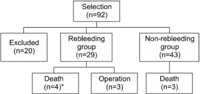
We retrospectively reviewed consecutive 92 patients
who underwent BAE due to hemoptysis. Twenty cases
were excluded in which hemoptysis was not associated
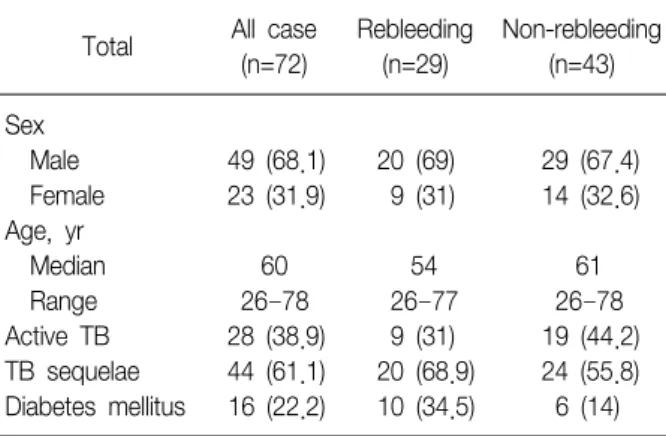
Table 1. Clinical characteristics of groups
Total All case(n=72)
Rebleeding (n=29)
Non-rebleeding (n=43) Sex
Male 49 (68.1) 20 (69) 29 (67.4)
Female 23 (31.9) 9 (31) 14 (32.6) Age, yr
Median 60 54 61
Range 26–78 26–77 26–78
Active TB 28 (38.9) 9 (31) 19 (44.2) TB sequelae 44 (61.1) 20 (68.9) 24 (55.8) Diabetes mellitus 16 (22.2) 10 (34.5) 6 (14) Values are presented as number (%).
TB: tuberculosis.
Table 2. Analysis of bleeding recurrence after immediate hemostasis
n (%) Overall immediate hemostasis 67 (93.1)
Recurrence rate after BAE 29 (40.3)
Time interval to rebleeding after hemostasis
<1 mo (early onset) 13 (18.1)
>1 mo or <1 yr 14 (19.4)
<1 yr 27 (37.5)
>1 yr 2 (2.8)
BAE: bronchial artery embolization.
with activity or sequelae of TB: metastatic tumor lesion (n=1), emphysema (n=1), leukemia (n=1), lung mass (n=4), pneumonia (n=1), and equivocal cases (n=12).
The patients consisted of 72 (49 men, 68.1%; median age, 60 years; range, 26 to 78 years) (Table 1).
2. Characteristics of rebleeding
The mean amount of bleeding at initial presentation was 210 mL (range, 20 to 2,000 mL). Overall immediate success rate of BAE was 93.1% (67 of 72). The rebleed- ing group consisted of 29 (40.3%) and the non-rebleed- ing group 43 patients (59.7%). The patients with suc- cessful BAE have undergone follow-up for median 13 months (range, 1 to 63 months). Among 29 patients of the rebleeding group, 13 patients experienced re- currence within 1 month, and 14 patients between 1 month to 1 year. Only two cases showed rebleeding be- yond 1 year after initial hemostasis by BAE: bron- chiectasis (n=1) and TB destroyed lung (n=1) managed by conservative medication and BAE, respectively. So, rebleeding after BAE was shown in 27 of 72 cases with- in 1 year (Table 2).
3. Rebleeding risk factors
Active TB patients were 38.9% (n=28) including MDR TB (n=10). Post-TB sequelae included bronchiectasis, TB destroyed lung, aspergilloma, or fibrotic scar change (Table 3). Among five patients who failed to stop bleed-
ing by BAE immediately, two patients died: one patient with active MDR TB showed massive rebleeding died despite second BAE within 30 days after first BAE; an- other patient with active non-MDR TB had massive re- bleeding and expired despite second BAE within 60 days after BAE. In subgroup of 28 patients with active TB, MDR TB showed significantly higher rebleeding than non MDR TB (odds ratio [OR], 7.5; 95% confidence interval [CI], 1.276 to 44.085; p<0.035). Active TB group excluding MDR TB was significantly associated with non-rebleeding (OR, 0.240; 95% CI, 0.061 to 0.949;
p=0.044).
The rate of recurrence based on the underlying pul- monary disease was 40% (10 of 25) for bronchiectasis, 51.6% (16 of 31) for destroyed lung, 100% (6 of 6) for aspergilloma, 36.7% (11 of 30) for fibrotic scar change, and 32.1% (9 of 28) for active TB. Active TB was not associated with risk of rebleeding (OR, 0.57; 95% CI, 0.21 to 1.53; p>0.05). Bronchiectasis, TB destroyed lung, fibrotic change, cavity, age, sex, or amount of he- moptysis were not associated with the risk of recurrence of bleeding (Table 3).
Recurrence rates in the suspected bleeding site based on direct and/or indirect sign in angiographic finding were 50% (15 of 30) for right lung, 21% (4 of 19) for left lung, and 39% (7 of 18) for both lung (p=0.315).
Shunt and neovascularization of angiographic finding were significantly different between rebleeding and non-rebleeding group (OR, 2.91; 95% CI, 1.09 to 7.74;
p<0.031 and OR, 3.07; 95% CI, 1.06 to 8.91; p<0.036,
Table 3. Analysis of risk factors affecting rebleeding after immediate bleeding control with bronchial artery embolization
Etiology Case (n=72)
n (%)
Rebleeding (n=29) n (%)
Non-rebleeding (n=43)
n (%) Odds ratio (95% CI) Sex
Male 49 (68.1) 20 (69) 29 (67.4) 1.07 (0.39–2.95)
Female 23 (31.9) 9 (31) 14 (32.6)
Active TB 28 (38.9) 9 (31) 19 (44.2) 0.57 (0.21–1.53)
MDR TB 10 (13.9) 6 (20.7) 4 (9.3) 2.54 (0.65–9.97)
Non-MDR TB 18 (25) 3 (10.3) 15 (34.9)
TB sequelae
Bronchiectasis 25 (34.7) 10 (34.5) 15 (34.9) 0.98 (0.37–2.64)
TB destroyed lung 31 (43.1) 16 (55.2) 15 (34.9) 2.29 (0.88–6.02)
Aspergilloma 6 (8.3) 6 (20.7) 0 (0) 2.87 (2.06–3.99)
Fibrotic scar change 30 (41.7) 11 (37.9) 19 (44.2) 0.77 (0.29–2.02)
Cavity 16 (22.2) 8 (27.6) 8 (18.6) 1.67 (0.54–5.11)
Angiographic finding
Shunt 36 (50) 19 (65.5) 17 (39.5) 2.91 (1.09–7.74)
Hypervascularity 59 (86.8) 19 (76.0) 40 (93.0) 0.24 (0.05–1.05)
Extravasation (direct sign) 9 (13.2) 6 (24.0) 3 (7.0) 4.21 (0.95–18.68)
Neovascularization 36 (53.7) 17 (70.8) 19 (44.2) 3.07 (1.06–8.91)
Diabetes mellitus 16 (22.2) 10 (34.5) 6 (14) 3.81 (1.11–13.12)
Amount of bleeding
<200 mL 43 20 (69) 23 (53.5)
>200 mL 29 9 (31) 20 (46.5) 1.93 (0.72–5.19)
CI: confidence interval; TB: tuberculosis; MDR: multi-drug resistant.
Table 4. Risk factors of recurrence after BAE in multi- variate logistic regression analysis
Odds ratio 95% CI p-value
Sex 1.293 0.40–4.18 0.668
Age 1.025 0.98–1.07 0.260
Shunt 3.650 1.06–12.62 0.041*
Aspergilloma 3.557 1.16–10.87 0.026*
DM 4.351 1.19–15.86 0.026*
*Statistically significant with p<0.05.
BAE: bronchial artery embolization; CI: confidence interval; DM:
diabetes mellitus.
respectively). The findings of nonbronchial systemic ar- tery in angiographic examination were not risk factors for rebleeding. Findings of extravasations as direct sign in angiography (the pathognomonic sign of hemor- rhage)
3were not associated with rebleeding after BAE (OR, 4.21; 95% CI, 0.95 to 18.68).
There were no significant factors which can influence on difference between early-onset group and late-onset group among active TB, destroyed lung, fibrotic change, aspergilloma, diabetes mellitus, shunt, age, sex, amount of bleeding, and embolization material.
Diabetes mellitus (DM) was the factor significantly as- sociated with a risk of rebleeding (OR, 3.81; 95% CI, 1.11 to 13.12). Of 16 patients with DM, 10 patients (62.5%) showed recurrent bleeding: 6 patients (60%) within 1 month and 4 (40%) after 1 month. Among these 10 patients, 4 patients (40%) had active TB, 2 (20%) destroyed lung, 2 (20%) fibrotic change, 1 (10%) bronchiectasis, and 1 (10%) aspergilloma.
Aspergilloma were significantly associated with re-
bleeding after BAE (p=0.003). Aspergillom, DM and ex- istence of shunt in angiographic finding were the risk factors of recurrence after BAE in multivariate logistic regression analysis (Table 4).
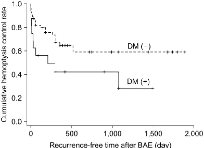
Cumulative hemoptysis non-recurrence rate after BAE were 84.7% for 1 month, 76.2% for 2 months, 71.4%
for 6 months, 59.4% for 12 months, 55.7% for 24
months, and 50.6% for 36 months. Recurrence-free time
Table 5. Independent factors of recurrence-free time after BAE in multivariate analysis using Cox's regressional haz- ards model
Hazard ratios 95% CI p-value
Shunt 1.85 0.83–4.13 0.134
Aspergilloma 4.76 1.78–12.70 0.002*
DM 2.35 1.04–5.27 0.039*
*Statistically significant with p<0.05.
BAE: bronchial artery embolization; CI: confidence interval; DM:
diabetes mellitus.