R E S E A R C H A R T I C L E
Open Access
Hemoptysis requiring bronchial artery
embolization in patients with
nontuberculous mycobacterial lung disease
Su Hwan Lee
1, Jin Hwa Lee
2, Jung Hyun Chang
2, Soo Jung Kim
2, Hee-Young Yoon
2,
Sung Shine Shim
3, Min Uk Kim
4, Sun Young Choi
3and Yon Ju Ryu
2*Abstract
Background: Although infections caused by nontuberculous mycobacteria (NTM) are increasing in prevalence, there are few data about hemoptysis in patients with NTM lung disease. This study investigated the characteristics and prognosis of hemoptysis secondary to NTM infection.
Methods: Following a retrospective review of cases managed between 2006 and 2016, 183 patients with NTM lung disease were enrolled and analyzed.
Results: Among 183 patients with NTM lung disease, Mycobacterium intracellulare (n = 64, 35%) was the major cause of NTM infection, followed by M. avium (n = 59, 32.2%) and M. abscessus complex (n = 40, 21.9%). Hemoptysis developed in 78 patients (42.6%), among whom 33 (42.3%) required bronchial artery embolization (BAE). Between patients with and without hemoptysis, there were no significant differences with respect to sex, radiographic manifestations, distribution over 3 lobes on chest computed tomography, history of pulmonary tuberculosis, antiplatelet or anticoagulation therapy, and species of NTM. However, mean age at diagnosis was significantly lower in the hemoptysis group in univariate and multivariate analyses (65.7 ± 12.8 vs. 59.7 ± 11.8, P = 0.002, odds ratio: 0.969, 95% confidence interval: 0.944–0.996). Among patients with hemoptysis, those requiring medical therapy and those requiring BAE were not significantly different in terms of demographic characteristics, radiographic manifestations, and distribution over 3 lobes. All patients who received BAE showed immediate clinical improvement, no procedure-related complications, and none of them died during the period under review.
Conclusions: NTM lung disease patients commonly experienced hemoptysis without specific risk factors except for relatively young age. Although some patients with hemoptysis needed BAE, the success rate of BAE was high, and there were no serious complications associated with BAE.
Keywords: Mycobacterium infections, nontuberculous, Tuberculosis, Hemoptysis, Embolization, therapeutic Background
The prevalence of human infection with nontuberculous mycobacteria (NTM), an emerging cause of chronic pul-monary disease, is gradually increasing worldwide [1,2]. Most NTM have low pathogenicity, and they are widely distributed in natural environments, such as soil and water sources [3]. It is difficult to distinguish among
NTM colonization, contamination, and infection. The American Thoracic Society (ATS) and the Infectious Disease Society of America (IDSA) reported diagnosis criteria for NTM infection with composite and complex contents in 2007 [4].
There are more than 150 known NTM species, and new species have recently been discovered [3]. Although there are regional differences, Mycobacterium avium complex, Mycobacterium kansasii, and Mycobacterium abscessus complex (MABSC) are known major pathogens associated with pulmonary diseases [5,6]. The initiation of treatment for NTM disease depends on various factors, such as
© The Author(s). 2019 Open Access This article is distributed under the terms of the Creative Commons Attribution 4.0 International License (http://creativecommons.org/licenses/by/4.0/), which permits unrestricted use, distribution, and reproduction in any medium, provided you give appropriate credit to the original author(s) and the source, provide a link to the Creative Commons license, and indicate if changes were made. The Creative Commons Public Domain Dedication waiver (http://creativecommons.org/publicdomain/zero/1.0/) applies to the data made available in this article, unless otherwise stated.
* Correspondence:[email protected]
2Division of Pulmonary and Critical Care Medicine, Department of Internal
Medicine, College of Medicine, Ewha Womans University, 1071 Anyangcheon-ro, Yangcheon-gu, Seoul 07985, Republic of Korea Full list of author information is available at the end of the article
individual clinical course, comorbidities, and adverse ef-fects of antimycobacterial therapy. Furthermore, long-term treatment is required after initiation [4]. Hemoptysis is among the various potential clinical features of NTM disease, regardless of treatment [3].
The severity of hemoptysis ranges from blood-tinged sputum to massive hemoptysis, and it can be life-threatening depending on the cause and severity [7]. In Korea, pulmonary tuberculosis is known to be the lead-ing cause of hemoptysis, and there are many studies on pulmonary tuberculosis and hemoptysis [8,9]. However, there are few data about hemoptysis among patients with NTM lung disease in Korea as well as in other countries. This study investigated the characteristics and prognosis of NTM lung disease with hemoptysis. Methods
Study design and study subjects
This study was performed through a retrospective review of medical records at Ewha Womans University Mokdong Hospital, a tertiary referral hospital in Seoul, Korea. We reviewed 272 patients who had at least one NTM isolation in the sputum or bronchial lavage specimen culture be-tween January 2006 and December 2016. Among them, 207 patients were diagnosed with NTM lung disease ac-cording to the ATS/IDSA diagnostic criteria [4]; 183 pa-tients with available medical records were included in the final analysis. The enrolled patients were divided into two groups according to the occurrence of hemoptysis. Pa-tients with hemoptysis received medical management when the amount of hemoptysis was low or vital signs were stable, and further management—such as bronchial artery embolization (BAE) and surgery—was considered if the hemoptysis persisted or the vital signs were unstable. We evaluated the development rate of hemoptysis, the characteristics of hemoptysis patients, and the complica-tions that occurred among patients who underwent BAE.
Data and definitions
Data for all enrolled patients were collected from the hos-pital’s electronic medical records. We recorded various demographic and comorbidity data, including sex, age, and body mass index, as well as the presence of diabetes mellitus (DM) and hypertension. We also noted the ad-ministration of antiplatelet and anticoagulation medica-tion, which may be associated with hemoptysis. All enrolled patients underwent chest X-ray and chest com-puted tomography (CT) scans, which were reviewed retro-spectively by the same chest radiologist (SS Shim). For the purposes of this study, we evaluated chest CT images from when hemoptysis was present for patients with hemoptysis; for patients without hemoptysis, we used the most recent follow-up chest CT images. Radiologic find-ings were classified as either nodular bronchiectatic type
or fibrocavitary type. The extent of lung involvement was classified as either localized disease with involvement of 1 to 3 lobes or extensive disease with involvement of 4 to 5 lobes. Potential comorbidities were compared using the Charlson Comorbidity Index [10].
Hemoptysis was defined as the patient who underwent chest CT scan for hemoptysis evaluation. We excluded patients who did not undergo CT scan when defining hemoptysis. BAE was defined by actual embolizations; this definition did not include simple angiographies. The analysis included 33 consecutive patients who under-went BAE performed by two interventional radiologists (SY Choi, MW Kim). Medications, including antitus-sives, antibiotics, and tranexamic acid, were used for pa-tients who had hemoptysis but did not undergo BAE.
Statistical analysis
Results for all continuous variables are reported as mean ± standard deviation, and all categorical variables are re-ported as numbers and percentages. Between groups, con-tinuous variables were analyzed using t-tests, and categorical variables were analyzed using the χ2test and Fisher’s exact test. Multivariate analysis was performed using the results of initial univariate analysis and logistic regression. All statistical analyses were performed using SPSS version 23.0 (IBM Corp., Armonk, NY, USA). In all comparisons, a P-value of < 0.05 was considered statisti-cally significant.
Results
Baseline characteristics
The baseline characteristics of enrolled patients from January 2006 to December 2016 are summarized in
Table 1. Among 183 patients, there were 74 males
(40.4%), and the mean age at diagnosis was 62.9 ± 12.7 years. Mycobacterium intracellulare (n = 64, 35%) was the most frequent cause of NTM infection, followed by
Mycobacterium avium (n = 59, 32.2%), MABSC (n = 40,
21.9%). Fiberoptic bronchoscopy was conducted on 53% of patients for the diagnosis of NTM infection or evalu-ation of hemoptysis. Patients with the nodular bronch-iectatic type of NTM disease were more common than those with the fibrocavitary type (n = 155, 84.7% vs. n = 28, 15.3%, respectively). Of the 155 patients with nodular bronchiectatic NTM disease, 113 had localized disease, and 42 had extensive disease. Of the 28 patients with fibrocavitary NTM disease, 12 had localized disease, and 16 had extensive disease. During a mean follow-up period of 48.7 ± 37.8 months, 78 patients (42.6%) devel-oped hemoptysis, and 33 of 78 (42.3%) develdevel-oped hemoptysis requiring BAE. No patients underwent sur-gery to treat hemoptysis.
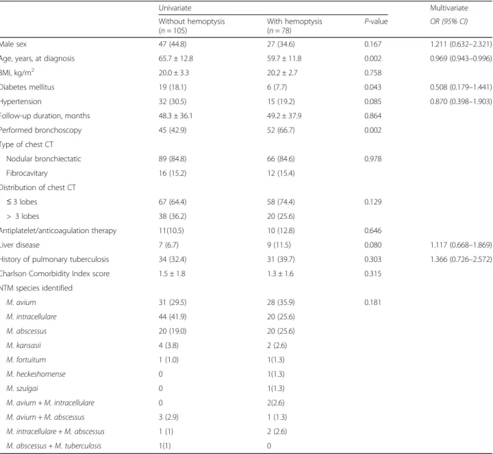
Comparison of groups
For comparison, enrolled patients were divided into two groups according to the occurrence hemoptysis (Table2). There was no significant difference between the groups in terms of follow-up duration, the proportion of male sex, radiologic finding classification, extent of lung involve-ment, presence of pulmonary tuberculosis history, anti-platelet/anticoagulation therapy, and species of NTM. However, patients in the hemoptysis group tended to be
younger than those without hemoptysis (mean age 65.7 ± 12.8 vs. 59.7 ± 11.8 years, P = 0.002). Additionally, patients without hemoptysis had a significantly higher DM preva-lence than those with hemoptysis (18.1% vs. 7.7%, P = 0.043). In the multivariate analysis including sex, the pres-ence of a pulmonary tuberculosis history, and variables with P-values < 0.1 from the univariate analysis, only age at diagnosis showed a statistically significant between-group difference (odds ratio: 0.969, 95% confidence inter-val: 0.944–0.996, P = 0.023).
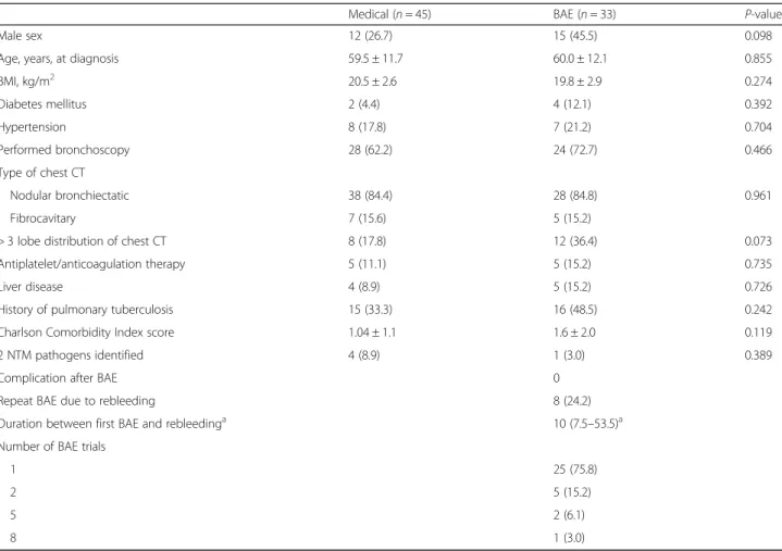
Analysis of group with hemoptysis
Of 78 patients with hemoptysis, 33 patients received BAE, and 45 patients received medical treatment (Table 3). All patients who received BAE showed immediate clinical im-provement, and there were no patients who required sur-gery for hemoptysis. Among patients with hemoptysis, those requiring medical therapy and those requiring inter-vention therapy with BAE were not significantly different in terms of demographic characteristics, radiologic find-ings, extent of lung involvement, and comorbidity index. Additionally, there were no procedure-related complica-tions or deaths, and there was no hemoptysis-related mortality. Eight of the 33 patients who underwent BAE subsequently underwent repeat BAE due to rebleeding, and the median interval between first and second BAE was 10 months (interquartile range 7.5–53.5 month). Four pa-tients experienced recurrent hemoptysis within 12 months after the first BAE, and three of them required more than two more BAEs (total 8, 5, and 5 BAEs, respectively). On the other hand, 12 out of the 45 patients who received
medical therapy experienced recurrence of their
hemoptysis. Recurrent hemoptysis in both groups was mainly the nodular bronchiectatic type. However, patients in the BAE group were mainly infected with MABSC, and patients who received medical treatment were mainly
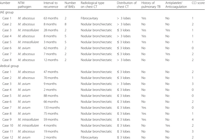
in-fected with Mycobacterium avium complex. Table4shows
the clinical characteristics among recurrent hemoptysis pa-tients after BAE and medical therapy.
Discussion
This study investigated the clinical characteristics, the de-velopment rate of hemoptysis requiring BAE, and the treatment of hemoptysis among NTM lung disease pa-tients. Many patients with NTM lung disease experienced hemoptysis (42.6%) regardless of amount, and 18% of NTM lung disease patients required BAE. No significant risk factors for hemoptysis were determined other than relatively young age. Many patients (33/78, 42.3%) re-ceived nonmedical treatment for their hemoptysis, and BAE was preferred to surgical treatment. Furthermore, BAE was not associated with significant adverse events, and patients who experienced recurrence within 12 months were more likely to require BAE treatment.
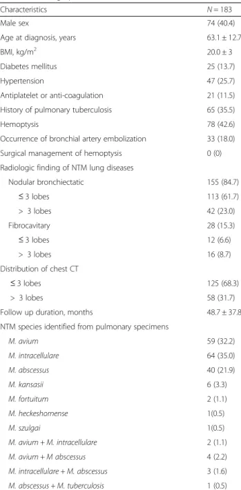
Table 1 Demographic and clinical characteristics
Characteristics N = 183 Male sex 74 (40.4) Age at diagnosis, years 63.1 ± 12.7 BMI, kg/m2 20.0 ± 3 Diabetes mellitus 25 (13.7) Hypertension 47 (25.7) Antiplatelet or anti-coagulation 21 (11.5) History of pulmonary tuberculosis 65 (35.5) Hemoptysis 78 (42.6) Occurrence of bronchial artery embolization 33 (18.0) Surgical management of hemoptysis 0 (0) Radiologic finding of NTM lung diseases
Nodular bronchiectatic 155 (84.7) ≤ 3 lobes 113 (61.7) > 3 lobes 42 (23.0) Fibrocavitary 28 (15.3) ≤ 3 lobes 12 (6.6) > 3 lobes 16 (8.7) Distribution of chest CT ≤ 3 lobes 125 (68.3) > 3 lobes 58 (31.7) Follow up duration, months 48.7 ± 37.8 NTM species identified from pulmonary specimens
M. avium 59 (32.2) M. intracellulare 64 (35.0) M. abscessus 40 (21.9) M. kansasii 6 (3.3) M. fortuitum 2 (1.1) M. heckeshornense 1(0.5) M. szulgai 1(0.5) M. avium + M. intracellulare 2 (1.1) M. avium + M abscessus 4 (2.2) M. intracellulare + M. abscessus 3 (1.6) M. abscessus + M. tuberculosis 1 (0.5)
Results are presented as n (%) or mean ± standard deviation, unless otherwise indicated
BMI body mass index, NTM nontuberculous mycobacteria, CT computed tomography, M. Mycobacterium
Until now, NTM lung disease was known to cause hemoptysis through pulmonary parenchymal infection
[11]; however, it is not considered a major cause of
hemoptysis. Although the causes of hemoptysis vary from country to country—bronchitis, bronchogenic carcinoma, bronchiectasis, Paragonimus westermani in-fection, and pulmonary tuberculosis are known to be common causes [8, 12–14]. However, our findings dem-onstrated that many NTM lung disease patients experi-ence hemoptysis requiring BAE.
Recently, the prevalence of NTM lung disease has been gradually increasing worldwide, and its onset is known to be associated with old age [1, 15, 16]. As the
global population continues to age, and many countries already have aging or aged societies [17], the proportion of NTM lung disease is expected to increase, and the frequency of NTM lung disease-associated hemoptysis is expected to increase.
In the multivariate analysis, relatively young age was revealed as an independent risk factor. Unfortunately, this observation is difficult to explain. However, regard-ing pulmonary tuberculosis, some studies have reported that hemoptysis was significantly more common among younger people [18,19]; the assumption is that the more aggressive immune response among younger patients may be more likely to induce hemoptysis [20]. Similarly,
Table 2 Comparison of groups with and without hemoptysis
Univariate Multivariate Without hemoptysis (n = 105) With hemoptysis (n = 78) P-value OR (95% CI) Male sex 47 (44.8) 27 (34.6) 0.167 1.211 (0.632–2.321) Age, years, at diagnosis 65.7 ± 12.8 59.7 ± 11.8 0.002 0.969 (0.943–0.996) BMI, kg/m2 20.0 ± 3.3 20.2 ± 2.7 0.758
Diabetes mellitus 19 (18.1) 6 (7.7) 0.043 0.508 (0.179–1.441) Hypertension 32 (30.5) 15 (19.2) 0.085 0.870 (0.398–1.903) Follow-up duration, months 48.3 ± 36.1 49.2 ± 37.9 0.864
Performed bronchoscopy 45 (42.9) 52 (66.7) 0.002 Type of chest CT Nodular bronchiectatic 89 (84.8) 66 (84.6) 0.978 Fibrocavitary 16 (15.2) 12 (15.4) Distribution of chest CT ≤ 3 lobes 67 (64.4) 58 (74.4) 0.129 > 3 lobes 38 (36.2) 20 (25.6) Antiplatelet/anticoagulation therapy 11(10.5) 10 (12.8) 0.646 Liver disease 7 (6.7) 9 (11.5) 0.080 1.117 (0.668–1.869) History of pulmonary tuberculosis 34 (32.4) 31 (39.7) 0.303 1.366 (0.726–2.572) Charlson Comorbidity Index score 1.5 ± 1.8 1.3 ± 1.6 0.315
NTM species identified M. avium 31 (29.5) 28 (35.9) 0.181 M. intracellulare 44 (41.9) 20 (25.6) M. abscessus 20 (19.0) 20 (25.6) M. kansasii 4 (3.8) 2 (2.6) M. fortuitum 1 (1.0) 1(1.3) M. heckeshornense 0 1(1.3) M. szulgai 0 1(1.3) M. avium + M. intracellulare 0 2(2.6) M. avium + M. abscessus 3 (2.9) 1 (1.3) M. intracellulare + M. abscessus 1 (1) 2 (2.6) M. abscessus + M. tuberculosis 1(1) 0
Results are presented as n (%) or mean ± standard deviation, unless otherwise indicated
in our study, NTM lung disease may have also caused more hemoptysis among relatively young individuals via a more aggressive immune response. Additionally, our study showed that the occurrence and severity (requiring BAE or not) of hemoptysis among NTM lung disease pa-tients was not associated with chest CT patterns, severity of involvement, or species of NTM pathogen. Although patients with cavities on chest CT were expected to ex-perience severe hemoptysis as well as recurrence, there was no difference compared to those with bronchiectatic CT features. This may be because the great majority of the study sample did not have cavitary lung disease, or be-cause bronchiectasis itself is a major risk factor for hemoptysis [9]. Therefore, clinicians need to consider the possibility of hemoptysis in young patients with NTM re-gardless of severity and extent of lung involvement.
BAE has been used as the first-line therapy, or a bridge to surgical treatment, for massive hemoptysis [14]. With im-proved technology, the overall success and immediate clin-ical success rates of BAE have been reported to be as high as over 90 and 73% to 99% [14]. Although several guide-lines and studies have reported that NTM lung disease can
cause hemoptysis, the treatment of hemoptysis has not been extensively detailed, with some studies briefly men-tioning surgical treatment [3,4,21–23]. However, this study demonstrated that BAE is a good treatment option for hemoptysis in NTM lung disease, like other diseases which can cause hemoptysis [7]. There was immediate clinical success and no serious complications for all patients who underwent BAE during the period under review.
About one-quarter of BAE patients developed recurrent hemoptysis in this study. Some studies have reported re-currence in 10 to 20% patients over follow-up periods between 6 and 12 months [24–26]. Considering the dur-ation (median 10 months) between first BAE and rebleed-ing in this study, BAE for hemoptysis in NTM lung disease showed a similar recurrence rate to BAE for hemoptysis in other diseases [24–26]. Causes of rebleed-ing include incomplete embolization, revascularization, or recanalization [24–26]. Some studies have found pulmon-ary tuberculosis and aspergillus to be risk factors for the recurrence of hemoptysis [27,28]. However, although we did not perform the relevant statistical analysis due to the small sample size, the presence of lung damage from
Table 3 Comparison of BAE and non-BAE subjects with hemoptysis
Medical (n = 45) BAE (n = 33) P-value Male sex 12 (26.7) 15 (45.5) 0.098 Age, years, at diagnosis 59.5 ± 11.7 60.0 ± 12.1 0.855 BMI, kg/m2 20.5 ± 2.6 19.8 ± 2.9 0.274 Diabetes mellitus 2 (4.4) 4 (12.1) 0.392 Hypertension 8 (17.8) 7 (21.2) 0.704 Performed bronchoscopy 28 (62.2) 24 (72.7) 0.466 Type of chest CT Nodular bronchiectatic 38 (84.4) 28 (84.8) 0.961 Fibrocavitary 7 (15.6) 5 (15.2)
> 3 lobe distribution of chest CT 8 (17.8) 12 (36.4) 0.073 Antiplatelet/anticoagulation therapy 5 (11.1) 5 (15.2) 0.735 Liver disease 4 (8.9) 5 (15.2) 0.726 History of pulmonary tuberculosis 15 (33.3) 16 (48.5) 0.242 Charlson Comorbidity Index score 1.04 ± 1.1 1.6 ± 2.0 0.119 2 NTM pathogens identified 4 (8.9) 1 (3.0) 0.389 Complication after BAE 0
Repeat BAE due to rebleeding 8 (24.2) Duration between first BAE and rebleedinga 10 (7.5–53.5)a Number of BAE trials
1 25 (75.8)
2 5 (15.2)
5 2 (6.1)
8 1 (3.0)
Results are presented as n (%) or mean ± standard deviation, unless otherwise indicated
BAE bronchial artery embolization, BMI body mass index, CT computed tomography, NTM nontuberculous mycobacteria, M. Mycobacterium
a
previous pulmonary tuberculosis did not appear to affect recurrence in this study. It was notable that most rebleed-ing patients were infected with MABSC and had nodular bronchiectatic radiologic findings on chest CT. Although this finding is hard to generalize due to the small number of patients with recurrent hemoptysis, considering that MABSC is relatively difficult to treat and can also progress with a fulminant course and that bronchiectasis features on chest CT include hypertrophy and tortuosity of the bronchial arteries [14,29], these may be suggested as risk factors for the recurrence of hemoptysis.
This study had some limitations, including its retrospect-ive design and that it was performed at a single center. Therefore, in our study, data regarding the rate of hemoptysis, hemoptysis volume, indication for BAE, and the techniques used for BAE—including late hemoptysis during the follow-up period after diagnosis—were not always avail-able and may not be accurately reported. Additionally, we could not review the first presentations among the NTM lung disease patients or the role of bronchoscopy among these patients. This study did not reveal how NTM treat-ment affected the occurrence or recurrence of hemoptysis because we could not find consistent indications for NTM
treatment in this study. Finally, subjects in this study had a higher prevalence of pulmonary tuberculosis history com-pared with other studies because Korea has a high preva-lence of tuberculosis [1, 8, 30]. It is, therefore, difficult to generalize the results of this study to countries with low rates of tuberculosis. However, this study is meaningful be-cause, to our knowledge, it is the first study of hemoptysis in NTM lung disease. As the prevalence of NTM disease in-creases, prospective studies about hemoptysis in NTM lung disease may be needed.
Conclusion
NTM lung disease patients commonly experienced hemoptysis without specific risk factors except for relatively young age. Some patients with hemoptysis required BAE, the success rate of BAE was high, and there were no serious complications associated with BAE. Therefore, BAE could be considered a suitable treatment option for hemoptysis in NTM lung disease.
Abbreviations
ATS:American Thoracic Society; BAE: Bronchial artery embolization; CT: Computed tomography; DM: Diabetes mellitus; IDSA: Infectious Disease
Table 4 Clinical characteristics of recurrent hemoptysis patients after BAE
Number NTM pathogen Interval to recurrence Number of BAEs Radiological type on chest CT Distribution of chest CT History of pulmonary TB Antiplatelet/ Anticoagulation CCI score BAE group
Case 1 M. abscessus 63 months 2 Fibrocavitary > 3 lobes Yes No 1 Case 2 M. abscessus 8 months 8 Nodular bronchiectatic > 3 lobes No No 2 Case 3 M. intracellulare 28 months 2 Nodular bronchiectatic ≤ 3 lobes Yes Yes 1 Case 4 M. abscessus 8 months 5 Nodular bronchiectatic > 3 lobes Yes No 1 Case 5 M. intracellulare 3 months 5 Nodular bronchiectatic ≤ 3 lobes Yes No 1 Case 6 M. avium 62 months 2 Nodular bronchiectatic ≤ 3 lobes No No 0 Case 7 M. abscessus 7 months 2 Nodular bronchiectatic ≤ 3 lobes No Yes 2 Case 8 M. abscessus 12 months 2 Nodular bronchiectatic > 3 lobes No No 2 Medical group
Case 1 M. abscessus 47 months Nodular bronchiectatic ≤ 3 lobes No No 2 Case 2 M. abscessus 70 months Nodular bronchiectatic ≤ 3 lobes No No 1 Case 3 M. avium 9 months Nodular bronchiectatic > 3 lobes No No 3 Case 4 M. avium 2 months Nodular bronchiectatic ≤ 3 lobes No No 0 Case 5 M. avium 88 months Nodular bronchiectatic ≤ 3 lobes No No 0 Case 6 M. avium 66 months Nodular bronchiectatic ≤ 3 lobes No No 2 Case 7 M. avium 133 months Nodular bronchiectatic ≤ 3 lobes Yes No 0 Case 8 M. avium 75 months Nodular bronchiectatic ≤ 3 lobes Yes No 1 Case 9 M. intracellulare 59 months Nodular bronchiectatic ≤ 3 lobes Yes No 4 Case 10 M. intracellulare 4 months Nodular bronchiectatic ≤ 3 lobes No Yes 2 Case 11 M. abscessus 19 months Nodular bronchiectatic ≤ 3 lobes No No 3 Case 12 M. avium 2 months Fibrocavitary ≤ 3 lobes No No 2
BAE bronchial artery embolization, NTM nontuberculous mycobacteria, CT computed tomography, TB tuberculosis, CCI Charlson Comorbidity Index, M. Mycobacterium
Society of America; MABSC: Mycobacterium abscessus complex; NTM: Nontuberculous mycobacteria
Acknowledgements None.
Author contributions
SHL and YJR take full responsibility for the content of this manuscript, including its data and analysis. SHL and YJR made substantial contributions to the conception and design of the study. SHL, SJK, HYY, JHL, JHC, and YJR made substantial contributions to the analysis and interpretation of data. S.S.S. interpreted the radiological images. MUK and SYC interpreted and analyzed bronchial artery embolization images. SHL and Y.J.R. drafted the initial manuscript. All authors discussed the results and reviewed the manuscript. All authors read and approved the final manuscript.
Funding
This research did not receive any funding from agencies in the public, commercial, or not-for-profit sectors.
Availability of data and materials
The datasets generated and/or analyzed during the current study are not publicly available due to our IRB policy but are available from the corresponding author upon reasonable request.
Ethics approval and consent to participate
The protocol for this study was approved by the institutional review board of Ewha Womans University Mokdong Hospital in Korea (IRB FILE No: EUMC 2018–10-032). All methods were performed in accordance with the relevant guidelines and regulations. Informed consent was waived by the institutional review board because of the study’s retrospective nature.
Consent for publication Not applicable. Competing interests
The authors declare that they have no competing interests. Author details
1Division of Pulmonology, Department of Internal Medicine, Severance
Hospital, Yonsei University College of Medicine, Seoul, Republic of Korea.
2Division of Pulmonary and Critical Care Medicine, Department of Internal
Medicine, College of Medicine, Ewha Womans University, 1071 Anyangcheon-ro, Yangcheon-gu, Seoul 07985, Republic of Korea.
3
Department of Radiology, College of Medicine, Ewha Womans University, Seoul, Republic of Korea.4Department of Radiology, Seoul National
University Boramae Medical Center, Seoul, Republic of Korea.
Received: 26 December 2018 Accepted: 19 June 2019
References
1. Adjemian J, Frankland TB, Daida YG, Honda JR, Olivier KN, Zelazny A, et al. Epidemiology of nontuberculous mycobacterial lung disease and tuberculosis, Hawaii, USA. Emerg Infect Dis. 2017;23(3):439–47. 2. Stout JE, Koh WJ, Yew WW. Update on pulmonary disease due to
non-tuberculous mycobacteria. Int J Infect Dis. 2016;45:123–34.
3. Johnson MM, Odell JA. Nontuberculous mycobacterial pulmonary infections. J Thorac Dis. 2014;6(3):210–20.
4. Griffith DE, Aksamit T, Brown-Elliott BA, Catanzaro A, Daley C, Gordin F, et al. An official ATS/IDSA statement: diagnosis, treatment, and prevention of nontuberculous mycobacterial diseases. Am J Respir Crit Care Med. 2007; 175(4):367–416.
5. Ryu YJ, Koh WJ, Daley CL. Diagnosis and treatment of nontuberculous mycobacterial lung disease: Clinicians’ perspectives. Tuberc Respir Dis (Seoul). 2016;79(2):74–84.
6. Hoefsloot W, van Ingen J, Andrejak C, Angeby K, Bauriaud R, Bemer P, et al. The geographic diversity of nontuberculous mycobacteria isolated from pulmonary samples: an NTM-NET collaborative study. Eur Respir J. 2013; 42(6):1604–13.
7. Jean-Baptiste E. Clinical assessment and management of massive hemoptysis. Crit Care Med. 2000;28(5):1642–7.
8. Kim SW, Lee SJ, Ryu YJ, Lee JH, Chang JH, Shim SS, et al. Prognosis and predictors of Rebleeding after bronchial artery embolization in patients with active or inactive pulmonary tuberculosis. Lung. 2015;193(4):575–81. 9. Hwang HG, Lee HS, Choi JS, Seo KH, Kim YH, Na JO. Risk factors influencing
Rebleeding after bronchial artery embolization on the Management of Hemoptysis Associated with pulmonary tuberculosis. Tuberc Respir Dis (Seoul). 2013;74(3):111–9.
10. Sundararajan V, Henderson T, Perry C, Muggivan A, Quan H, Ghali WA. New ICD-10 version of the Charlson comorbidity index predicted in-hospital mortality. J Clin Epidemiol. 2004;57(12):1288–94.
11. Larici AR, Franchi P, Occhipinti M, Contegiacomo A, del Ciello A, Calandriello L, et al. Diagnosis and management of hemoptysis. Diagn Interv Radiol. 2014;20(4):299–309.
12. Hirshberg B, Biran I, Glazer M, Kramer MR. Hemoptysis: etiology, evaluation, and outcome in a tertiary referral hospital. Chest. 1997;112(2):440–4. 13. Prasad R, Garg R, Singhal S, Srivastava P. Lessons from patients with
hemoptysis attending a chest clinic in India. Ann Thorac Med. 2009;4(1):10–2. 14. Sopko DR, Smith TP. Bronchial artery embolization for hemoptysis. Semin
Intervent Radiol. 2011;28(1):48–62.
15. Ringshausen FC, Wagner D, de Roux A, Diel R, Hohmann D, Hickstein L, et al. Prevalence of nontuberculous mycobacterial pulmonary disease, Germany, 2009-2014. Emerg Infect Dis. 2016;22(6):1102–5.
16. Adjemian J, Olivier KN, Seitz AE, Holland SM, Prevots DR. Prevalence of nontuberculous mycobacterial lung disease in U.S. Medicare beneficiaries. Am J Respir Crit Care Med. 2012;185(8):881–6.
17. Beard J, Suzman R. Global health and aging: preface. 2011.http://www.who. int/ageing/publications/global_health/en. Accessed 1 Mar 2018.
18. Perez-Guzman C, Vargas MH, Torres-Cruz A, Villarreal-Velarde H. Does aging modify pulmonary tuberculosis?: a meta-analytical review. Chest. 1999; 116(4):961–7.
19. Rizvi N, Shah RH, Inayat N, Hussain N. Differences in clinical presentation of pulmonary tuberculosis in association with age. J Pak Med Assoc. 2003; 53(8):321–4.
20. Achkar JM, Joseph G. Independent Association of Younger age with hemoptysis in adults with pulmonary tuberculosis. Int J Tuberc Lung Dis. 2012;16(7):897–902.
21. Haworth CS, Banks J, Capstick T, Fisher AJ, Gorsuch T, Laurenson IF, et al. British Thoracic Society guidelines for the management of non-tuberculous mycobacterial pulmonary disease (NTM-PD). Thorax. 2017;72(Suppl 2):ii1–ii64. 22. van Ingen J. Treatment of pulmonary disease caused by non-tuberculous
mycobacteria. Lancet Respir Med. 2015;3(3):179–80.
23. Wassilew N, Hoffmann H, Andrejak C, Lange C. Pulmonary disease caused by non-tuberculous mycobacteria. Respiration. 2016;91(5):386–402. 24. Osaki S, Nakanishi Y, Wataya H, Takayama K, Inoue K, Takaki Y, et al.
Prognosis of bronchial artery embolization in the management of hemoptysis. Respiration. 2000;67(4):412–6.
25. White RI Jr. Bronchial artery embolotherapy for control of acute hemoptysis: analysis of outcome. Chest. 1999;115(4):912–5.
26. Menchini L, Remy-Jardin M, Faivre JB, Copin MC, Ramon P, Matran R, et al. Cryptogenic haemoptysis in smokers: angiography and results of embolisation in 35 patients. Eur Respir J. 2009;34(5):1031–9.
27. Katoh O, Kishikawa T, Yamada H, Matsumoto S, Kudo S. Recurrent bleeding after arterial embolization in patients with hemoptysis. Chest. 1990;97(3):541–6. 28. Lee S, Chan JW, Chan SC, Chan YH, Kwan TL, Chan MK, et al. Bronchial
artery embolisation can be equally safe and effective in the management of chronic recurrent haemoptysis. Hong Kong Med J. 2008;14(1):14–20. 29. Lee MR, Sheng WH, Hung CC, Yu CJ, Lee LN, Hsueh PR. Mycobacterium
abscessus complex infections in humans. Emerg Infect Dis. 2015;21(9):1638–46. 30. Huang HL, Cheng MH, Lu PL, Shu CC, Wang JY, Wang JT, et al.
Epidemiology and predictors of NTM pulmonary infection in Taiwan - a retrospective, five-year multicenter study. Sci Rep. 2017;7(1):16300.
Publisher’s Note
Springer Nature remains neutral with regard to jurisdictional claims in published maps and institutional affiliations.