www.earticle.net
동융자원연구 21(2):82-87 (2010)
Annals of Animal Rcsouπ、s Scicnæs 21α:):82-87 (2010)
소 난자의 체외성숙 시 는댄세포와의 공동배앙이 체외수정 후 난자의 발육에 미치는 영향
전혜레나1 청회태2
.OJ-부근1 박춘근,.
강원대학교 동붕생명과학대학 강원대학교 수의과대학2
Effects of Co-cuIture with Cumulus Cells on bz Vitro Maturation
뻐d [농velopment
of Bovine OocytesHy~Le-Na
Jeon1, Hee-Tae Oteong2
,’
ßo<>.K에n
Yang’ and G1oon-Keun Part·때l쩡
01Alli"찌
/ife Scie"æs, Kilngux:m Natiol1o/미liversity,
‘ Co/l,앵'e of Veterinnry Medicine, Kangt
‘
xm Nation.a1 UniversityABSfRACf
The 0따tive of this study 뼈 !。 뼈uate the effects of ∞。J1tureof bovine oocytes with αm띠 us cells on in vitro maturation arνj development followîng in vitro fertilization în bovine oocytes, Bovine cum띠u&<JOCyte
∞뼈,Iexes (,α:G) 때 denuded oocytes (00) were co-rultl뼈 with the cumulus cells in 1CM199 for 2(• 22 tu-, and evaluated the nudear type of αx:yte. After i" vitro maturatior、 oocyte;
‘
wrt' coio::ubated for j" t,;fro fertilization with frozen-thawed s야rπ\atozoa selected by 65% 야π011 in DM-H,야)il.rtn aI、d DM-Caffeine for 15-18 tu-. Prεωnptive zygotes W'eI"e 떼 tured for 48 hr în 0깅aa ,’, uitro culture IT생ium with 10% FB5, ar녕 ev피uated the deavage rates. 1he results confirmed that the hi영-.,;1 떠"('ffitage of rret:ap띠", 11 α4미 stage was observed in ax::s (30.H3.5%, 24.2:1:1.8%) as ∞mpared to 0。σ1:tl.3%, 17.4:tl3.9%) (p에뼈 In addition. the incr얹혀c1eavage rates were φtained from ax::s (69.6:t21%, 75.6:t2.9%) ‘이m ∞mp'"떠 to DO (.잉 6û.5%, 29.5:!:l26%)
@에 α)). In ∞nclusion, th잉 study suggested that αmulus cells 앙8언ed positive fac
‘
ors during íll vitromaruration of oocytes and early embryonic development after ;11 vitro fertilization of bovine (x양e
(Key words: Bovine oocytes, Co-c띠bure, 0ωm매 us cells, ItJ vitro maturatioπ Embryo development)
[
서론
2αη년부터 소고기 이력제가 시앵핑에 따라 이력추척을
위해 영확한 개제정보가 핑요하게
되었고 이러한 개제식영 시스댐용 통해 우수 유전자원의 정보흩 활용하고 있다 기존
얘 개쩨 정보가 영확하지 않았댄 도축왼 압소로부터 채집한 난소흉 。l용항 정우 영확하지 않은 유전정보흉 사용하재 되 므로 우수형질올 활용할 수 없다 한연 영확한 유전정보릉 가진 우수형쟁의 앙소가 도축휠 때 그 개체의 우수행정용황용할 수 있고 우수형질융 사용하기 위
해서는 체외수갱용 통한 수정란 생산기술의 영요성이 커지고 있냐 그러나 하나
의 개체에는 두 개의 난소뿐이", 하나의 난소에서 얻용 수
있는 난자의 개수는 재한적이다 이를 난^fOII서 우수 난
자흉선병하여 이용원 수 있는 난자의 수는 급격히 떨어지게 되 고 선별한 난자
내에서도 인공수정에 쓰이는 수정란으후 빵 랄하는 난사는
얘우척다 따각서 체외수갱란올 수갱란이식 에 도입.j.는데
문재갱융안견하였고 개채앵 채외수정란 생 산성 향상과 이용 중대률
위한 안정적인 체외수갱랑 생산은 정상쩍인 셰내 조건과 같은 만촉할만한 수준이1 도당하지 옷 하였다 이려한 문제훌 극복하기 위해 난포액이나 혹」F액퉁과 같은 체액의 이원'CoIlas 둥"
1991),난굉장피세포.(Eyestone과
First, 19t:에/난구세포(GoI。 등
IS1'l8)등과같은 공동배양이
• αI'TCSponding author: Choon-Kcun Park, D:!α 이 Animal Biotcchnology, collcge of A찌"'" 미c SciCOC'CS, Kan양NOn Nati Oi때 University, Dlundleon '2f.μπn, Korea. Tl언 +82-33녕;()&;2]. ε”‘lil: fWi<.ck@kang“~.ιk
[Provider:earticle] Download by IP 118.70.52.165 at Monday, December 20, 2021 8:06 PM
www.earticle.net
황항하게 이루어지고 있다 。l전의 실험올 흥혜 채외성숙시 기에 셰세포와 공동배양이 돼지, 개 등의 난자 핵 성숙에 긍 갱척인
효과가 었다고 뺑졌다κatska 둥
1995; Abeydeern동., 11})8; Hewitt와 En잉 and, 1999; Bureau 둥" 200);
&썽jo!,。 둥" 2(x)2; μßsa 둥, 2002; Kid∞n 둥, 2003) 체세포 의
종류인 난판상피세포와 난자와의
공동배양용제외에서 왜지 난자 성숙과 체외 배
생산효올올 향상시키고(Ooen
퉁, 20(6)
다정자수정 6낸용
낮추oj,자용 전핵단계에
도당하는 난자의 비융율 중가시켰다ρ,buc과
5때'" 1995)또
한 난구애포는 정자 가능올 향상시키고(G씨kin 퉁, 1m;Boatrnan과 Rol쳐05, 1991), 다갱자 수정을 제여하며
(Overstreet 퉁., 1978; Cununiæ와 Yanagimachi, 1쨌), 수갱
시 정자률 낭사 쪽으로 유도한대Austin과
Brnden, 1952)한충 더 나아가
난구세 g는수정이 이루어지는 낭관 앵대부
의환정에시쳐령
갱자에게비슷한
직용을한다따dford와 Ki,π
19Ç에 따라서 본 연구는 소 난구세포올 회수, 배양하 여 체내 난포~정과 비슷한 환정 죠성올 통혜 난구세포와 공동애양시 난자의 성숙율파 배 발달융에 미치는 영양올
검토하였다IL
재료및 방법
1 뇨맘새포의 준비
도상
직후적충원 소의 난소로부터 채취한 난지-난구 복 합체를 인위켜으로 툴리켜얀 아창을 이용해 난자와 난구세
포률분리시킨 후 낭구써포만용 수집하였다 수짐한
난구세포률
10% FBS가챙기핀 TCM-I99배양액으로
2-3회 에척후 세포수융
1-2x1ifcell/ m1의 농도로 조정하여
4“~Il<tish에 배양액과 항께 500 이썩 분주하였으며 38.5'(;, 5%
CD,의
조건으로
얘양하였다 24시간애양 후 상충액융 재거 하고 새로운 배양액올 갱가하여 체외애양에 이용하였다
미성숙 난포란올 채취하었다 채취한 미성숙 난자 중 난구 세포가 치얻하고 3층 이상이여 세포갱이 균칭한 난포란 만
을
선멸하여난지~난구옥함에툴 대조군으로 난구세포툴 풀 리척인 방법으로 재거한 남자훌 처리군으로 나누어
TCM.199배양액에 10% FB5, 0.1% fGF읍 정가하여 조성원 배양 액에서 2-3회 세척하였다 이를 난자를 난구세포가 당총으 로 작성왼 4써성l 이sh에 옹겨 500 띠의 배양액올 분주한 :f
20-22시간동안 38.5'(;, 5% αi가 유지되는 인큐베이터에서 공동배양하였다
2)
난자의 성숙융분석
난자의
성숙윷온mæm염색옹 이용하여 분석하였다 체 외생숙원 낭자릅 난구세포흘 완벽히 제거한 후
211개썩슬라 이드 글라스
위에옳긴 후 커버글라스훌 덮어
acetate9J.이hanol이 1 대 3.91 비융로 혼합한 고정액에서 2-4일 동안
고정시켰다
그후 난자는
1%acetic-αæm으로
10분동안 영씩한 후" aceto-잉y=이로
세척하였다 영색왼난
'''1볕현 미정하에서
액형올판찰함으흐써 성숙융올 분석하였다
3 제외수정
20- 22시간 체외성숙이 유도왼 난자를 난구세포가 화장왼
난포란용 선별하여
제외수정 배양액인 0.1 g ffiA가 청가왼 α싸k뼈rinE.후3회
세혀후
157H의 난x←즘 % 띠의체외
배양 소적으로 융긴 후 보관하였다 동결정액올 37'(;의 항 용 수조에서 45호간 융해 후 1 m!의 65% 어mU에 운주해 3(0) ')'m에서 10분간 원심분리하였다 운동성이 있는 정자수가 2xl
<f개1m!가 되도록 하여 채외수정 배양액
DMζaffeine과 혼항해 재외배양 소척에 50 씨씌 분주한 후,
38.5 '(;, 5%
CD,가
유지되는 인큐베이터얘서 15-18시간 수정시켰다
4 체외배앙
수정배양액으로부터 수정원 난지융 회수하여 난구세포와
2
난자의 핵 싱숙 갱자용 제거 후
10%FBS가 청가왼 。갱 aa 얘양액으로 3회
새척
:f5O띠의 셰외배양 소객에 용겨 48시간 배양하였다
1) 난포란외 희수 및 예외성숙도촉'lNl서 희수한 년소릎 38'(;의 멸균
생리식염수로 5 통계분석 1-2회 새척한 다용 칭척하여 실험실로 운반하였다 난포란의
채취는
18-gauge의주사칭이 장작된 주사기로
2-7 mm온 연구얘서 얻어진 경파를은
SA5 9.1 Duncan다중검정
의난포로부터 난포액과 함께 난포란율 홈입하는 땅엄율
사으로 통계
분석하였jl, p<O.05 야하의유의성 경과를올 서
용하였대 난포액과 난포란의 혼합올얘 l객&PVA륭 회석해 '" 비교하였다[Provider:earticle] Download by IP 118.70.52.165 at Monday, December 20, 2021 8:06 PM
www.earticle.net
84 용융자원연구 제 21 킨 Z호 (201이
m.
결과및 고찰
셰외성숙 시
난구세포와 공동얘양의 유무에
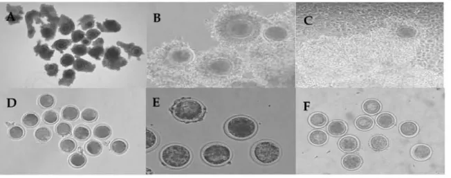
여흔 난자의 형태학척 변화오습용 Fig. 1 에 나타내었다난^f.난구쩍←합채를
난구세포와 공동배양
하였올 때큰 차
이는 나타내지않았으나 공동배양하지 않온 난지-닌구복합 체에
비해난구세포 앵창현상이
더크게
나타냈다 20-22시간 성숙시킨 난구셰포 제거 난자는 난구셰포와 공동배양하
여성숙시킨
갱우예 비해 세포질의용축, 흐릿혜진
또는 어두워지는 현상이 더 많이 관창되었다 이전에 실험에서 난 구세포는 소(Liu 둥, 1‘195와
깅~ng 둥, 1995)와버팔로σ)as 풍i
199끼 난자의 세포질그리고 핵의 성숙에 휠요하다고
보B
고되었다 아러한 결과흉 통해 난구세포홍 재거해 성숙시켰 용 경우 낭자의 세포갱이 현저히 떨어징이 판한핑얘 따라
셰외에서 성숙이 되었다고 해도 질 좋은 수정란 생산응
툴가능할 것으료 생각되며, 또한 남구세포의 존재는
핵성숙
완 아니라 난세포질에도 영향용 미치는 것으로 추측되었다 제외생숙시 난구세포와 공동배양의 유우에
따흔난자의
성숙정도를 Table 1에 나타냈다체외생숙 후 남자의
핵형용 관창한경파 수정이
가농한성
숙낭자 단계인 M-U단계에서 난À}- 난구욕합체그훌(24.2:!:1.8%, 30.1>3.5%)이 난구세포률 재거한 난자그룹(l.1>1.3%,
17.4:!:l3.9%)보다 성숙융이 유의책A로 높게 나타났다
@예 05)
또한 공동배양하지 않온
난자내에서
난구세포룡E 훌’·
• • ~훌
Fig. 1. Morphol야~ of bovine oocytes during in vitro maturation
" CIJCS (Cum버잉 Oocyte ∞nψlexes) before maturation,. B: cocs 하ter rnaturatiOl、 " CIJCS 따ter maturatiOll co-cultured with rumulus α:11, D: 00 (denud띠 Ch:ytes) bcfore maturation. E: ∞ 따ter maturation, F: D。따tcr maturatlon ∞αJtured WÎth
마mul야 " ,11
Tablc 1. Nuclcar typc of bovinc 。‘>cytcs c<H:ulturcd with cumulus cclls during in νitro maturation Group No. of Nuc1"" ψ1"' of
@"""
αgruSEM. %)00ni\e QI P-I M-I A-I T-I p.I1 wι11
CIJCS 66 0 11 10 1 4
2
‘
16(0'이
06.7t3이i (l5.2i4외(1.5t1 끼
‘ (6.1t。이 (21.7'21) (24.2:tl.8)ab=“
?꺼 (L4:t1 l.2){13.7i2.피i
10 (6.IHl5 끼 (1.4:t1 l.6)" ('.1tU3 3) (잉 5:t3.1) 31 (잉1>3.5)" 깅DO 70 5 깅 10 8 7 U 3
(7.1:1:21) (329I9.에. 04.3t5 이 (11.4:1:1에‘ (10:1:4.6) (17.1:1:5 씨 σ l:tl.3)"
CO-D。 46 1 6 3 1 10 17 8
(22t3끼
(13.0:1:5.이b
(65:1:2에 (22)28)' (21.7:1:21) (37.0:1:14.1) (17.4:t13 의@CIJCS, cum비야 oocψe Complexes, CD-OXS: cxxs roαltured ‘Nith cum띠us ælls, ro. D:muded 。∞_CO-oo D。
∞αltuæd with rumulus ccll, GV: Genninal Vesiclc, p.[: Prop벼'" 1, 샤1: Metaphase 1, A.I: Anaphase 1, T.I: Tcl。뼈.se 1, M-II
M-ta"""'"
11AA\<" Va1ues ‘Mth 이hπnt letters within colWTll""lS are signilicantly dj떠'erent (p<O.ffi)
[Provider:earticle] Download by IP 118.70.52.165 at Monday, December 20, 2021 8:06 PM
www.earticle.net
Table 2 Oeavage of bovine embryos co-cu1tured with cumu1us cells during ;', vitro maturation
Group No 。이
C1 cavagl이%) N。 이 embroys developed (Mean;tSEM,%)
αX'yte 2 cell 4 cell 8-16 cell Deg‘
COCs 110 69‘6%2.1' 20 20 32 33
(20.0::t3 끼 (20.9:t2.4t (29.H3.0)"
(30.0소
2.1)bco-ccεs 155 75.6:t2.9" 38 30 48 39
(24.5::t3 이 (19.4:t2히. (31.0::t1.5)" (25.2소O.9)b
α〕 76 21.6!7.Sb 11 4 1 60
(14.5::t3.1) (5.3:t2.9)b (1.3::tl꺼b (78.9::t7.5)"
CO-OO 73 29.5::tl2.6b 12 10 1 %
(16.4::t6 이 (13.7:t6.2)‘ (1.4::tl.4)b (68.5! 12.6)"
O:DC cun 띠잉 。ocyte Complexes, CO-COCS: cocs co-cultured with αm띠잉 cells, 00: 다:!nuded Ch:ytes, CO-OO: 00
∞αltured with cumulus cell, I:kg.: IR양’lE!ration.
..., Va\ues wilh differcnl lclters wilhln ∞lumns are significantly 이fferent ψ얘뼈
제거히여 성숙사켰융 경우 이성숙난지 단계인 A-I (11.4::t 1 에단계가 유의척으로 높게 나타났고 같옹 미성숙난자 단 계인 P-I (329i9.8%)단계에서 공동배양하지 않옹
난구세포
제거낭자가 공동 배양한남구세포
재거낭자{13.0::t 5.6%)보 다유의격으로
높제 나타났다@이뼈이성숙
난자의 비융 이높아짐으로서
낮옹성숙융에 따용
쩨외수정또한
낮옹 수정융올보일것이라고
추측왼다본
실험응 난자의 높응 수정란 생산올 옥표도 하기에 체외성숙 시 난구세쪼의 필요가 용가피하다고
생각된다 본 연구훌 통해 남구세포 유우에 따라 성숙난사 단계까지의 성숙융이 높아지는 것용 확인할수 있었고 난구에포와 공동배양함에 따라 생숙융에 긍정걱
인 효과가 있응용 확인하였다
체외성숙
시 난구써포외
유우에 따라 성숙시킨난자외
제 외수정 후 분할용용 Table 2에나타냈다
수정 후 운할율을 판창환 경과 공용배양한 낭자난구북합 체 그흉{75.6소29%) 의 분항융이 일반객인 방엄으흐 체외생 숙시킨 난자난구복합체의 경우,69.6>2.1%)보다 높은 것으 호 나타났지만 유의직인 자이는
없었다
또한 닝구세포를 제거 .f여 낭구세포와 공동 배양환 경우,29.5<126%)와 대조 구j21.6<7.5%)에서제외
성숙시킨 난자와도 유의적차이가
냐타나지 않았다그러나 난자-난구복합체용 성숙시켰흘
때의 분할융(69.6.
21%,
75.6>2.9%)이 난구세포용 제거혜
성숙시킨 난자의분
할융ρ1.6::t7.5%, 29.5::tl26%) 에 비해 유의껴으로 높게 나타났대P<O,뼈, 이러한
결파는 성숙시키기 션 남구세포의 유무가
수갱얘영향융
끼지는 것으로 생각된다이전의
실험에서는
취의경우
난구세포흉 제거한 난자룡 쳐|외에서배양 하었올
때 투영대가 정화되고(De f,마.e과 Siracusa. 1982), 투영대의 경화는 수갱 비율의 갑소와 판언이 있다 (Gianfortoni 와 C띠yas, 198.되고 보고되었다 소에서는 채외수갱 이전에
난구세포 제거의 해로훈 효파가
관찰되었대'Co, 동., 1993 zhang둥,
1995: Fatehi 웅, 2002) 수갱시 남구세포는 성숙 원 난자의
수정비융을
향상시키고.(Co' 둥" 1993), 소 α.enz 둥, 198잉, 엠스터(Bavister, 1982), 얀간βock 둥" 1989)의 경 우 난구세포의 몇몇요소을온
정자의 청제반용올 측진시킨 다 또한 체외수정 이진에 남구세포의 쩌거는 세포에 의해 분비되는 요인의 손실로 인해 분할율에 해로운 영향을 끼친 다{Fa핸j 둥, 2002) 이전의 실험파 본 설험올 통해 난구세포의 유무에 따를 수정 에 는 여허 가지 요얀에 의해 와우핍
올 확인할 수 있었Jl, 난구세포가 난사의 분할융훌 높이는
데
효과가 있융올 확인하였다본
연구의
결과로부터 닝구세포는소
남자의 체외성숙과 셰외수정 후의 초기 애 말육에 긍정적인 효과가 았으며/ 추 후 이에 따릉 난구써포의 효과에 대한 얘카니즘옹 규명할 수 있도록 추가실험이
필요하다고생각완다
W
요약
용 연구는
소 난사의
체외생숙시 낭구새포와의
공동배양。1 체외수정 ~
낭자의
발육에미치는
영향을 검토하였다 난.~~난구복합체와 난구세포용 채거한 난자로 나누어 TCM 199배양액에서 앙22시간동안난구세포와
공동배양시킨 후 。rrem
영씩올 실시해 난자의 빽형옹 운석하였다 성숙배양
시킨후
O싸야wπ1배양액으흐
낭자흩 보관하고 65%며℃이l로 운려한 풍결-융해왼 정자를 DM<affeine 배양액과
[Provider:earticle] Download by IP 118.70.52.165 at Monday, December 20, 2021 8:06 PM
www.earticle.net
86 용융자원연구 제 21 킨 Z호 (201이
혼합해 15-18 시간수정시켰다 。l툴 난자는 α1 혀 배양액에 서 48시간 배양시켜 용항융을 완창하었다 난자의 성숙융융
관창한 결과
M-I딴계에서난자난구복합체가(3O.H3.5%,
24.2.1.8%)가 난구세포훌 제거한 남자(7.1.1.3%, 17.4.13.9%)
보다 성숙융이 유의척으료 높게 나타났으며ψ<0뼈‘ 또한 공동배양하지 않옹 난자 내에서 난구세포용 쩌거하여 성숙 시켰올 정우 P-I β,29.9.8%) 이 유의적으로 높게 나타났다
(p<OO5)
분할용용 관창한 결과
난자-난구복합체률성숙시
컸을
때의분할융{f:8.6:t2.1%, 75.6:t29%)이 난구세포률 재거
해 성숙시킨 난자의 분할융{21.6.7.5%, 29.5t126%)에 "1해유의적으로 높게 나타났다@이 O히 온 연구의 절과로부터 난구세포는 소
난자의채외성숙파 체외수정 후의 초기 배 방육에 효과객인 울질의
분비가 있는것으로 추측되며, 추
후 배반¥모의 방당을 지켜보는 추가싱험이 훨요하다고 생각핑다
사사
본
연구는 농링수산식풍부 농링기술개벨사업(Grant N。
1α>007-3)의 지왼에
의해
이추어진것이여, 연구의 수앵 시
소 정액의 보존파 분석을 위해 협조한 강원대학교 동율자원공동연구소에 감사드립니다
V.
인용문현
1. Abeyd",πa, L R, Wang, W. H., Cantly, T. C, Rieke, A, Prnther, R 5. and Day, 8. N. 1~. Oxulture ‘이th
follìcular shell pìeces can enhar‘:e the developrrental
。ompeten:e of pig QO(γte 따ter in uitro fertìlization:
relevance to intracellular glutathione. B 이 Reprod. 58 213-218
2 Austirι C R ar녕 Braden. W. H 1952 p,잉osage of the spenn and the pmetration of tl
‘
e egg în mammals.Nature. 170:919얘21
3. &visU!T, B. D. 1962 Evidt!lU! for a rolt! uf
α"'-ov띠atory cum띠w; ∞mponents in suppαtmg fertili깅ng ability of hamiter sperπlatozoa. J. A찌rOI 3,365-372
4.&잉fom, J. M and Klln, H. H. 1
‘
193. Cumulus ∞야",rus"" a """"' 잊때Jestering deηce, in vivo. J. Exp. Zool
265:321-328
5 "갱h이0, L, 2edda, M T., Ledda, 5., Leoni, G., Naitana, S. and Pau, S. 2002. lnOueoce of ∞-cuI ture with
。,viductal epitt에 ial cells on in uitro maturation of canine oocytes. Reprod. Nutr. D?v. 42:265-273
6. Boattnan, D. E ar녕 Rob이"LS, R R 1991. [농tection of a
50Iu비 e acrosorre reaction-ir‘ducing factor, different from 앙um albumin,. a용ooated with the ovulated
야잉ß<um띠떠 complex. MoI. RepJ여 [농V. 3fr.39tμm
7. Bureau, M, Bailey, J. L and Sirard, M A 2cαl νtfl uen:e of oviducta1 cells and ∞쩌jtioned m::d.ium on porrine 링JTEteC;.. Zigote. 8:139-144.
s.Ü>en, X.Y., 니 Q. W., Zhang, S. 5., Han, Z 5., zl‘aα κ Wu, 5. Y. and Huang, J. 'lf1Jl 타fros 이 ovarian rortex æll αl-CU lture during i’! vitro πlaturation on porcine oocytes maturatioπ f얀tili깅 tion ar녕 a빼'}'O
eveloprrent. Animal R매πxluction Sdence. 99::n>-316 9. CoIl잉, P. and Robl, J. M 1991. R아ationship 야hν""
nuclear rem:xl.eling and dev,리얘rænt in nuclear
h퍼lSplant rabbit embryos. BioL Reprod. 45:4!잊냐65
10. coκ J. F., Hormazabal, J. and San어 Maria, A 1옛영 티ìect 야 the 。띠nulus 00 in uifro fertilization of bovine
mahu혀 oocytes. lheri야껑、。 ogy.4α12땅1267 11. Cummins, J. M and Yar녕19imachi, R 1982. Spenn-egg
rnti∞ and the site of the acrα'ìO!re reaction during ù
,
vitro fertilization in the hamster. Gamete Res. 5
239약;6.
12 Das, S. K., Qauhaπ M 5., Palta, P. and Torrer, 0. S.
19\η lnfluence of cum띠us cells on in lJitro maturation of denuded bu뻐l。 αx:ytes. Vet. Rec. 141:522-523 13. D? f,잉 ici, M and Siracusa, G. 1~ ’5pontaneous'
hardening of the wna pellucida of rrouse 0αyte;
durir잉 in vitro culture. Grurete Res. 6:107-113 14. Dubuc, A and Sirard, M A 1995. Effect of
cocu1turing spermatozoa with ovîductal c티~ on the incidence of polyspermy in pig in vitro fertilization.
M:lL Reprod. IRv. 41:360-367.
15. Eyestone, W. H. and First, N. L 1969. co-。이]ture of
않rly cattle embryos 10 the 버""'0<γ51 stage with oviducal tissue or in COi녕itioned medium. J. Reprαj Fert. 85 기5-72ll
16. Fatehi, A N., Zeinstra, E C, KI。이, R V., Colenbrar녕er, B. and Bevers, M M 2002 Eifect
(‘
[Provider:earticle] Download by IP 118.70.52.165 at Monday, December 20, 2021 8:06 PM
www.earticle.net
cumulus c잉15 remo\i띠 。 f in VÎtro matured bo이ne
αr꺼es priOT to in vitro fertilization on subseqllent deavage rate 까-.en야g、。,logy. 57(4)'1347-55.
17. G이'0, K , f에 ihara, Y., Kasaka, 5., Koba, M., Nakanish.
Y.ru、d 。링‘써 K 19E였 Pregnarries after αxu1 ture of
cum띠 us cells with bo、깨ne embryos derived from in vitro fertilization of in vitro mahlred follicular α>eYtES
j. Reprod. Fertil. 83 연:1-758.
18. Gianfortoni, J. G. and G비yas, B. J. 1~. The effects of short-tenn iκubation (.야9"잉 이 mω.se oocy이:es on rn
uitro fertilization. zor딩 에ubility, and embryonic development. Gamete Res. 11:59-68
19. G“latkin, R B. L, Andersα、 。F. and Hutchiso", C F. 1m.. Capocitation of hamster spermatozoa in vitro the role of cumu1us ro찌"'"'잉1ι J. Repr,여 r<ertil. 30:
잊19-394
21.1 Hewitt, D. A and En잉and. G. C W. 1잊19. Synthetic
。.viducta1 fluid and 。‘ ductal cell coculture for canine
αqte maturation i
’
I vitro. Anim Repπx1. Sci. 55,63-75
21. Ka야a, L , Rynska, B. and Srrorag, Z 1995. 까'" effect
이 ∞c띠 ture system on developmental ca어0에 。f
bovine IVMjlVF oocytes. Theri야전@ 야;y. 43,85%70 22 Kidsor、 A., 닮lOeVers, E., lan양’찌1<, P., Verheiiden,.
J., Colenbrru녕er, B. ar녕 Bevers, M 21X13. The effect of oviductal epithelial cell ∞αlture during in vitro maturation on sow αx.ytes iOOrpoology, fertilization and embryo development. 1heri gonol아;y. 5'}1889- 1903
23. Lenζ R W., Aκ R L, Grure1ι H. J. and First, N. L
1~. prot∞영yεm from b。이 ne folliClIIar f1uid
σIhances on aCfO!!잉'ITe reaction în bovîne sperπlatozoa
Biophys. Rε Commun. 1~:1092-1098
24. Lîu, Y., Holyoak, G. κ Wang, 5. and Burrl、 T.D.
1995. The impαtance of cumulus cells on the
’
11 vitroproduction of bovîne oocytes. Theri야엠",1야;y. 43267 25 μ.ûsa, 8., Maria, T. z., Sergiα L, Giovanni, L,
Salvatore, N. and Salvatore, P. 2002. 1r삐uer<e of
C'O<Ulture with 。、riductal epithelial celIs on in uitro maturation of canine α>eytES. ~야:mxl Nutr. Dev 잉;
265-7-끼3
26. J\.1ahi-Browπ C A. ru녕 Yana잉machi, R 1983
Pamme떠"s influencing ovum pickup by the oviductal fimbria în the golden 뼈m;ter. Garre‘e Res. 8:1-10 27. OverstTeeι j. W" C∞per, G. W. and Katι D. F. 1978.
Sperm tr밍1Sport in 야le reproductive tract of the female rabbit. 1I. n、e sustained 야'\aSe of transport BioL Repr여 19:115-132
28 잊ock. C E., Bates, R, Lîndsay, K 5., Edmonds, D. K
m、d Fraser, L R 1989. Human oocyte-cωn띠 us
oomplexes stimu1ate the human a。α:;ome reaction. J Refrod. Fe며1. 86π:1-730
29. zt‘ang, L, Lîang, 5., Wozniak. P. J., Yang, X. and
Gαike, RA1잊l5. Cumu1us cell functjon during bovine oocyte maturation. fertilization and er빼')'0 deve1때nent În VÎtro. MoL Reprod. ~. 4O::X양.344
(투고일 외10.12~ 수정일 2010.1213 판정일 2010.1214)
[Provider:earticle] Download by IP 118.70.52.165 at Monday, December 20, 2021 8:06 PM