ISSN 0378-6471 (Print)⋅ISSN 2092-9374 (Online)
https://doi.org/10.3341/jkos.2019.60.1.55
Original Article
벤잘코니움의 장기 노출이 섬유주세포의 노화에 미치는 영향
Effect of Chronic Benzalkonium Chloride Exposure on Senescence in Trabecular Meshwork Cells
박지혜1,2⋅이수진1,2⋅이규원1,2⋅김재찬1,2
Jihae Park, MD1,2, Soo Jin Lee1,2, Kyoo Won Lee, MD, PhD1,2, Jae Chan Kim, MD, PhD1,2
제일안과병원 안의학연구소1, 제일안과병원2 Cheil Eye Research Institute1, Daegu, Korea
Cheil Eye Hospital2, Daegu, Korea
Purpose: To determine the possible effects of chronic exposure of low dose benzalkonium chloride (BAK) on trabecular mesh- work cells, and to characterize the pathways involved in the effects.
Methods: Trabecular meshwork cells were treated with 0.0005%, 0.00075%, 0.001%, and 0.0025% BAK for 10 minutes; then, the cells were transferred to a new medium for 24 hours. This process was repeated three times. Cell survival was assessed us- ing the MTT assay to determine the non-apoptotic BAK concentration. Senescence-associated (SA)-β-gal staining was per- formed to compare quantitatively the cellular senescence of BAK-treated cells with the control group. Cells treated with BAK were analyzed by western blot to determine whether the expressions of cell cycle regulators were affected.
Results: Two concentrations (0.0005% and 0.00075%) showed persistent cell viability and were chosen for further experiments.
After SA-β-gal staining, cells treated with 0.0005% and 0.00075% BAK showed 28% (± 2.08), 37% (± 2.08) increases in cellular senescence expression, respectively, when compared with control cells (p < 0.05). To identify the molecular pathways involved in cell cycle arrest via BAK, western blot analysis was performed on trabecular meshwork cells, resulting in decreased ex- pressions of cyclin E/CDK2, and increased expressions of the upper stream control molecules, p53 and p21.
Conclusions: Chronic exposure to low dose BAK accelerated cell senescence through cell cycle arrest. Because senescent cells of the trabecular meshwork can inhibit its outflow pathway function and ultimately worsen the glaucomatous process, long-term usage of topical glaucoma medications containing BAK should be conducted with caution.
J Korean Ophthalmol Soc 2019;60(1):55-61
Keywords: Benzalkonium chloride, Senescence, Trabecular meshwork
■Received: 2018. 8. 14. ■ Revised: 2018. 9. 14.
■Accepted: 2018. 12. 20.
■Address reprint requests to Jae Chan Kim, MD, PhD Cheil Eye Hospital, #1 Ayang-ro, Dong-Gu, Daegu 41196, Korea
Tel: 82-53-960-0214, Fax: 82-53-959-1758 E-mail: [email protected]
* This work was supported by the National Research Foundation of Korea (NRF) grant funded by the Korea government (MSIT) (No. NRF-2017R1A2A2A05001128).
*Conflicts of Interest: The authors have no conflicts to disclose.
ⓒ2019 The Korean Ophthalmological Society
This is an Open Access article distributed under the terms of the Creative Commons Attribution Non-Commercial License (http://creativecommons.org/licenses/by-nc/3.0/) which permits unrestricted non-commercial use, distribution, and reproduction in any medium, provided the original work is properly cited.
녹내장은 만성적인 질환으로 상승한 안압을 하강시키는 것이 치료의 핵심이기 때문에 대부분의 환자들은 여러 종 류의 안압하강제를 장기적으로 사용한다.1,2 약제에 포함된 대표적인 보존제인 benzalkonium chloride (BAK)가 안구 표면에 급성 괴사와 세포 사멸, 및 산화 스트레스를 유발하 여 안구표면질환을 일으키는 것에 대해서는 이미 여러 연 구가 보고되었다.3-8 그러나 일반 녹내장 환자의 전방각과 같은 환경에서 저농도의 BAK가 세포사멸을 일으키지 않 고 방수 내 장기간 존재할 경우, 그에 노출된 세포에 어떤 유해한 변화가 일어나는지에 대하여는 아직 국내에서 발표
된 바 없다. 실제로 BAK의 친유화적(lipophilic) 성질로 인 해 장기간 사용했을 때 안구 내 섬유주뿐 아니라 맥락막과 같은 깊은 조직까지 침투하여 오랜 기간 축적될 수 있다고 알려졌으며,9,10 수술 전 BAK에 노출된 정도가 많을수록 섬 유주절제술의 실패율이 증가하여 두 인자 사이에 유의한 상관관계가 보고된 연구가 있다.11 또한 개방각녹내장의 특 징적 소견 중 하나로 세포노화가 있는데12 세포노화는 세포 주기가 비가역적으로 억제된 상태로 조직의 기능 감소에 기여하며 그 기전에는 p53 경로가 중요한 것으로 알려졌 다.13 그 외에도 BAK가 녹내장을 악화시킬 수 있다는 최근 연구 결과들이 보고되었다.1,14 이런 보고들을 바탕으로 BAK 의 만성적인 노출이 섬유주세포에 유발하는 변화와 그 기 전을 알아보고자 하였다. 본 연구에서는 1) 일반 녹내장 환 자의 안구와 유사하게 저농도의 BAK를 지속적으로 처리 한 섬유주세포의 노화 정도를 노화특이적인 senescence- associated (SA)-β-gal 염색을 통해 정량적으로 측정하고 비 교 분석하였으며, 2) 노화 촉진 기전을 세포주기 억제와 관 련된 단백질 표지자인 cyclin E/CDK2와 cyclin D1/CDK4, 그리고 그 상위기전 조절인자인 p53, p21, p16의 발현성을 통해 세포 주기와의 연관성을 확인하였다.
대상과 방법
1. 세포배양
섬유주세포(primary human trabecular meshwork cells, HTM cells)는 ScienCell Research Laboratories로부터 구매 후 5% CO2를 함유한 37°C 배양기에서 2% 우태아혈청(FBS;
HyClone, Logan, UT, USA)과 trabecular meshwork cell growth supplement (TMCGS, ScienCell Research Laboratories, Carlsbad, CA, USA)를 포함한 Trabecular Meshwork Cell Medium (TMCM, ScienCell Research Laboratories)으로 배 양하였다.
2. 약물처리
Benzalkonium chloride (BAK, Sigma Aldrich, St. Louis, MO, USA)의 지속적인 노출이 섬유주세포에 미치는 영향 을 확인하기 위해 배양 접시에 일차 배양한 섬유주세포를 6 well plate에 1.7×10⁵ cells/mL로 분주한 후, 5% CO2를 함 유한 37°C 배양기에서 24시간 동안 배양하였다. 이후 배지 를 흡입 제거하고 2% 우태아혈청만 첨가한 배양액으로 4시 간 배양한 후, BAK를 0.00075%, 0.0005%를 10분간 처리 하여 배지를 제거하고 새로운 배양액을 넣은 후 5% CO2를 함유한 37°C 배양기에서 24시간 동안 배양하였다. 이 과정 을 3회 반복하였다.
3. 세포생존율 측정
세포의 생존에 대한 효과는 세포생존과 세포독성의 screening test로 이용되고 있는 발색검사법의 일종인 MTT (3-[4, 5-dimethylthiazol-2-yl]-2, 5-diphenyltetrazolium bro- mide, Sigma Aldrich, St Louis, MO, USA) assay를 이용하 였다. MTT assay는 96 well plate에 5×10⁴ cells/mL로 분주 한 뒤, 5% CO2를 함유한 37°C 배양기에서 24시간 동안 배 양하였다. 그 후 BAK를 처리한 세포의 배지에 MTT 시약을 각 well 당 20 μL씩 투여한 후 2시간 동안 정치배양한 배양 액을 제거한 다음 dimethylsulfoxide (DMSO, Sigma Aldrich) 를 각 well당 100 μL씩 넣고, 10분 이상 흔들어 spectropho- tometer (SpectramaxTM 340PC384 microplate photometer, Spectra Max Molecular Devices, Sunnyvale, CA, USA)로 570 nm에서 흡광도를 측정하였다. 이때 세포의 생존 정도 는 실험군의 값을 약물처리를 하지 않은 대조군의 비로 나 누어 백분율로 나타내었다.
4. 섬유주세포의 노화
노화된 세포에서 특징적으로 발현되는 β-galactosidase의 활성을 측정하기 위해 상용의 senescence-associated β -galactosidase kit (SA-β-gal, Cell Signaling Technology, Danvers, MA, USA)를 이용하였고 제조사에서 제공되는 실 험 방법에 따라 실험을 진행하였다. BAK 처리가 된 세포들 의 배지를 흡입 제거하고 염류용액으로 세척한 후 세포를 고 정한 다음 SA-β-gal 염색액을 넣어 37°C에 18시간 동안 반 응시켰다. 이때 노화된 세포는 β-galactosidase를 발현하여 푸른 색을 나타내므로 광학현미경으로 β-galactosidase 양성 인 푸르게 염색된 노화된 세포수와 전체 세포수를 세어서 노 화된 세포의 정도를 백분율로 표시하였다.
5. Western blot analysis
BAK가 처리된 섬유주세포를 PRO-PREP™ (iNtRON Biotechnology Inc., Seongnam, Korea)으로 용해한 뒤 얼음 위에서 30분 동안 반응시킨 후, 13,000 rpm에서 10분 동안 원심분리한 후 상등액을 얻었다. Bicinchoninic acid (BCA) (Pierce™ BCA Protein Assay Kit, Pierce Biotechnology, Inc., Rockford, IL, USA)법으로 단백질 농도를 측정한 후 30 μg의 단백질을 sodium dodecyl sulfate-polyacrylamide gel electrophoresis (SDS-PAGE)로 분리한 뒤 nitrocellulose membrane (Pall Corporation, Port Washington, NY, USA) 에 옮겼다. Membrane을 상온에서 5% 탈지분유를 포함한 1X TBST (50 mM NaCl, 20 mM Tris, 0.05% Tween 20)로 블리킹(blocking)한 후, 4°C에서 항체 p53, p27, p16 CDK2, CDK4, cyclin D1, cyclin E (1:1,000, Cell Signaling Technology)
Figure 1. Cell viability was measuresd by MTT assay. BAK
decreased the survival of trabecular meshwork cells sig- nificantly from concentrations 0.001% to 0.0025% (*p <0.05, **p < 0.001). Veh = vehicle; MTT assay = 3-[4, 5-di- methylthiazol-2-yl]-2, 5-diphenyltetrazolium bromide assay;
BAK = benzalkonium chloride.
Figure 2. Senescence detection by senescence-associated (SA) β-galactosidase staining. Photograph of SA-β-gal positive senescent
trabecular meshwork cells (blue color) after being exposed to 0.0005%, 0.00075% of BAK. Both 0.0005% and 0.00075% BAK showed concentration depedent increase of blue stained senescent cells compared to non-exposed control (×200). Veh = vehicle;BAK = benzalkonium chloride.
와 β-actin (1:2000, Sigma Aldrich)을 overnight으로 반응시 켰다. 세척 후, membrane을 horseradish peroxidase가 붙은 항 mouse IgG 항체 또는 항 rabbit IgG 항체(1:2000, Santa Cruz Biotechnology, Santa Cruz, CA, USA)와 반응시킨 뒤, chemiluminescence Western Blotting detection kit (Pierce Biotechnology, Inc.)를 실시하였다.
6. 통계적 처리
모든 실험은 3계대에서 5계대 사이의 세포를 이용하였고 한 세트씩 3회 이상 반복하여 시행하였다. 모든 실험에서 음성 대조군은 BAK 적용을 제외한 나머지 과정을 동일하 게 시행하였으며, 실험군과 대조군의 비교는 Student's t-test 를 사용하였으며 유의수준은 p<0.05로 정하였다.
결 과
1. 섬유주세포의 생존에 미치는 영향
96 well plate에 배양한 섬유주세포에 0.0005%, 0.00075%, 0.001%, 0.0025% BAK를 10분간 처리한 후, 배지를 제거 하고 새로운 배양액을 넣은 다음 24시간 배양하였으며 이 전 과정을 3회 반복하였다. 대조군에 비하여 BAK는 0.0005%, 0.00075%에서는 섬유주세포의 생존에 영향을 미치지 않았 으나, 0.001%와 0.0025%의 농도에서는 각각 26% (p<0.05)와 86% (p<0.001)로 세포의 생존을 유의하게 감소시켰다(Fig. 1).
본 실험에서는 지속적인 BAK의 처리가 섬유주세포의 노 화에 미치는 영향을 확인하기 위해 세포 생존에 영향의 미 치지 않는 농도인 0.0005%와 0.00075%의 BAK를 사용하 여 실험을 진행하였다.
2. BAK에 의한 섬유주세포의 노화에 미치는 영향 6 well plate에 배양한 섬유주세포에 0.0005%, 0.00075%
BAK를 10분간 처리한 후 배지를 제거하고 새로운 배양액 을 넣고, 24시간 배양하였으며 본 과정을 3회 반복하였다.
노화된 섬유주세포는 SA-β-gal에 의해 푸르게 염색되었다 (Fig. 2). 음성 대조군에 비해 BAK의 농도 0.0005%와 0.00075%를 처리한 군에서 섬유주세포의 노화가 촉진되었 고, 0.0005%에 비해 0.00075% BAK를 처리한 군에서 노화 된 세포가 9% 더 유의하게 증가되었다(p<0.05) (Fig. 3).
3. BAK에 의한 세포주기에 미치는 영향
BAK에 의한 세포주기억제를 생화학적으로 분석하기 위 하여 세포주기 조절에 관련된 단백질 표지자의 발현 정도 를 확인하였다. 음성대조군과 비교하여 0.00075% BAK를 처리한 군에서 G1기와 S기의 활성인자인 CDK2와 cyclin E의 발현이 억제되는 것으로 관찰되었으며, CDK4와 cy-
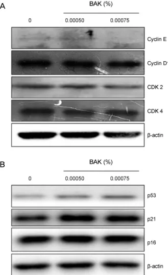
Figure 4. Effect of BAK on cell cycle regulator molecules of tra-
becular meshwork cells. β-actin, also known as a “housekeeping"protein, acts as a loading control. (A) Expressions of cyclin E and CDK2 are decreased with 0.00075% BAK while expressions of cyclin D1 and CDK4 seem to be unaffected. (B) The ex- pressions of phosphorylated p53 and p21 show concentration dependent increase with 0.0005% and 0.00075% BAK. BAK
= benzalkonium chloride; CDK = cyclin-dependent kinase.
Figure 3. Effect of BAK on senescence of trabecular mesh-
work cells. BAK concentrations more than 0.0005% increased cell senescence significantly (*p < 0.05). Veh = vehicle;BAK = benzalkonium chloride.
clin D1의 발현의 변화는 관찰되지 않았다(Fig. 4A). 상위 기전 분석을 위해 종양억제 유전자인 인산화된 p53, CDK inhibitor인 p21, 세포주기 억제자인 p16의 단백질 발현을 확인하였고 그 결과, 섬유주세포에 BAK를 처리한 군에서 인산화된 p53과 p21의 단백질 발현은 증가하였으나, p16의 발현의 변화는 없었다(Fig. 4B).
고 찰
개방각녹내장은 안압 증가로 인한 영구적인 시신경 손상 을 초래하는 만성적인 질환으로 여러 가지 기전 중의 하나 로 다양한 메커니즘에 의해 변성된 섬유주로 인한 방수유 출로의 저항 증가가 있다.5,12,15 섬유주의 변성을 설명하는 특징적인 소견으로 세포외기질의 증가, 세포소실, 세포골격 의 변화, 세포 조기 노화가 있는데, 그중 세포노화를 유발 할 수 있는 요인에 대하여 알려진 바는 많지 않다.14,16 세포 노화는 세포 스스로 수차례의 세포증식 또는 다양한 외부 자극 요인들(stress-induced premature senescence, SIPS)에 의해 유도될 수 있는데,17-19 섬유주세포 자체의 세포주기 회전율이 매우 낮은 것을 고려하면 복제 잠재력의 고갈보 다 외부 자극에 의한 세포 조기 노화 유발의 가능성이 더 크다고 볼 수 있다.14,20
세포분열은 CDK와 cyclin의 결합에 의해서 조절되며 cy- clin E/CDK2 복합체는 노화에 관여하는 작용 회로도에서 노화의 주요 활성인자로 알려진 p53 경로의 S기 활성인자 로 작용한다.21,22 인산화에 의해 활성화된 p53은 p21의 발 현을 유도하여 cyclin E와 CDK2를 억제하게 되는데 이는 결국 S기를 진행시키는 전사인자 E2F를 억제하여 노화를 유도하게 된다. 따라서 세포노화를 유도하기 위해서는 인 산화된 p53과 p21의 발현 증가, 그리고 cyclin E/CDK2의
발현 감소와 같이 각 관문에서 조절인자들의 상대적인 비 율이 중요하다.13,23-25 세포 분열이 정상적으로 조절되지 못하 는 세포는 노화에 빠지게 된다. 노화된 세포는 크기가 커지고 편평해지며 조직 수복, 재생, 대사순환을 할 수 없고, 이러한 세포가 축적되면 결국 조직의 기능이 저하된다.24,26-28
녹내장 환자는 대부분 여러 종류의 안압하강제를 장기간 사용하기 때문에 점안약에 포함된 대표적인 보존제인 BAK 에 안구가 만성적으로 노출되는 것을 피할 수 없다. BAK 는 현재 시판 중인 안과용 점안액의 약 70%에 포함되어 있 고, 다른 보존제를 사용하는 점안액은 10% 정도 밖에 이르 지 않는다.29 BAK가 안구 표면 및 전방 내에서 세포사멸 및 산화 스트레스를 유발하는 것은 이미 많은 연구를 통해
A
B
증명되었으며,30-34 특히 최근 연구에 의하면 BAK는 개방각 녹내장안의 섬유주와 같은 더 깊은 조직에도 축적되어 있 고, 장기간 사용 시 녹내장을 악화시킬 가능성이 있다고 발
표되었다.3,9,14 Yu et al5의 연구 결과에 의하면 BAK와 함께
산화 스트레스를 처리한 섬유주세포에서 세포노화가 유발 되었으나, 본 실험보다 높은 농도의 BAK를 사용하여 세포 사멸도 함께 나타났으며 노화를 유발하는 기전에 대해서는 아직 많은 연구가 필요하다고 제기하였다. 본 연구에서는 녹내장 환자의 섬유주에서 증가되어 관찰되는 세포의 조기 노화가 일반적인 녹내장의 병리적인 과정에 의한 것보다는 저농도의 BAK의 지속적인 노출에 의해 유발될 수 있다는 가설을 세웠고, 세포노화가 유발되는지, 그리고 노화가 일 어난다면 어떤 기전과 연관성이 있는지 알아보고자 하였다.
본 연구에서는 실제 녹내장 환자의 안구와 같은 환경을 조성하기 위하여 세포사멸이 일어나지 않고 섬유주세포의 생존이 유지되는 농도군인 0.0005%, 0.00075%의 BAK에 노출시킨 뒤, 노화세포 특이적 SA-β-gal 염색을 통하여 대 조군에 비해 세포의 조기노화가 유도되는 것을 확인할 수 있었다.35 세포노화의 정도는 BAK의 농도와 비례하여 증가 하였는데, 이러한 결과는 매우 낮은 농도의 BAK (0.00002%) 에 처리했을 때 섬유주세포의 밀도 감소 및 증식을 억제한 Samples et al36의 연구 결과와도 상응하였다. 노화에 빠지 게 되는 기전과 관련된 경로를 설명하기 위하여 세포 주기 에서 중요한 역할을 하는 p53, p21, p16 CDK2, CDK4, cy- clin D1, cyclin E 분자들의 발현성을 western blot를 통해 확인하였다.37,38 음성대조군과 비교했을 때 BAK를 처리한 군에서 세포주기 G1기에서 S기로 전환을 촉진하는 CDK2 와 cyclin E의 발현이 0.00075%에서 의미 있게 억제되었으 나 0.0005%에서 증가한 결과는 여러 가지 실험 조건이나 기간에 따른 좀 더 상세한 연구가 필요할 것이라 생각된다.
Cyclin D1/CDK4의 발현은 두 농도에서 모두 변화를 보이 지 않았지만 cyclin E/CDK2의 발현은 0.00075%에서 의미 있게 감소함을 보여준다. 이런 실험 결과는 cyclin E/CDK2 복합체가 관여하는 p53 의존적 경로가 BAK에 의한 섬유주 세포의 조기노화와 상관관계가 있는 반면, cyclin D1/CDK4 가 관여하는 p16 의존적 경로는 섬유주의 세포노화에는 의 미 있게 관여하지 않다는 점을 시사한다.13,39
결론적으로 BAK의 지속적인 처리는 p53 경로를 통해 세포분열을 억제하여 섬유주세포의 조기노화를 유발하는 것으로 보인다. 개방각녹내장안의 섬유주에 노화된 세포가 쌓이면 방수 유출이 잘 이루어지지 않아 안압이 오르게 되 고, 이를 개선하기 위해 안압하강 점안제를 사용하면 약제 에 포함되어 있는 BAK로 인해 오히려 노화가 더 촉진되는 악순환이 진행될 것이다. 이런 현상이 약으로 안압이 잘 조
절되는 환자임에도 불구하고 질환의 진행이 계속되는 상황 과도 연관이 있을 것으로 사료된다. 현재 녹내장의 많은 치 료용 점안약에는 BAK가 대부분 포함되어 있기 때문에 본 연구 결과에 따르면 장기간 치료를 할수록 세포노화가 유 도되어 약을 통해 얻을 수 있는 치료적 효과에는 어느 정도 한계가 있을 것으로 생각된다. 한편 BAK 자체의 살균 역 할을 고려하면 그 중요성 또한 간과할 수 없으므로 장기 치 료로 인해 얻는 비용 및 이익 또한 고려해야 한다. 그러므 로 현 치료에서 섬유주세포의 조기노화를 억제하며 치료적 효과를 극대화할 수 있는 새로운 치료제의 개발에 대한 연 구가 수행되고 위에 제기한 문제에 대한 더 다양한 연구가 진행되어야 할 것이다.
REFERENCES
1) Rasmussen CA, Kaufman PL, Kiland JA. Benzalkonium chloride and glaucoma. J Ocul Pharmacol Ther 2014;30:163-9.
2) Abu-Hassan DW, Acott TS, Kelley MJ. The trabecular meshwork:
a basic review of form and function. J Ocular Biol 2014;2.
https://www.ncbi.nlm.nih.gov/pmc/articles/PMC4209746/.
Accessed June 20, 2018.
3) Baudouin C, Denoyer A, Desbenoit N, et al. In vitro and in vivo ex- perimental studies on trabecular meshwork degeneration induced by benzalkonium chloride (an American Ophthalmological Society thesis). Trans Am Ophthalmol Soc 2012;110:40-63.
4) Chang C, Zhang AQ, Kagan DB, et al. Mechanisms of benzalko- nium chloride toxicity in a human trabecular meshwork cell line and the protective role of preservative-free tafluprost. Clin Exp Ophthalmol 2015;43:164-72.
5) Yu AL, Fuchshofer R, Kampik A, Welge-Lussen U. Effects of oxi- dative stress in trabecular meshwork cells are reduced by prosta- glandin analogues. Invest Ophthalmol Vis Sci 2008;49:4872-80.
6) Fechtner RD, Godfrey DG, Budenz D, et al. Prevalence of ocular surface complaints in patients with glaucoma using topical intra- ocular pressure-lowering medications. Cornea 2010;29:618-21.
7) Katz G, Springs CL, Craven ER, Montecchi-Palmer M. Ocular sur- face disease in patients with glaucoma or ocular hypertension treat- ed with either BAK-preserved latanoprost or BAK-free travoprost.
Clin Ophthalmol 2010;4:1253-61.
8) Rossi GC, Pasinetti GM, Scudeller L, et al. Risk factors to develop ocular surface disease in treated glaucoma or ocular hypertension patients. Eur J Ophthalmol 2013;23:296-302.
9) Brignole-Baudouin F, Desbenoit N, Hamm G, et al. A new safety concern for glaucoma treatment demonstrated by mass spectrome- try imaging of benzalkonium chloride distribution in the eye, an experimental study in rabbits. PLoS One 2012;7:e50180.
10) Desbenoit N, Schmitz-Afonso I, Baudouin C, et al. Localisation and quantification of benzalkonium chloride in eye tissue by TOF-SIMS imaging and liquid chromatography mass spectrometry.
Anal Bioanal Chem 2013;405:4039-49.
11) Boimer C, Birt CM. Preservative exposure and surgical outcomes in glaucoma patients: the PESO study. J Glaucoma 2013;22:730-5.
12) Kim JW, Kang SH, Lee KW. Effect of ascorbic acid against the oxi-
dative stress-induced cellular senescence in trabecular meshwork cells. J Korean Ophthalmol Soc 2013;54:490-5.
13) Childs BG, Durik M, Baker DJ, van Deursen JM. Cellular sen- escence in aging and age-related disease: from mechanisms to therapy. Nat Med 2015;21:1424-35.
14) Liton PB, Challa P, Stinnett S, et al. Cellular senescence in the glaucomatous outflow pathway. Exp Gerontol 2005;40:745-8.
15) Park CH, Kim JW. Effect of advanced glycation end products on oxidative stress and senescence of trabecular meshwork cells.
Korean J Ophthalmol 2012;26:123-31.
16) Lütjen-Drecoll E. Morphological changes in glaucomatous eyes and the role of TGFbeta2 for the pathogenesis of the disease. Exp Eye Res 2005;81:1-4.
17) Hayflick L, Moorhead PS. The serial cultivation of human diploid cell strains. Exp Cell Res 1961;25:585-621.
18) Toussaint O, Medrano EE, von Zglinicki T. Cellular and molecular mechanisms of stress-induced premature senescence (SIPS) of hu- man diploid fibroblasts and melanocytes. Exp Gerontol 2000;35:
927-45.
19) Toussaint O, Royer V, Salmon M, Remacle J. Stress-induced pre- mature senescence and tissue ageing. Biochem Pharmacol 2002;
64:1007-9.
20) Rohen JW, Lutjen-Drecoll E. Biology of the trabecular meshwork.
In: Lutjen-Drecoll E, ed. Basic Aspects of Glaucoma Research, 1st ed. Stuttgard; Schattauer FK, 1982;141-66.
21) Sherr CJ. Mammalian G1 cyclins. Cell 1993;73:1059-65.
22) Xiong Y, Hannon GJ, Zhang H, et al. p21 is a universal inhibitor of cyclin kinases. Nature 1993;366:701-4.
23) Roninson IB. Tumor cell senescence in cancer treatment. Cancer Res 2003;63:2705-15.
24) Campisi J, d'Adda di Fagagna F. Cellular senescence: when bad things happen to good cells. Nat Rev Mol Cell Biol 2007;8:729-40.
25) Gu Z, Cao X, Jiang J, et al. Upregulation of p16INK4A promotes cellular senescence of bone marrow-derived mesenchymal stem cells from systemic lupus erythematosus patients. Cell Signal 2012;24:2307-14.
26) Hayflick L. The limited in vitro lifetime of human diploid cell strains. Exp Cell Res 1965;37:614-36.
27) Drummond-Barbosa D. Stem cells, their niches and the systemic environment: an aging network. Genetics 2008;180:1787-97.
28) Chang BD, Broude EV, Dokmanovic M, et al. A senescence-like phenotype distinguishes tumor cells that undergo terminal pro-
liferation arrest after exposure to anticancer agents. Cancer Res 1999;59:3761-7.
29) Freeman PD, Kahook MY. Preservatives in topical ophthalmic medications: historical and clinical perspectives. Expert Rev Ophthalmol 2009;4:59-64.
30) Pisella PJ, Debbasch C, Hamard P, et al. Conjunctival proin- flammatory and proapoptotic effects of latanoprost and preserved and unpreserved timolol: an ex vivo and in vitro study. Invest Ophthalmol Vis Sci 2004;45:1360-8.
31) Guenoun JM, Baudouin C, Rat P, et al. In vitro comparison of cyto- protective and antioxidative effects of latanoprost, travoprost, and bi- matoprost on conjunctiva-derived epithelial cells. Invest Ophthalmol Vis Sci 2005;46:4594-9.
32) Ammar DA, Noecker RJ, Kahook MY. Effects of benzalkonium chloride-preserved, polyquad-preserved, and sofZia-preserved top- ical glaucoma medications on human ocular epithelial cells. Adv Ther 2010;27:837-45.
33) Brignole-Baudouin F, Riancho L, Liang H, et al. In vitro com- parative toxicology of polyquad-preserved and benzalkonium chloride-preserved travoprost/timolol fixed combination and lata- noprost/timolol fixed combination. J Ocul Pharmacol Ther 2011;
27:273-80.
34) Stevens AM, Kestelyn PA, De Bacquer D, Kestelyn PG.
Benzalkonium chloride induces anterior chamber inflammation in previously untreated patients with ocular hypertension as meas- ured by flare meter: a randomized clinical trial. Acta Ophthalmol 2012;90:e221-4.
35) Dimri GP, Lee X, Basile G, et al. A biomarker that identifies sen- escent human cells in culture and in aging skin in vivo. Proc Natl Acad Sci U S A 1995;92:9363-7.
36) Samples JR, Binder PS, Nayak S. The effect of epinephrine and benzalkonium chloride on cultured corneal endothelial and tra- becular meshwork cells. Exp Eye Res 1989;49:1-12.
37) Aliouat-Denis CM, Dendouga N, Van den Wyngaert I, et al.
p53-independent regulation of p21Waf1/Cip1 expression and sen- escence by Chk2. Mol Cancer Res 2005;3:627-34.
38) Beauséjour CM, Krtolica A, Galimi F, et al. Reversal of human cel- lular senescence: roles of the p53 and p16 pathways. EMBO J 2003;22:4212-22.
39) Rodier F, Campisi J. Four faces of cellular senescence. J Cell Biol 2011;192:547-56.
= 국문초록 =
벤잘코니움의 장기 노출이 섬유주세포의 노화에 미치는 영향
목적: 섬유주를 저농도의 벤잘코니움(BAK)에 장기간 노출하였을 때, 섬유주세포에 어떤 변화가 유발되는지 알아보고, 그와 관련된 경로를 분석하였다.
대상과 방법: 섬유주세포에 0.0005%, 0.00075%, 0.001%, 0.0025% BAK를 10분간 처리한 후, 새로운 배양액에 24시간 배양하는 과정 을 3회 반복하였다. MTT = 3-[4, 5-dimethylthiazol-2-yl]-2, 5-diphenyltetrazolium bromide assay를 통해 세포생존율을 측정하여 세포사멸이 일어나지 않는 농도를 결정하였다. BAK를 처리한 후 섬유주세포를 노화특이적인 Senescence-associated (SA)-β-gal 염색을 통해 푸르게 염색된 세포 수를 음성 대조군과 비교하였고, Western blot를 통해 세포주기 조절 관련된 표지자들의 발현 정도를 확인하였다.
결과: 섬유주세포의 생존에 영향을 미치지 않는 BAK 농도는 0.0005%와 0.00075%로 확인하였고, 이 농도들을 실험에 사용하였다.
섬유주세포에 SA-β-gal 염색을 하였을 때 세포노화는 음성대조군에 비해 0.0005% BAK 처리군에서 28% (± 2.08), 0.00075% BAK 처리군에서 37% (± 2.08)로 유의하게 증가하였다(p<0.05). BAK에 의한 세포주기 조절인자의 분자적 분석을 위해 시행한 Western blot에서 cyclin E/CDK2의 발현이 억제되었고, 그 상위기전 조절인자인 p53와 p21의 발현이 증가하였다.
결론: 저농도 BAK의 만성적인 처리는 세포주기 억제를 통한 세포의 조기노화를 유발할 수 있을 것이다. 노화된 섬유주는 방수유출 기능이 저하되어 녹내장을 악화시킬 수 있기 때문에, 녹내장 환자에게 BAK가 포함된 안약을 장기적으로 사용할 시 이를 고려해야 할 것이다.
<대한안과학회지 2019;60(1):55-61>
박지혜 / Jihae Park
제일안과병원 안의학연구소 Cheil Eye Research Institute