혈청과 난포액 및 성선자극호르몬 첨가가 염소 난자의 체외성숙에
미치는 영향
이상훈, 전다연, 이진욱, 이성수, 김승창, 김찬란, 김관우
*농촌진흥청 국립축산과학원 가축유전자원센터
Influence of blood serum, follicular fluid and gonadotropin on in
vitro maturation for goat oocytes
Sang-Hoon Lee, Dayeon Jeon, Jinwook Lee, Sung-Soo Lee,
Seungchang Kim, Chan-Lan Kim, Kwan-Woo Kim
*Animal Genetic Resources Research Center, National Institute of Animal Science, RDA
요 약 본 연구는 염소 난자의 체외배양 시 혈청과 호르몬 첨가의 효과를 알아보기 위하여 체외성숙 효율, 유전자발현
및 체외수정 후 배발달율을 조사하였다. 본 실험에서는 기본 체외성숙 배양액에 10% gBS, 10% gFF를 첨가하여 혈청
유무에 따른 효과와 FSH를 조합하여 체외성숙 효율을 조사한 결과, 처리구 모두 대조구보다 유의적으로 높은 성숙률을
보였다. 특히 gBS와 FSH를 조합하였을 때 처리구 중에서도 77.8%로 유의적으로 가장 높은 체외성숙 결과를 보였다.
또한, 각 처리구로부터 얻은 성숙 난자를 이용하여 체외수정 배발달 효율을 조사한 결과 모두 유의적으로 향상되었으며
특히 gBS와 gFF에 FSH를 혼합 처리한 두 그룹이 우수한 결과를 보였다. 난자의 성숙과 관련된 BMP15와 GDF9의
mRNA 발현량을 비교한 결과, 처리구간 특이점이나 유의적 차이가 나타나지 않았다. 본 연구의 결과 염소 난자의 체외
성숙에 관한 추가적인 실험이 필요할 것으로 생각되나, gBS, gFF 및 FSH 첨가가 성숙 효율 향상에 도움이 됨을 알 수
있었다.
Abstract This study investigated the effects of goat blood serum (gBS), goat follicular fluid (gFF) and
gonadotropin (FSH) on the in vitro maturation, fertilization and development of Korean native black goat
oocytes. Our results indicate that the gBS combined with FSH treated group showed significantly higher
maturation rate than the other groups. Furthermore, blastocyst formation rate was significantly increased
in all treated groups, and gBS and gFF combined with FSH treated groups were higher than other groups.
However, gene expression levels of BMP15 and GDF9 in COC, both oocyte maturation related genes,
remained unaffected after 24 h maturation. The results of the present study indicate that
supplementation of the maturation medium with gBS, gFF and FSH is efficacious in improving the in
vitro maturation, fertilization and development of Korea native black goat oocytes.
Keywords : Goat, Blood Serum, Follicular Fluid, Gonadotropin, In Vitro Maturation
본 논문은 농촌진흥청 연구사업(PJ01431502)의 지원과 국립축산과학원 전문연구원 과정 지원 사업에 의해 수행되었음.
*Corresponding Author : Kwan-Woo Kim (National Institute of Animal Science, RDA) email: [email protected]
Received July 19, 2019 Revised August 5, 2019 Accepted September 6, 2019 Published September 30, 2019
1. 서론
최근 고령화와 건강에 대한 관심이 커지면서 과거 약
재용 또는 일부 식용으로 공급되었던 염소 고기가 한우,
돼지, 닭과 더불어 일반인들에게 인기를 얻고 있다. 이에
따라, 국내에서도 염소의 번식, 사양, 영양 및 유전적 특
성과 개량을 위한 연구가 진행되고 있다 [1~6]. 가축의
번식을 위한 기술로는 정액동결, 인공수정, 과배란처리,
발정동기화, 체외수정란 생산 및 수정란이식 등 다양한
분야의 연구가 있으며, 이와 같은 번식관련 기술을 이용
한다면 염소의 개체증식을 통해 개량을 가속화할 수 있
을 것이다. 그 중에서 염소의 수정란 이식에 관한 연구는
1980년 말부터 시작되었으나 발정주기나 동기화, 수정
란 이식에 관한 단편적인 연구가 일부 수행되었으며, 체
외수정란 생산이나 배발달 향상에 관한 기초적인 연구가
절대적으로 부족하여 소의 방법을 기본으로 삼아, 응용하
고 적용하고 있는 실정이다.
포유동물 난자의 체외성숙은 도축장에서 난소를 운반
하는 시간 및 온도와 같은 조건, 배지의 구성성분, 성장인
자 및 공배양 등 다양한 요인에 의해 영향을 받는다. 하
지만 도축장에서 채취한 난소의 경우 각 개체마다 발정
주기나 영양상태 등의 영향으로 난소의 상태가 서로 다
른 문제점이 있다. 따라서 체외성숙에 이용되는 난자들이
동일한 조건에서 성숙을 유도하는 것은 적합하지 않을
수 있어, 핵 성숙과 더불어 세포질 성숙이 충분이 이뤄질
수 있는 체외성숙 조건을 탐색해야할 것이다 [7].
난자의 핵 성숙과 세포질 성숙에 있어 transforming
growth factor-beta (TGF-b) superfamily는 난자와
난구세포간의 양방향 로컬 커뮤니케이션에 주요한 역할
을 하며, 스테로이드 합성, gonadotropin 수용체 발현,
난포발달, 난자의 성숙, 배란 및 황체 형성 등에 관여한다
고 보고된 바 있다 [8-10]. Growth differentiation
factor 9 (GDF9)과 bone morphogenic protein 15
(BMP15)는 난자에서 분비되는 TGF-b superfamily로
포유류 난자 성숙을 조절하는 중요한 요소들이다 [11].
포유동물의 미성숙 난자는 일반적으로 혈청과 호르몬
이 첨가된 tissue culture medium-199 (TCM-199)
배지에서 성숙시키며 [12-14], 현재 소의 경우엔 90%
이상이 성숙되고 이중 약 75%가 정상적인 수정란이 된
다고 알려져 있다 [15]. 소와 돼지와 같은 산업동물에서
는 현재까지도 체외성숙이나 체외수정 등 수정란 생산
효율 향상을 위한 연구가 많이 진행되어 있다 [16-19],
하지만, 염소의 수정란 생산과 관련된 연구가 많이 부족
한 상황이며, 체외배양 시스템 역시 소의 방법을 적용한
것이기 때문에 적절한 체외성숙 배양방법의 확립이 필요
한 실정이다.
따라서 본 연구에서는 염소의 체외성숙 효율 향상을
위해 체외성숙용 배지에 혈청과 호르몬의 첨가에 대한
효과를 확인하기 위하여 체외 성숙률, mRNA 발현량 및
체외수정 후 배발달율을 조사하였다.
2. 재료 및 방법
2.1 시약
본 연구에서 특별히 명시하지 않은 화학물질들은
Sigma-Aldrich Korea (St. Louis, MO, USA)에서 구
입하여 사용하였다.
2.2 난소 채취 및 난자 회수
체외수정란 생산을 위해 도축장에서 도축된 염소의 난
소를 적출하여 50 ㎍/ml Gentamicin 0.9%가 함유된
32℃ 내외의 생리식염수에 담아 1~2시간 내에 실험실로
운반하였다. 운반된 난소는 항생제가 첨가된 새로운
32~35℃ 생리식염수로 3~4회 세척하였다. 세척된 난소
는 21게이지 바늘이 장착된 10 ml 멸균주사기를 이용하
여 난포액을 회수하여 15 ml 튜브에 모아 32~35℃ 항
온수조에 정치하였다. 정치된 난포액의 상층액을 제거하
고, 침전물은 10mM HEPES, 0.013mM kanamycin이
포함된 TCM-199 으로 2회 세정하고 현미경을 통해 난
자를 회수하여 체외성숙 실험에 이용하였다.
2.3 체외성숙
난자의 체외성숙은 기본적으로 3층 이상 난구세포로
둘러싸인 Cumulus oocyte complex (COC)를 골라
10mM HEPES, 0.013mM kanamycin, 0.2mM
sodium pyruvate, 1㎍/ml Epidermal Growth
Factor (EGF)이 포함된 TCM-199 (IVM medium)으로
3회 세정하고, 500㎕의 IVM medium이 채워진 4-well
dish에 옮겨 38.5℃의 5% CO
2배양기에서 24시간 배양
하였다. 이때 사용된 IVM medium은 배양기에서 20시
간 전후로 전배양하여 사용하였다. 체외성숙 효율 비교실
험을 위해 염소의 난포액 (gFF, goat follicular fluid)과
발정주기에 있는 염소의 혈액에서 혈청 (gBS, goat
blood serum)을 분리하여 IVM medium에 10% 첨가
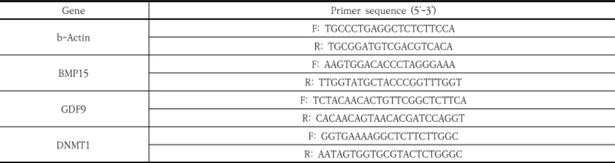
Gene Primer sequence (5’-3’)
b-Actin F: TGCCCTGAGGCTCTCTTCCA
R: TGCGGATGTCGACGTCACA
BMP15 F: AAGTGGACACCCTAGGGAAA
R: TTGGTATGCTACCCGGTTTGGT
GDF9 F: TCTACAACACTGTTCGGCTCTTCA
R: CACAACAGTAACACGATCCAGGT
DNMT1 F: GGTGAAAAGGCTCTTCTTGGC
R: AATAGTGGTGCGTACTCTGGGC Table 1. Primers used for real-time PCR
Treatments No. of examined oocytes No. of matured oocytes Maturation rate (% ± SE)
Control (Non serum) 146 66 44.65 ± 3.12 a
gBS 149 95 62.02 ± 3.58 b
gBS+FSH 146 115 77.80 ± 2.42 c
gFF 143 88 60.73 ± 1.77 b
gFF+FSH 145 104 70.58 ± 2.31 d
a-d Different letters with in a column are significantly different (P<0.05).
Table 2. Effects of different serum and FSH on the nuclear maturation of goat.
하였고, 1㎍/ml의 Follicular Stimulating Hormone
(FSH, Folltropin-V, Bioniche Co., Canada) 첨가도
비교하였다. 체외성숙 배양 24시간 후에 COC는 qPCR
을 위해 일부 –80℃ 동결하여 보관하였고, 체외수정을
위해 0.1% hyaluronidase에서 난구세포를 제거하고 제
1극체의 유무로 난자의 성숙을 판단하였다.
2.4 체외수정 및 체외배양
체외수정을 위해 국립축산과학원 가축유전자원센터에
서 생산된 염소 동결정액과 114mM Nacl, 3.1mM KCl,
25mM NaHCO
3, 0.4mM NaH
2PO
4·2H
2O, 15mM sodium
lactate, 2mM CaCl
2·2H
2O, 0.5mM MgCl
2·6H
2O,
0.5mM sodium pyruvate, 8㎎/ml bovine serum
albumin (BSA), 0.75㎍/ml kanamycin으로 구성된 체
외수정 배지를 이용하였다. 성숙된 난자는 2x10
6의 정자
와 38.5℃의 5% CO
2배양기에서 6시간 동안 공배양 하
였다. 이후 0.1% hyaluronidase를 이용하여 투명대에
붙어있는 정자를 제거하고 103mM Nacl, 7.2mM KCl,
1.2mM KH
2PO
4, 5.6㎖/L Na-lactate, 0.13mM
kanamycin, 25mM NaHCO
3, 0.3mM Na-pyruvate,
0.5mM MgCl
2·6H
2O, 1.7mM CaCl
2·2H
2O, 1.5mM
D-glucose, 2% essential amino acids, 1% non-essential
amino acids, 1mM L-glutamin, 10ng/ml EGF, 1%
BSA로 구성된 mSOF 배지로 옮겨 6일간 배양하여 배발
달을 확인하였다.
2.5 Quantitative Polymerase Chain Reaction
(qPCR)
RNA는 RNeasy Micro Kit (Qiagen, USA)을 이용
하여 제조사의 매뉴얼에 따라 추출을 진행하였다. RNA
의 농도와 순도는 OPTIZEN NanoQ (Mecasys, South
Korea)로 측정하였고, gDNA Eraser (Takara, Japan)
와 PrimeScript™ RT reagent Kit (Takara)을 이용하
여 제조사의 매뉴얼에 따라 cDNA를 합성하였다. 프라이
머 시퀀스 (Table 1)는 NCBI 데이터베이스를 이용하여
디자인하였고, 2X SYBR Premix Ex Taq Ⅱ(Takara)을
이용하였다. Real-time PCR은 CFX-96™ Real-Time
System (BIO-RAD, USA)을 통해 95℃에서 30초동안
반응 후 95℃에서 5분, 60℃에서 30초를 40회 반복하여
진행하였고, 이를 통해 얻은 결과값(Ct, Cycle of
thresholds)을 이용하여 b-actin에 대한 ΔΔCt으로 표
준화하였다.
2.6 통계분석
본 연구에서 얻은 모든 백분율 데이터는 평균±평균의
표준오차 (SEM)로 나타내었다. 실험 결과의 통계학적 분
Fig. 1. Quantitative real-time PCR analysis about oocyte maturation related genes in COC at 24 h after IVM.
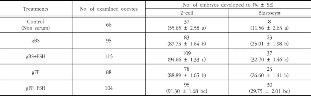
Treatments No. of examined oocytes No. of embryos developed to (% ± SE)
2-cell Blastocyst
Control
(Non serum) 66 37
(55.65 ± 2.58 a) 8
(11.56 ± 2.63 a)
gBS 95 83
(87.73 ± 1.64 b) 23
(25.01 ± 1.98 b)
gBS+FSH 115 109
(94.66 ± 1.33 c) 37
(32.70 ± 1.46 c)
gFF 88 78
(88.89 ± 1.65 b) 23
(26.60 ± 1.41 b)
gFF+FSH 104 95
(91.30 ± 1.68 bc) 30
(29.75 ± 2.01 bc) a-c Different letters with in a column are significantly different (P<0.05).
Table 3. Effects of different serum and FSH on
in vitro
development ofin vitro
fertilized goat embryos.석을 위하여 SAS (Statistical Analysis System) package를
이용하였으며, GLM (General Linear Models)
procedure를 적용하여 각 요인별 least square means
를 구하여 처리구간 유의성을 검정하였다. 본 연구에서
나타난 결과들에 대한 통계적 유의차는 p<0.05 이하인
것만 표기하였다.
3. 결과
3.1 혈청과 호르몬이 체외성숙에 미치는 효과
염소 난자의 체외성숙 배지에 10% 발정주기 혈청
(gBS)과 10% 난포액 (gFF)을 첨가하고, 추가로 FSH의
조합 효과를 조사하였다. 그 결과, 대조구(Non serum)
과 비교하였을 때 gBS, gFF 모두 난자의 성숙률이 유의
적으로 증가한 결과를 보였다 (Table 2). 또한 gBS, gFF
와 함께 FSH를 조합하여 첨가하였을 때, gBS, gFF만 첨
가한 것 보다 유의적으로 성숙률이 향상되는 것을 확인
하였고 gBS와 FSH를 같이 처리한 그룹이 모든 처리구보
다 유의적으로 높은 성숙률을 보였다 (Table 2).
3.2 혈청과 호르몬 첨가와 COC에서 성숙관련 유전
자 발현
각 처리군별 COC를 real-time PCR 분석을 통해 난
자의 성숙과 관련된 BMP15와 GDF9의 mRNA 발현량
을 비교하였다. 그 결과는 Figure 1과 같으며, 처리군간
특이점이나 유의적 차이가 나타나지 않았다.
3.3 혈청과 호르몬 첨가로 성숙된 염소 난자의 체외
수정 및 배발달
각 처리구를 통해 얻은 성숙 난자를 이용하여 체외수
정을 통해 배발달을 확인한 결과, 성숙률이 가장 낮았던
대조구에서 난할률 (2-cell)과 배반포 (Blastocyst) 형성
률 역시 낮게 나타났다. 처리구의 결과에서는 gBS, gFF
첨가로 성숙된 난자들이 난할률과 배반포 형성률이 유의
차 있게 향상됨을 확인할 수 있었다. FSH 조합된 처리구
에서는 체외성숙 결과와 비슷하게 gBS와 FSH 처리구에
서 가장 좋은 결과를 보였으며, gFF와 FSH 처리구에서
는 gBS, gFF 보다 조금 향상된 경향은 보였으나 유의차
는 나타나지 않았다.
4. 고찰
본 연구는 염소 난자의 체외배양에 있어 gBS, gFF와
같은 혈청과 호르몬의 첨가의 효과를 알아보기 위하여
체외성숙효율, 유전자발현 및 체외수정 후 배발달율을 조
사하였다. 난자의 체외성숙 또는 체외배양에 이용되는
gBS, gFF와 같은 혈청에는 호르몬, 성장인자, 영양소,
cytokine 및 각종 확인되지 않은 여러 물질이 포함되어
있어 난자의 성숙 및 배발달에 중요한 역할을 한다고 알
려져 있다 [20]. 또한 난포액은 난모세포가 성숙되며 수
정 및 발생 능력을 획득하는 환경을 제공하며 [21], 대부
분의 성분이 혈장에서 유래하고 난포막에서 유래된 스테
로이드 호르몬이 존재하고 있다 [22]. 과거 소와 돼지 등
의 미성숙 난자 체외성숙 시 배양액에 Fetal bovine
serum (FBS)이나 난포액을 첨가하여 효율을 향상시키는
연구가 많았다 [23-28]. 그간 연구 결과를 바탕으로 많은
곳에서 체외성숙 배양액에 10% FBS나 10% 난포액을 첨
가하는 방법을 적용하고 있다. 본 실험에서는 기본 체외
성숙 배양액에 10% gBS, 10% gFF를 첨가하여 혈청 유
무에 따른 효과와 이와 더불어 FSH를 조합하여 체외성
숙 효율을 알아보았는데, 처리군 모두 대조구 보다 유의
적으로 높은 성숙률을 보였다. 그 중에서도 gBS와 FSH
를 조합하였을 때 처리구 중에서도 77.8%로 유의적으로
가장 높은 체외성숙 결과를 보였다 (Table 2). 또한, 각
처리구를 통해 얻은 성숙 난자를 이용하여 체외수정 배
발달 효율을 알아보았을 때도 모두 유의적으로 향상된
결과를 보였다 (Table 3). 이는 gBS나 gFF 또는 FSH를
조합하여 체외성숙 배지에 첨가하는 방법 모두 염소 난
자의 체외성숙에 있어 핵 성숙과 세포질 성숙 모두 도움
을 주는 결과로 보이며, 특히 gBS과 FSH를 조합하여 첨
가하는 것이 가장 효율이 좋게 나타났다. 이는 체외성숙
용 배지에 성선자극호르몬인 FSH의 첨가는 난구세포 팽
창 [29], 핵 성숙률 향상 [30] 및 웅성전핵형성 촉진 [31]
등의 효과가 과거 연구를 통해 확인되었는데, 본 연구의
결과인 Table 2과 Table 3에서 체외성숙 배지에 gBS나
gFF를 단독으로 처리하는 것보다 FSH를 조합하여 첨가
하는 것이 효율 향상에 더욱 도움 되는 이유로 생각된다.
난자의 핵 성숙과 세포질 성숙에 있어 transforming
growth factor-beta (TGF-b) superfamily는 난자와
난구세포간의 양방향 로컬 커뮤니케이션에 주요한 역할
을 하며, 스테로이드 합성, gonadotropin 수용체 발현,
난포발달, 난자의 성숙, 배란 및 황체형성 등에 관여 한다
고 알려져 있다 [8-10]. GDF9과 BMP15는 난자에서 분
비되는 TGF-b superfamily로 포유류 난자 성숙을 조절
하는 중요한 요소들이다 [11]. 본 연구에서 24시간 체외
성숙 배양이 완료된 각 처리구별 COC에서 GDF9과
BMP15의 발현양을 비교하였지만, 유의적 차이를 나타
내진 않았다. 이는 처리군별 난자들의 핵성숙이 거의 완
료되어 mRNA 발현이 비슷한 수준을 보인 것으로 생각
되나, 성숙과정에 있어서는 처리군별 발현이 달랐을 것으
로 추측된다.
결론적으로 gFF 처리구에서 gBS와 비교하여 핵 성숙
률과 배발달은 차이가 없었으나 (Table 2, 3), FSH와 혼
합하여 처리하였을 때 핵 성숙률이 가장 우수했던
gBS+FSH 처리구와 비교하여 gFF+FSH 처리구가 배발
달 효율이 유의하게 유사한 수준을 보였다 (Table 3). 이
는 gFF와 FSH의 혼합처리에서 난포액이 난자의 핵 성숙
을 억제한다는 연구 결과를 토대로 [32] gFF가 난자의
핵성숙을 억제하여 FSH를 통해 충분한 세포질 성숙을
유도하는 것으로 생각된다. 본 연구의 결과가 염소 난자
의 체외성숙 조건으로 충분하다고 할 수는 없으나, gBS
나 gFF 및 FSH 첨가만으로도 성숙효율을 상당히 향상시
킴을 알 수 있었다.
References
[1] L. L. Mamuad, S. H. Kim, S. S. Lee, K. K. Cho, C. O.
Jeon, S. S. Lee, “Characterization, metabolites and gas formation of fumarate reducing bacteria isolated from Korean native goat (Capra hircus coreanae)”. Journal of Microbiology, Vol.50, No.6, pp.925-931, 2012.
DOI: https://doi.org/10.1007/s12275-012-2497-3 [2] B. Y. Jung, M. G. Seo, S. H. Lee, J. W. Byun, J. K. Oem,
D. Kwak, “Molecular and serologic detection of Coxiella burnetii in native Korean goats (Capra hircus coreanae)”, Veterinary Microbiology, Vol.173, No.1-2, pp.152-155, 2014.
DOI: https://doi.org/10.1016/j.vetmic.2014.06.029 [3] S. W. Kim, S. B. Park, M. J. Kim, D. H. Kim, D. G. Yim,
“Effects of different levels of concentrate in the diet on physicochemical traits of Korean native black goat meats”, Korean J Food Sci Anim Resour, Vol.34, No.4,
pp.457-463, 2014.
DOI: https://doi.org/10.5851/kosfa.2014.34.4.457 [4] A. M. M. T Reza, S. Shiwani, N. K. SinghEmail author,
J. D. Lohakare, S. J. Lee, D. K. Jeong, J. Y. Han, D.
Rengaraj, B. W. Lee,“Keratinocyte growth factor and thiazolidinediones and linolenic acid differentiate characterized mammary fat pad adipose stem cells isolated from prepubertal Korean black goat to epithelial and adipogenic lineage”, In Vitro Cellular and Developmental Biology. Animal, Vol.50, No.3, pp.
194-206, 2014.
DOI: https://doi.org/10.1007/s11626-013-9690-5 [5] S. Y. Jang, E. K. Kim, J. H. Park, M. R. Oh, Y. J. Tang,
Y. L. Ding, H. J. Seong, W. H. Kim, Y. S. Yun, S. H.
Moon, “Effects of physically effective neutral detergent fiber content on dry matter intake, digestibility, and chewing activity in Korean native goats (Capra hircus coreanae) fed with total mixed ration”, Asian-Australasian Journal of Animal Sciences, Vol.30, No.10, pp.1405-1409, 2017.
DOI: https://doi.org/10.5713/ajas.16.0868
[6] A. Bakhsh, I. Ismail, Y. H. Hwang, J. G. Lee, S. T. Joo,
“Comparison of blood loss and meat quality characteristics in Korean black goat subjected to head-only electrical stunning or without stunning”, Korean J Food Sci Anim Resour, Vol.38, No.6, pp.1286-1293, 2018.
DOI: https://doi.org/10.5851/kosfa.2018.e64
[7] M. A. Sirard, H. M. Florman, M. L. Leibfried-Rutledge, F. L. Barnes, M. L. Sims, N. L. First, “Timing for nuclear progression and protein synthesis necessary for meiotic maturation of bovine oocytes”, Biol. Reprod., Vol.40, No.6, pp.1257-1263, 1989.
DOI: https://doi.org/10.1095/biolreprod40.6.1257 [8] N. Kaivo-oja, L. A. Jeffery, O. Ritvos, D. G. Motter,
“Smad signalling in the ovary”, Reprod Biol Endocrinol. Vol.4, No.21, pp.1-13, 2006.
DOI: https://doi.org/10.1186/1477-7827-4-21 [9] D. J. Trombly, T. K. Woodruff1, K. E. Mayo, “Roles for
Transforming Growth Factor Beta Superfamily Proteins in Early Folliculogenesis”, Semin Reprod Med. Vol.27, No.1, pp.14-23, 2009.
DOI: https://doi.org/10.1055/s-0028-1108006 [10] S. A. Pangas, “Regulation of the ovarian reserve by
members of the transforming growth factor beta family”, Mol Reprod Dev. Vol.79, pp.666–679, 2012.
DOI: https://doi.org/10.1002/mrd.22076
[11] F. Otsuka, K. McTavish, S.Shimasaki, “Integral Role of GDF-9 and BMP-15 in Ovarian Function”, Mol Reprod Dev. Vol.78, pp.9-21, 2011.
DOI: https://doi.org/10.1002/mrd.21265
[12] P. Lonergan, T. Fair, “Maturation of Oocytes in vitro”, Annu Rev Anim Biosci. Vol.4, pp.255-68, 2016.
DOI:
https://doi.org/10.1146/annurev-animal-022114-1108 22
[13] J. S. Lopes, A. Canha-Gouveia, E. París-Oller, P. Coy,
“Supplementation of bovine follicular fluid during in vitro maturation increases oocyte cumulus expansion, blastocyst developmental kinetics, and blastocyst cell number”, Theriogenology. Vol.126, Vol.1, pp.222-229, 2019.
DOI:https://doi.org/10.1016/j.theriogenology.2018.12.010 [14] B. K. Redel, L. D. Spate, R. S. Prather, “In Vitro Maturation, Fertilization, and Culture of Pig Oocytes and Embryos”, Methods Mol Biol. Vol.2006, pp.93-103, 2019.
DOI: https://doi.org/10.1007/978-1-4939-9566-0_6 [15] A. J. Watson, P. Sousa, A. Caveney, L. C. Barcroft, D.
Natale, J. Urquhart, M. E. Westhusin, “Impact of bovine oocyte maturation media on oocyte transcript levels, blastocyst development, cell number, and apoptosis”, Bio. Reprod., Vol.62, Vol.2, pp.355-364. 2000.
DOI: https://doi.org/10.1095/biolreprod62.2.355 [16] N. A. R. Sá, L. A. Vieira, A. C. A. Ferreira, J. Cadenas,
J. B. Bruno, C. Maside, F. G. C. Sousa, F. W. S. Cibin, B. G. Alves, A. P. R. Rodrigues, J. H. Leal-Cardoso, E.
L. Gastal, J. R. Figueiredo, “Anethole Supplementation During Oocyte Maturation Improves In Vitro Production of Bovine Embryos”, Reprod Sci. Vol.26, 1933719119831783, 2019.
DOI: https://doi.org/10.1177/1933719119831783 [17] M. V. O. Santos, L. E. Nascimento, É. A. Praxedes, A.
A. Borges, A. R. Silva, L. M. Bertini, A. F. Pereira,
“Syzygium aromaticum essential oil supplementation during in vitro bovine oocyte maturation improves parthenogenetic embryonic development”, Theriogenology. Vol.128, No.1, pp.74-80, 2019.
DOI: https://doi.org/10.1016/j.theriogenology.2019.01.031 [18] Z. Cao, D. Gao, X. Tong, T. Xu, D. Zhang, Y. Wang,
Y. Liu, Y. Li, Y. Zhang, Y, Pu, “Melatonin improves developmental competence of oocyte-granulosa cell complexes from porcine preantral follicles”, Theriogenology. Vol.133, No.15, pp.133:149-158, 2019.
DOI: https://doi.org/10.1016/j.theriogenology.2019.05.003 [19] J. D. Yoon, S. U. Hwang, M. Kim, Y. Jeon, S. H. Hyun,
“Growth differentiation factor 8 regulates SMAD2/3 signaling and improves oocyte quality during porcine oocyte maturation in vitro”, Biol Reprod. Vol.101, No.1, pp.63-75, 2019.
DOI: https://doi.org/10.1093/biolre/ioz066
[20] D. K. Gardner, “Mammalian embryo culture in the absence of serum or somatic cell support”, Cell Ciol Int., Vol.18, No.12, pp.1163-1179, 1994.
DOI: https://doi.org/10.1006/cbir.1994.1043
[21] K. P. McNatty, D. M. Smith, A. Markris, R.
osathanonch, K. J. Ryan, “The microenvironment of the human antral follicle: interrelationships among the steroid levels in antral fluid, the population of granulosa cells, and the status of the oocyte in vivo and in vitro”, J. Clin. Endocrinol. Metab., Vol.49, pp.851-860, 1979.
[22] KP McNatty and RS Sawers, “Relationship between the endocrine enviroment within the Graafin follicle and the subsequent rate of progesterone secretory human granulosa cells in vitro”, J. Endocrinol. Vol.66, pp.391-400, 1975.
[23] M. L. Leibfried-Rutledge, E. S. Critser, N. L. First,
“Effects of fetal calf serum and bovine serum albumin on in vitro maturation and fertilization of bovine and hamster cumulus-oocyte complexes”, Biol Reprod. Vol.35, No.4, pp.850-857, 1986.
[24] M. Del Collado, N. Z. Saraiva, F. L. Lopes, R. C.
Gaspar, L. C. Padilha, R. R. Costa, G. F. Rossi, R.
Vantini, J. M. Garcia, “Influence of bovine serum albumin and fetal bovine serum supplementation during in vitro maturation on lipid and mitochondrial behaviour in oocytes and lipid accumulation in bovine embryos”, Reprod Fertil Dev. Vol.28, No.11, pp.1721-1732, 2015.
DOI: https://doi.org/10.1071/RD15067
[25] B. Avery, L. Strøbech, T. Jacobsen, I. B. Bøgh, T.
Greve, “In vitro maturation of bovine cumulus-oocyte complexes in undiluted follicular fluid: effect on nuclear maturation, pronucleus formation and embryo development”, Theriogenology. Vol.59, No.3-4, pp.
987-999, 2003.
DOI: https://doi.org/10.1016/S0093-691X(02)01139-1 [26] G. Puri, S. S. Chaudhary, V. K. Singh, A. K. Sharma, “Effects
of fetal bovine serum and estrus buffalo serum on maturation of buffalo (Bubalus bubalis) oocytes in vitro”, Vet World. Vol.8, No.2, pp.143-146, 2015.
DOI: https://doi.org/10.14202/vetworld.2015.143-146 [27] Y. S. Zheng, M. A. Sirard, “The effect of sera, bovine
serum albumin and follicular cells on in vitro maturation and fertilization of porcine oocytes”, Theriogenology. Vol.37, No.4, pp.779-790. 1992.
DOI: https://doi.org/10.1016/0093-691X(92)90041-O [28] Y. Ducolomb, H. González-Márquez, R. Fierro, I.
Jiménez, E. Casas, D. Flores, E. Bonilla, Z. Salazar, M.
Betancourt, “Effect of porcine follicular fluid proteins and peptides on oocyte maturation and their subsequent effect on in vitro fertilization”, Theriogenology. Vol.79, No.6, pp.896-904. 2013.
DOI: https://doi.org/10.1016/j.theriogenology.2013.01.024 [29] Y. H. Choi, E. M. Carnevale, G. E. Seidel, E. L. Squires,
“Effects of gonadotropins on bovine oocytes matured in TCM-199”, Theriogenology. Vol.56, No.4, pp.661-670, 2001.
DOI: https://doi.org/10.1016/S0093-691X(01)00597-0 [30] A, Ali, M. A. Sirard, “Effect of the absence or presence
of various protein supplements on further development of bovine oocytes during in vitro maturation”, Biol.
Reprod., Vol.66, No.4, pp.901-905, 2002.
DOI: https://doi.org/10.1095/biolreprod66.4.901 [31] S. Kito, B. D. Bavister, “Male pronuclear formation
and early embryonic development of hamster oocytes matured in vitro with gonadotropins, amino acid and
cysteamine”, J. Reprod. Fertil., Vol.110, No.1, pp.35-46. 1991.
DOI: https://doi.org/10.1530/jrf.0.1100035
[32] M. A. Sirard, “Temporary inhibition of meiosis resumption in vitro by adenylate cyclase stimulation in immature bovine oocytes”, Theriogenology. Vol.33, No.4, pp.757-767. 1990.
DOI: https://doi.org/10.1016/0093-691X(90)90811-7
이 상 훈(Sang-Hoon Lee) [정회원]
• 2004년 8월 : 경상대학교 대학원 응용생명과학부 (이학석사)
• 2007년 8월 : 경상대학교 대학원 응용생명과학부 (이학박사)
• 2008년 1월 ~ 2014년 12월 : 농 촌진흥청 국립축산과학원 농업연 구사
• 2015년 1월 ~ 현재 : 농촌진흥청국립축산과학원 농업연 구관
<관심분야>
분자육종, 염소유전체
전 다 연(Dayeon Jeon) [정회원]
• 2016년 2월 : 건국대학교 동물생 명대학 동물자원과학과 (농학학사)
• 2019년 8월 : 충남대학교 대학원 축산학과 (농학석사)
• 2016년 10월 ~ 현재 : 농촌진흥청 국립축산과학원 농업연구사
<관심분야>
가축번식, 가축육종
이 진 욱(Jinwook Lee) [정회원]
• 2015년 2월 : 전북대학교 축산학 과 (농학석사)
• 2016년 10월 ~ 현재 : 농촌진흥청 국립축산과학원 농업연구사
<관심분야>
가축영양, 반추미생물
이 성 수(Sung-Soo Lee) [정회원]
• 1998년 2월 : 제주대학교 대학원 축산학과 (농학석사)
• 2010년 8월 : 제주대학교 대학원 축산학과 (농학박사)
• 1993년 8월~2012년 6월 : 농촌 진흥청 국립축산과학원 농업연구 사
• 2012년 7월 ~ 현재 : 농촌진흥청 국립축산과학원 가축유 전자원센터장
<관심분야>
가축번식, 염소개량
김 승 창(Seungchang Kim) [정회원]
• 1999년 2월 : 전남대학교 대학원 생물학과 (이학석사)
• 2009년 2월 : 전남대학교 자연과 학대학원 생물학과 (이학박사)
• 2018년 2월 ~ 현재 : 농촌진흥청 국립축산과학원 농업연구사
<관심분야>
유전육종, 유전자원 관리
김 찬 란(Chan-Lan Kim) [정회원]
• 1999년 2월 : 서울대학교 수의과 대학 수의학과 (수의학학사)
• 2005년 3월 : 일본 기후연합대학 원 수의학과 (수의학박사)
• 2006년 7월 ~ 2014년 10월 : 농 림축산검역본부 수의연구사
• 2014년 10월 ~ 현재 : 국립축산과 학원 수의연구사
<관심분야>
수의학, 예방의학, 친환경
김 관 우(Kwan-Woo Kim) [정회원]
• 2015년 2월 : 충남대학교 대학원 축산학과 (농학석사)
• 2018년 8월 : 충남대학교 대학원 축산학과 (농학박사)
• 2018년 8월 ~ 현재 : 농촌진흥청 국립축산과학원 전문연구원
<관심분야>
가축번식, 가축육종