I. 서론
여러 종류의 서로 다른 조직 대식세포군과 그 전구 세포인 혈액 단핵구를 합하여 단핵성 식세포계 (mononuclear-phagocyte system) 라고 한다1). 이 세 포들은 동일한 근원과 상호간의 형태적 유사성을 나 타내며 신속한 탐식작용을 비롯하여 동일한 기능을 지니고 있는 계로서, 거의 모든 조직에서 조직의 항 상성(homeostasis) 및 면역, 염증반응을 조절하는 역 할을 하고 있다. 대식세포가 매우 많이 분포되어 있 는 조직들로서는 간(kupffer cell), 허파(interstitial alveolar macrophage), serous cavity(흉강, 복강 대식 세포), 골조직(osteoclast), 뇌(microglial cell), 태반, 장벽(intestinal wall), 결체조직, 비장, 임파절, 골수, 모유 등이 있다2). 단핵구/대식세포는 pluripotent stem cell에서 myeloid stem cell을 거쳐 colony-form- ing unit for granulocyte-monocytes(CFU-GM) 단계 에서 proliferative pool을 떠난다3). 이 단계의 세포는 monoblast로 분화하여 이 series에서 처음으로 형태 학적 구분이 가능한 promonocyte로 된다1). 골수에 서 처음 단핵구 전구세포로부터 완전 성숙한 단핵구 로 변화하는데 약 6일이 소요된다. 국소적 염증병소 가 없는 상태라면 단핵구는 다양한 조직으로 무작위 적으로 이동하여 그곳에서 조직 특이적 형태와 기능 을 지닌 대식세포로 최종 분화한다. 단핵구/대식세
포의 다기능적 특성으로부터 유추할 수 있는 것은, 이들 세포가 미세하게 조정된 유전자 발현의 변화를 수반하는 매우 복잡한 분화과정을 거칠 것이라는 것 이다. 이 조직 특이적 최종 분화과정은 조직마다 특 이한 주변의 생물학적 활성 물질의 작용에 의한 결 과이며 이로 인하여 각 대식세포마다 독특한 표현형 을 나타내게 된다2).
생체내에 편재하는 1,25-dihydroxycholecalcifer- ol(1,25(OH)2D3)은 미분화 단핵구 세포주인 THP-1 을 부분적으로 분화 유도한다고 보고되어4)자연적 인 단핵구/대식세포 분화유도물질로 알려져 있다5). 그러나 이것은 인위적 단핵구/대식세포 분화유도물 질인 TPA6)와는 완전히 다른 세포내 신호전달체계를 통하여 분화를 유발한다. 즉, 1,25(OH)2D3는 세포핵 내의 steroid 수용체를 통하여, 그리고 TPA는 protein kinase C를 통한 신호전달체계를 거쳐 분화를 유도 한다7). 한편 IL-1, IL-2, IFN-γ, TNF, GM-CSF와 같은 cytokine중 어느것도 단독으로는 단핵구의 분화를 유도하지는 않았지만 이들중 몇 가지를 조합하여 적 용시키면 TPA에 의한 분화와 거의 유사한 결과를 나 타내기도 한다고 한다7). 생물학적 활성을 지닌 vita- min D3의 주 대사산물인 1,25(OH)2D3는 그 수용체 가 매우 다양한 조직에서 발견된다8, 9). 경조직 대사 와 관련된 1,25(OH)2D3의 고전적 역할 외에도, T 세 포 활성화와 lymphokine 생산의 조절과 같은 면역 대한치주과학회지 : Vol. 28, No. 4, 1998
단핵구의 분화/활성화 유도물질이 세균내독소에 의해 유도된 MHC ClassII 분자와 Integrin의 발현 및
Cytokine 생산에 미치는 영향
최병선·이 호·오귀옥·김형섭 전북대학교 치과대학 치주과학교실 전북대학교 구강생체과학연구소
조절 호르몬으로서의 기능도 가지고 있으며10, 11), 대 식세포로의 분화를 유도하는 것으로도 알려져 있다5,
12, 13). 이와 같은 1,25(OH)2D3의 효과는 이 자체가 단
핵구/대식세포로부터 생산되는 cytokine이라는 사실 로부터 거슬러 유추하여 밝혀지게 된 것이다14, 15). 대 식세포가 활성화될 때 25-hydroxycholecalciferol (25-OHD3)을 1,25(OH)2D3로 변환시키는 효소인 1 α -hydroxylase 효소활성이 증가된다. 대식세포의 1 α- hydroxylase는 전신적 1,25(OH)2D3의 공급처인 신장 에 존재하는 1α-hydroxylase와는 달리, IFN-γ나 내독 소(endotoxin, LPS)에 의하여 효소활성이 증가되며 부갑상선 호르몬(parathyroid hormone)에 의하여서 는 효소활성이 증가하지 않는다14, 15).
치주염, 류마티스 관절염 및 다발성 경화증(multi- ple sclerosis)과 같은 만성 염증병소에서는 침윤염증 세포와 거주세포(resident cell)간에 간격이 긴밀해질 수 밖에 없으므로, cytokine 같은 수용성 인자를 매개 로 하는 방법 외에도, 대식세포 및 다형핵 백혈구와 T 임파구의 직접적인 세포간 접촉에 의하여서도 세 포간 상호 연락이 이루어질 것으로 추측된다16). 이러 한 가설은 T 임파구가 단핵구와 직접 접촉하거나 IFN-γ와 같은 cytokine을 분비함으로써 이들이 단핵 구의 IL-1β분비를 촉진한다는 사실이 밝혀짐으로써 증명되었다17-22). 세포간 직접 접촉으로 인하여 IL-1 β 분비와 같은 대식세포의 기염증성 효과기 작용 (proinflammatory effector function)이 유도되는 것 은, 활성화된 T 임파구와 대식세포의 세포표면 유착 (adhesion) 분자들간의 상호관계 때문인 것으로 밝 혀졌다22). 대식세포의 MHC class II 분자와 함께 제 시된 항원에 T 임파구의 CD3/T cell antigen receptor (CD3/TCR) complex가 결합하는 것과 함께 양 세포 의 세포표면 유착 분자간의 costimulatory interaction 으로 이루어진 T-대식세포간 직접접촉 및 IFN - γ와 같은 T cell derived cytokine에 의하여 대식세포의 IL-1 β, TNF - α분비와 같은 효과기 작용이 유도된다
16, 23-25). T 임파구가 활성화될 때 분비되는 IFN - γ는
대표적인 단핵구의 활성화 물질로서, 세포형태의 변 화, 세포 표면항원의 발현26, 27), 항균작용의 상승28), 항암작용의 상승29), 및 항원 제시작용 증가30) 등의
작용을 나타내지만 단독으로는 단핵구의 분화를 유 도하지 못한다.
그람음성균의 세포외막 주성분인 lipopolysaccha- ride(LPS)는 세균 감염시 숙주 면역반응을 크게 자극 하는 인자로서 특히 단핵구/대식세포를 자극하여 TNF- α, IL-1 β, IL-6, IL-8, IL-10의 분비를 비롯한 기 염증성 효과기 작용을 나타내고31)면역반응을 활성 화시키는 작용을 가지고 있으나32), 과도한 반응, 특 히 TNF-α의 과도한 분비가 일어나게 되면 내독성 쇽 (endotoxic shock)을 유발하기도 하는 물질이다33,
34). 단핵구/대식세포가 LPS에 의해 생산된 cytokine 의 주된 공급원이기는 하나, 대식세포 분화와 LPS에 대한 반응능력과의 정확한 관계에 대하여는 아직 자 세히 알려진 바 없다35). 혈액 단핵구는 모세혈관으로 부터 혈관 밖 조직으로 이동하여 그곳에서 분화를 일으키며, 이러한 분화는 대식세포가 세균 내독소와 같은 외부 자극에 대하여 효과적인 기염증성, 또는 면역 조절 역할을 함으로써 숙주 방어를 적절히 할 수 있도록 하는데 필수적인 과정이다36).
본 연구는 IFN-γ에 의하여 priming이 되거나, 1,25(OH)2D3에 의하여 분화된 상태의 단핵구/대식 세포가 세균의 내독소에 노출되었을 때 효과기 반응 성에 있어 어떠한 차이가 있는지를 알기 위하여 시 행하였으며, 사람의 단핵구와 가장 유사한 표현형을 나타내는 promyelocyte 세포주 THP-1을 이용하여 MHC class II 분자와 intercellular adhesion mole- cule-1(ICAM-1; CD54), Mac-1(CD11b) 분자의 발현 과 IL-1β의 분비량을 면역화학적 방법으로 측정, 비 교함으로써, 단핵구/대식세포의 분화 및 활성화와 숙주 방어기전과의 관계를 규명하고자 하였다.
II. 실험재료 및 방법
1. 세포배양 및 약물 처리사람의 promyelocyte 세포주인 THP-1은 fetal calf serum 10%와 penicillin 100 U/ml, streptomycin (Sigma) 100 ㎍/ml이 포함된 RPMI1640(Gibco) 배양 액에서 5% CO2, 37℃의 1조건으로 배양하였다. 1,25
dihydroxycholecalciferol(1,25(OH)2D3, Sigma)는 10- 8 M, human interferon -gamma(hIFN-γ, Genzyme) 는 500 U/ml의 농도로 필요 시간동안 처리하였으며, lipopolysaccharide(LPS, Sigma)는 0.1 ㎍/ml 농도로 18시간 처리하였다.
2. MHC class II 분자와 integrin 발현 측정 을 위한 flow cytometry 분석
약물 처리된, 혹은 처리되지 않은 THP-1 세포를 phosphate buffered saline(PBS)으로 2회 세척하고 1% bovine serum albumin(BSA)이 포함된 PBS(PBS- BSA)에 2 × 10⁶/ml의 농도로 재부유 하여 eppen- dorf microcentrifuge tube에 1 ml를 옮겼다. 원침시 킨 후 상청액을 제거하고 세포괴(cell pellet)에 단일 클론항체(monoclonal antibody, mAb)를 1 : 100의 농도로 PBS-BSA 200 ㎕에 희석하여 각각 세포에 적 용시켰다. 본 실험에 사용한 단일클론항체는 mouse anti-HLA-D mAb(Pharmingen), mouse anti-ICAM- 1(CD54) mAb(Pharmingen), anti-Mac-1(CD11b) mAb(Pharmingen)이었다. 실온에서 tube를 회전시 키면서 30분간 반응시킨 후 PBS-BSA로 1회 세척하 고, fluorescein isothiocyanate(FITC) -conjugated goat anti-mouse IgG(Pierce)를 1 : 200으로 PBS-BSA 에 희석하여 20분간 tube를 회전시키면서 실온에서 반응시켰다. 세포를 PBS-BSA로 2회 세척한 후, 1%
paraformaldehyde(PFA)/PBS-BSA로 10분간 세포를 고정하고 1회 세척하여 250 ㎕의 PBS-BSA에 재부유 한 다음 fluorescence activated cell sorter(FACStar, Beckton Dickinson)로 분석하였다.
3. MHC class II 분자와 integrin의 형광 현 미경 관찰
THP-1을 조직배양용 8 well chamber slide(Nunc) 에서 80% confluent한 상태가 될 때까지 배양하여 PBS로 2회 세척하고 30분간 공기건조 시켰다. 2%
PFA/PBS로 10분간 고정한 후 PBS로 세척하고, anti- ICAM-1 mAb, anti-HLA-D mAb를 1 : 50으로 PBS-
BSA에 희석하여 각각 세포에 적용한 다음 30분간 실 온에서 반응시켰다. PBS로 2회 세척한 후 FITC-con- jugated anti-mouse IgG antibody를 1 : 50으로 희석 하여 20분간 반응시킨 다음 PBS로 3회 세척하였다.
Glycerol과 PBS가 9 : 1로 혼합된 mounting media를 slide 위에 떨어뜨려 cover glass로 덮어서 형광현미 경(Zeiss)으로 관찰하였다.
4. IL-1β분비량 측정을 위한 ELISA분석
IL-1β은 IL-1βPredicta kit(Genzyme)을 이용하여 제조회사의 방법에 따라 정량하였다. 즉, mouse anti-IL-1βmAb가 coating된 96 well microplate에 0
∼1024 pg/ml의 standard recombinant hIL-1 β, 혹은 THP-1 세포 배양상청액을 100 ㎕씩 첨가하여 37℃
로 30분간 반응시켰다. 세척액으로 5회 세척하고 rabbit polyclonal anti-hIL-1 βantibody를 첨가하여 37℃ 로 30분 간 반 응 시 키 고 5회 세 척 하 였 다 . Substrate로서 0.02% H2O2가 포함된 완충액과 tetramethylbenzidine / DMF를 1 : 1로 혼합하여 각 well에 첨가한 후 30분간 실온에서 반응시켰다. 그 후 1M H2SO4stop solution을 첨가하고 즉시 ELISA reader(Biorad)로 450nm 파장에서의 흡광도를 측정 하였다. 각 실험은 duplicate로 시행하였으며 측정치 흡광도에서 blank의 흡광도를 제한값을 이용하여 IL-1βstandard curve로부터 IL-1β농도를 환산하였 다.
III. 결과
1. 세포 표면항원 발현에 미치는 IFN - γ 와 1,25(OH)2D3의 효과
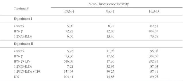
THP-1에 IFN-γ, 혹은 1,25(OH)2D3를 48시간 적용 하여 배양하였고, LPS의 경우에는 배양 이틀째 되는 날 배양액에 LPS를 첨가하여 하룻동안 세포에 적용 시켰다. 배양이 끝난 THP-1 세포를 각 세포 표면항 원에 대한 단일클론 항체로 처리하고, FACS 분석을 하여 mean fluorescence intensity 값을 비교하였다
(Table 1, Fig. 1, 2, 3). IFN-γ는 휴지(resting) 상태의 세포에 비하여 ICAM-1을 12배, 14배 발현 유도하였 고, HLA-D는 5배, 3배 가량 증가시켰다. Mac-1도 발 현이 약간 유도되기는 하였지만 그 정도가 매우 미 약하였다. 1,25(OH)2D3는 3가지 분자 중에서 Mac-1 에 대한 발현 유도가 비교적 뚜렷하여 휴지 상태보 다 1.5배∼3배 정도의 upregulation을 나타내었으나, HLA-D의 경우는 매우 적은 차이 이기는 하지만 재 현가능(reproducible)하게 감소하는 양상을 보였다.
LPS는 ICAM-1에서만 휴지 상태보다 20배 이상의 높 은 발현 유도를 나타내었으나, 나머지 두 분자에서는 뚜렷한 발현 유도 현상이 없었다. IFN-γ로 비교적 높은 발현 유도를 보였던 ICAM-1과 HLA-D를 fluo- rescence immunohistochemistry 방법으로 염색하여 형광현미경으로 관찰한 것이 Figure 5이다.
2. LPS의 세포 표면항원 발현작용에 미치 는 1,25(OH)2D3와 IFN-γ의 영향
IFN-γ로 전처치 한 경우 LPS에 의한 ICAM-1의 발 현이 휴지 상태보다 무려 120배 이상 증가되어 LPS
와 IFN-γ간에 상가적 관계를 나타내었다(Table 1, Fig. 4). 즉, IFN-γ로 priming 함으로써 THP-1의 LPS 에 대한 ICAM-1 발현 반응성이 크게 증가되었다고 볼 수 있다. 그러나 LPS단독으로 전혀 발현이 유도되 지 않았던 Mac-1과 HLA-D에서는 IFN-γ, 1,25 (OH)2D3와 LPS 사이에 상가적인 관계가 나타나지 않았다.
3. LPS의 IL-1 β분비작용에 미치는 IFN - γ와 1,25(OH)2D3의 영향
THP-1에 IFN-γ, 혹은 1,25(OH)2D3을 적용시켜 48 시간 배양하였으며, LPS의 경우에는 배양 이틀째 되 는 날 배양액에 첨가하여 하룻동안 적용시키고 그 배양 상청액으로 유리된 IL-1β의 양을 ELISA로 정량 하 여 비 교 하 였 다 (Table 2, Fig. 6). IFN-γ와 1,25(OH)2D3는 휴지 상태의 세포와 거의 같은 정도 로 밖에 IL-1β의 생산을 유도하지 못하였고, LPS도 측정 한계범위인 10 pg/ml에도 못미치는 매우 적은 양의 IL-1β를 생산 유도하였다. 그러나 IFN-γ로 priming한 경우에는 LPS에 반응하여 IL-1β생산량이 Mean Fluorescence Intensity
Treatmenta
ICAM-1 Mac-1 HLA-D
Experiment I
Control 5.98 8.77 82.31
IFN-γ 72.22 12.05 404.67
1,25(OH)2D3 6.50 13.46 73.55
Experiment II
Control 5.22 11.96 95.06
IFN-γ 73.36 17.63 304.56
IFN-γ+ LPS 616.09 17.30 292.91
1,25(OH)2D3 7.22 32.95 87.03
1,25(OH)2D3+ LPS 151.03 39.27 87.41
LPS 104.41 14.85 89.75
Table 1. Effects of IFN-γand 1,25(OH)2D3on the expression of integrins and MHC molecule by THP-1
aTHP-1 cells were treated for 48h with IFN - γ(500 U/ml) or 1,25(OH)2D3
(10-8M), and LPS was added at the second day of treatment
86.47±0.92 pg/ml의 높은 값을 나타내었으며, 1,25(OH)2D3를 전처치 한 후 LPS를 적용한 경우에는 LPS 단독보다 약간 증가하기는 하였으나 그 차이가 미약하였다.
4. LPS의 IL-1β 분비작용에 미치는 1,25 (OH)2D3와 IFN-γ의 동시효과
1,25(OH)2D3는 day 0 부터 day 4, IFN-γ는 day 1 부터 day 5, LPS는 day 4 부터 day 5까지 적용시켜서 IL-1β생산량을 비교하여 보았다(Table 3). Table 2에 서와 같이 IFN-γpriming된 세포는 LPS에 대하여 IL- 1β생산량의 큰 증가를 보였으며, 1,25(OH)2D3로 충 분한 분화를 미리 유도했던 경우에는 IFN-γpriming 된 세포의 LPS에 대한 반응성이 크게 증가하여, 1,25(OH)2D3를 적용하지 않는 경우(70.29±3.94 pg/ml)보다 거의 3.5배(252.7±14.34 pg/ml) 가까이 IL-1β생산량이 증가되었다.
IV. 고찰
단핵구/대식세포에 관한 연구를 시행함에 있어 가 장 어려운 점은 혈액 단핵구의 수가 제한되어 있다 는 것이나, 이러한 문제점은 HL-60, KG-1, U937 및 THP-1과 같은 수종의 사람 백혈병 세포주의 개발로 해결될 수 있었다37, 38). 이들 세포주는 서로 다른 분 화, 성숙의 단계에서 더 이상의 분화가 차단된 미분 화 단핵구세포주들로서 여러 자극들에 의하여 완전 한 대식세포로 분화할 수 있다. 또한 혈액 단핵구에 비하여 비교적 균질의 집단을 이루고 있으므로 생화 학적 연구 결과가 재현가능하다는 장점을 지니고 있
aTHP-1 cells were treated for 48h with IFN- γ(500 U/ml) or 1,25(OH)2D3(10-8M), and LPS was added at the second day of treatment
bMean±SD for duplicates from one of two experiments
cp〈0.001 when compared with control
dnot significant when compared with control
ep〈0.001 when compared with LPS treatment Treatmenta IL-1β(pg/ml)b
Control 1.63 ± 0.00
IFN - γ 2.72 ± 0.11c
1,25(OH)2D3 1.20 ± 0.03d IFN - γ+ LPS 86.47 ± 0.92c, e 1,25(OH)2D3+ LPS 12.02 ± 0.05c, e
LPS 7.45 ± 0.05c
Table 2. Effects of IFN-γand 1,25(OH)2D3on the IL-1β production by THP-1
Treatmenta
IL-1β(pg/ml)b
1,25(OH)2D3 IFN- γ LPS
- - - 10.29 ± 0.87
+ - - 16.15 ± 0.77
- + - 11.73 ± 0.38
- - + 14.14 ± 0.49
+ + - 15.87 ± 1.06
+ - + 12.50 ± 0.19
- + + 70.29 ± 3.94
+ + + 252.70 ± 14.34
day 0 1 2 3 4 5
a1,25(OH)2D3
IFN-γ LPS
bMean±S.D. for duplicates
Table 3. Effects of 1,25(OH)2D3on the IFN-γand/or LPS-induced IL-1βproduction by THP-1
어서, 특히 단핵구/대식세포 분화의 연구에 큰 가치 를 지니고 있다. THP-1은 이러한 세포주들 가운데 사람의 혈액에서 채취한 단핵구와 가장 유사한 표현 형(세포 형태, 분비 산물들, 종양 유전자 발현, 세포 표면항원 발현, 지질대사 관련 유전자 발현 등)을 지 니고 있는 세포로서 Tsuchiyama 등39)에 의하여 급성 단핵구성 백혈병을 앓고 있는 소년으로부터 처음 분 리되었다.
β2integrin family의 세포표면항원 CD11/CD18 복 합체는 세 종류의 heterodimer로 이루어져 있는데 공통적으로 같은 βsubunit(CD18)와 세 종류의 서로 다른 αsubunit(CD11a, CD11b, CD11c)가 비공유결
합으로 연결되어 있으며40), Mac-1은 이들중 하나인 CD11b/CD18 복합체의 α- subunit CD11b를 일컫는 다. β2integrin은 한 세포와 다른 세포 또는 세포와 세포외기질간의 부착을 강화시키는 역할을 한다41). 단핵구의 CD11a/CD18(leukocyte function associat- ed antigen-1, LFA-1)과 CD11b(Mac-1)/CD18은 lig- and가 ICAM-1으로서, 내피세포의 ICAM-1과 결합하 게 되면 혈액으로부터 transendothelial migration 하 기 전, 단핵구가 내피세포에 강하게 부착하도록 하는 역할을 하며42), 또 한편으로는. 대식세포가 T 임파구 에 항원을 제시할 때 T 임파구의 ICAM-1과 대식세포 의 CD11/CD18이 결합하여 항원제시의 costimula- Fig 1. Immunofluorescence analysis of the reactivity of mAb against HLA-D, Mac-1(CD11b), and ICAM-1(CD54) with IFN-γ
and/or LPS-treated THP-1 cells
ICAM-1 Mac-1 HLA-D
Control
IFN-γ
IFN-γ LPS
LPS
Log Fluorescence Intensity
Cell Number
tion 기능을 하기도 한다43, 44). β2 integrin에 대한 blocking mAb를 이용하여 THP-1과 T 세포의 접촉을 차단함으로써 T 세포 의존적으로 일어나는 THP-1의 활성화가 40 ∼ 60% 억제 되었다고 한 Vey 등21)의 보고를 통하여 단핵구-T 세포의 세포-세포간 접촉에 서 CD11/CD18 복합체가 costimulation 역할을 한다 는 것이 입증되었다. 또한 임파구의 CD11/CD18과 는 달리 단핵구에서는 이들 분자들이 정상적으로는 세포내 소포(vesicle)에 저장되어 있다가45)활성화될 때 자극에 의하여 빠르고도 선택적인 방식으로 upregulation됨으로써 염증 발달 과정에서 신속한 반 응성을 나타내게된다46). Limb 등47)에 의하면 cytokine 중 IL-2, IL-4, TNF-α, TNF-β도 이와 같은 신 속한 CD11c의 발현을 나타내었으나, IFN-γ는 CD11
family 분자의 발현에 변화를 일으키지 않는다고 하
였고48, 49), 본 연구에서도 IFN-γ는 Mac-1의 upregu-
lation을 일으키지 않았다. 혈액 단핵구는 시험관 내 에서 3 ∼ 7일 동안 배양하면 저절로 대식세포로 분 화하는데, 이때 모든 CD11의 발현이 증가한다고 Gessani 등36)은 보고하였다. 본 연구에서 단핵구 분 화물질로 알려져 있는 1,25(OH)2D3에 의하여 THP-1 세포의 Mac-1 발현이 증가했던 것은 THP-1이 조금 더 대식세포와 유사한 세포로 분화하였기 때문인 것 으로 사료된다. 본질적으로(constitutive) 발현되는 단핵구의 β2integrin은 정상적으로 매우 불활성한 상태로 존재하여, 매우 낮은 결합활성(avidity)으로 ligand에 결합한다. 세포가 활성화될 때 내외 신호 (inside-out signal)에 의하여 이러한 integrin의 결합 Fig 2. Immunofluorescence analysis of the reactivity of mAb against HLA-D, Mac-1, and ICAM-1 with 1,25(OH)2D3and/or
LPS-treated THP-1 cells
Control ICAM-1
Cell Number
Mac-1 HLA-D
1,25(OH)2D3
1,25(OH)2D3 LPS
LPS
Log Fluorescence Intensity
활성이 증가하여 ICAM-1과 같은 counterreceptor를 지닌 세포에 견고하게 부착하게 되는데, 이러한 현상 은 integrin의 수가 증가하지 않고도 일어날 수 있다
50). 즉, 본 연구에서 IFN-γ에 의하여 Mac-1의 숫적인 upregulation이 일어나지 않았다 할지라도 Mac-1에 의하여 매개되는 세포간 접촉 기능이 항진되지 않는 다고 속단하기는 어렵다.
단핵구/대식세포의 MHC II(HLA-D region 분자)는 T 세포에 대하여 단핵구/대식세포가 항원을 제시할 때 ICAM-1 및 CD18과 같은 costimulatory receptor와 더불어 중심 역할을 하는 수용체이다. 분화/활성화 함에 따라 항원제시세포의 제시능력이 상승 조절되 는 것은 숙주 방어기전을 향상시키기 위함이며, 따라 서 MHC modulation에 대한 연구보고가 상당히 많은 것은 놀라운 일이 아니다51, 52). Beller53)의 연구결과 에 의하면 생쥐에서 단핵성 식세포의 MHC II 항원 발현 수준과 항원 제시능이 숫적으로 비례한다는 것
이 증명되었다. 항원 제시나 임파구 활성화 등의 면 역반응은 단일 수용체-ligand 상호관계에 의하여 매 개되는 것이 아니라54)양 세포간에 많은 정보 교환과 신호들의 생산(소위 cross- talk)에 의하여 매개된다
55). 단핵구/대식세포의 MHC II에 upregulation을 유 발하는 것으로는 IFN-γ56, 57), IL-458)및 TNF - α59)등 이 있으며, LPS 역시 HLA-DR과 ICAM-1의 upregula- tion을 유발한다고 Heinzelmann 등69)은 보고하였다.
Vey 등21)의 결과와 마찬가지로 본 연구 결과에서도 IFN-γ처리로 MHC II 분자와 ICAM-1 분자의 발현이 크게 증가되었으나, LPS에 의하여서는 ICAM-1의 증 가만 뚜렷하였고, LPS에 의한 ICAM-1 발현 작용은 IFN-γ에 의하여 상가적으로 증가되었다. LPS 단독 처리만으로는 MHC II의 발현이 증가되지 않았던 것 은 사용했던 LPS의 양이 매우 소량(0.1 ㎍/ml)이었기 때문이거나, 혈액 단핵구와 암세포주(tumor cell line)인 THP-1과의 LPS에 대한 반응성 차이 때문일 Fig 3. Influence of IFN-γ, 1,25(OH)2D3, and LPS on the ICAM-1, Mac-1, HLA-D expression by THP-1 cells(solid profiles :
treated, open profiles : untreated control)
ICAM-1 Mac-1
Cell Number
HLA-D
IFN-γ
1,25(OH)2D3
LPS
Log Fluorescence Intensity
것으로 추측된다. 또한 1,25(OH)2D3는 미약하지만 재현가능하게 MHC II의 downregulation을 나타내었 는데 Rigby 등60)은 단핵구의 HLA-DR과 항원에 의하 여 유발되었던 T 세포 분열을 1,25(OH)2D3가 억제시 켰다고 하며 이는 1,25(OH)2D3가 MHC II를 감소시 킴으로써 나타난 현상으로 설명하였다. HLA II 분자 가 항원 인지과정에서 중심 역할을 한다는 것을 감
안할 때61, 62)1,25(OH)2D3는 단핵구에 직접 작용하거
나, IFN-γ생산을 감소 시킴으로써 간접적으로 항원 제시작용을 억제하여 면역 반응을 저하시킬지도 모
른다고 Rigby 등60)은 예측하였다. 이러한 억제작용 은 사람의 흑색종 세포에서도 보고되어63), 1,25(OH)
2D3에 의한 MHC II 발현 억제는 다른 모든 세포에서 도 일상적으로 일어나는 반응인 것으로 여겨진다.
따라서 1,25(OH)2D3는 granuloma를 한 편으로는 형 성하고 궁극적으로는 용해시키는 cytokine의 역할을 하며, 이를 이용하면 면역 억제제로서의 임상적 응용 도 가능할 것이라고 하였다60). ICAM-1의 기능은 ICAM-1이 결핍된“knockout”mice를 이용한 연구 결과에서 더욱 분명하게 알 수 있는데64), ICAM-1 Fig 4. Synergistic effect of IFN-γand LPS on the ICAM-1 expression by THP-1 cells. 1,25(OH)2D3-induced Mac-1 and IFN- γ
-induced HLA-D are not affected by LPS treatment ICAM-1
1. Control 2. IFN - γ 3. LPS 4. IFN - γ+ LPS
1. Control 2. 1,25(OH)2D3
3. LPS
4.1,25(OH)2D3+ LPS
1. Control 2. IFN - γ 3. LPS 4. IFN - γ+ LPS Mac-1
HLA-D
Log Fluorescence Intensity
Cell Number
(a) (b)
(d) (c)
Fig 5. Expression of ICAM-1(a) and HLA-D(c) by IFN-γ- treated THP-1 cells (b) : untreated control(anti-ICAM-1 mAb)
(d) : untreated control(anti-HLA-D mAb)
Fig 6. Synergistic effect of IFN-γand LPS on the IL-1 βproduction by THP-1 cells.
Control IFN-gamma 1,25(OH)
2D3
IFN+LPS 1,25(OH)
2D3 +LPS
LPS 100
90
80
20
10
0
IL-1beta(pg/ml)
*
* *
* *
*
knockout mice는 화학물질로 일으킨 복막염시에 호 중구의 유주(emigration)가 감소하였고65)이러한 작 용은 내피세포의 ICAM-1 결핍으로 일어난 결과라고 추측되었다. 또한 ICAM-1 결핍 mice는 광범위한 T 세포 활성화로 인하여 매개되는 내독소의 치사효과 에 대하여 저항성을 띄었다66).
세균의 LPS는 그람음성균 외막의 주성분으로서 숙 주면역반응의 중요한 조절인자로 알려져 있으며, 모 든 다세포 생물의 면역세포들에 의하여 LPS의 존재 가 인지된다67). 인지된 후 대사와 세포의 변화를 가 져와 숙주방어와 발병기전을 제공한다68). 또한 단핵 구의 HLA-DR과 ICAM-1의 발현을 증가시키며69), IL- 1, TNF-α, IL-6 등 면역 및 염증반응에서 중요한 역할 을 하는 cytokine들의 생산을 촉진하기도 한다32). 단 핵구가 시험관 내에서 분화되는 동안 CD11이나 CD14와 같은 세포 표면항원 분자가 upregulation되 며 CD11, CD14의 증가와 더불어 LPS에 의한 TNF - α, IL-6 분비도 비례하여 증가하는 것이 관찰되었는 데36), CD11, CD14는 LPS/lipid A의 수용체일 것으로 추측되고 있다70-72). LPS가 혈청단백질(LPS binding protein, LBP)과 결합하고, LPS-LBP 복합체가 단핵구 의 CD14 항원에 고친화도로 결합하였다고 Wright 등72)은 보고하였다. 본 실험에서는 1,25(OH)2D3에 의하여 Mac-1 발현이 upregulation 되기는 하였으나 LPS에 대한 반응성이 1,25(OH)2D3에 의하여 증가하 는 현상은 나타나지 않았다. 즉, LPS에 의한 IL-1β분 비나, MHC II, Mac-1, ICAM-1의 발현을 1,25(OH)2D3 가 상승시키지는 못하였다. 또 다른 단핵구세포주인 U937에서는 LPS 단독으로 IL-1β를 분비하게 할 뿐 아니라 1,25(OH)2D3가 LPS에 의한 IL-1β분비를 상승 시켰다고 하였는데, 같은 실험실에서 또 다른 U937 subline을 가지고 동일한 실험을 시행한 결과 LPS 단 독에 대하여도 반응이 없었고, 1,25(OH)2D3의 상승 작용도 나타나지 않았다고 Ucla 등73)은 보고하였다.
동일 세포주 일지라도 여러 세대가 지나면서 분화단 계가 약간씩 다른 subline들이 생겨날 수 있고 이것 이 위에서 처럼 LPS에 대한 반응성의 차이를 유발할 수 있다는 실험적 증거이다. THP-1 세포에 LPS를 처 리하였을 때 IL-1β유전자의 발현이 짧은 시간동안
유도되고, 곧 이어 IL-1βmRNA 전사(transcription)가 억제되었는데 이 clamping 효과는 전사억제인자 (transcription repressor factor) 때문이라고 Fenton 등74)은 보고하였다. IL-1 유전자 발현이 이렇게 신속 하게 억제되면 결과적으로 THP-1이 LPS에 대하여 반응할 때 IL-1의 burst 현상이 나타나게 된다74). 이 현상은 Ucla 등73)의 논문에서도 역시 보고되었는데 IFN-γ로 U937 세포주를 72시간 priming 함으로써 LPS에 의한 순간적 IL-1 발현이 적어도 16시간 이상 으로 연장되었다고 하였고, IFN-γpriming시 THP-1 세포에서도 역시 같은 현상이 관찰되었다고 하였다.
본 연구에서는 LPS 단독으로는 IL-1β의 분비를 유도 하지 않았으나 IFN-γ와 동시 처리하였을 경우 THP- 1의 IL-1β분비량의 급상승을 나타낸 것은 위와 같은 기전, 즉, IL-1 발현시간의 연장 때문인것으로 설명할 수 있다. IFN-γ는 대표적인 단핵구/대식세포 활성화 물질로서 단핵구의 산화성대사(oxidative metabo- lism)와 항균작용, 및 항암작용 등을 상승시킨다고 알려져 있다2, 75). 그러나 그 자체만 가지고는 사람의 단핵구에서 IL-1을 분비시키지 못하지만 LPS와 같이 또는 순차적으로 처리하면 IL-1 분비가 증가한다고 한 Gerrard 등76), Arenzana-Seisdedos 등77), 및 Ucla 등73)의 결과와 본 실험의 결과가 일치하였다.
1,25(OH)2D3는 IFN-γ와 마찬가지로 단핵구의 산화 성대사78)를 활성화시키고 IL-1 생산을 증가79)시킨다 고 하며, 1,25(OH)2D3단독으로뿐 아니라 T 임파구 에 의하여 발현된 인자들과 함께 적용하였을 때 단 핵구의 IL-1의 생산을 증가시킨다고 하였다80). 1,25(OH)2D3를 단독으로, 혹은 LPS와 함께 적용하였 을 때 THP-1의 IL-1β생산이 증가되지 않았던 본 실 험의 결과와 비교하면, 혈액 단핵구 및 U937 세포주 를 대상으로 하였던 실험은 결과에 있어 차이를 나 타내었다. Spear 등81)은 1,25(OH)2D3가 LPS에 의한 IL-1의 세포내 발현을 유도하는데 반하여 IFN-γ는 LPS에 의한 IL-1 분비를 유도하기 때문에 1,25(OH)
2D3, IFN-γ어느 한가지 만으로는 HL-60를 완전히 분 화시키지 못하므로, LPS에 노출되었을 때 HL-60로부 터 IL-1을 분비하도록 만들지는 못한다고 하였다. 본 실험의 결과에서 1,25(OH)2D3와 IFN-γ두가지를 모
두 처리하면 LPS에 대한 IL-1β분비 반응이 상가적으 로 증가하였는데 이는 Spear 등81)의 결과와 일치하 는 것이며, LPS에 의한 IL-1의 세포내 생산과 분비에 있어 1,25(OH)2D3와 IFN-γ이 서로 분리된 영향을 미 침으로써 LPS에 대한 반응성에 있어 상호 보완적인 효과를 나타낸 것으로 추측된다. 결론적으로, 본 실 험의 결과들로부터 단핵구/대식세포의 세포 표면항 원의 발현과 IL-1β의 분비가 분화/활성화 상태에 따 라 조절됨을 알 수 있었으며, LPS와 같은 외부 자극 물질에 대하여 효과적인 세포반응을 나타내어 적당 한 숙주 면역반응을 일으키기 위하여서는 세포의 분 화/활성화가 필수적인 조건인 것으로 사료된다.
V. 결론
단핵구/대식세포의 분화/활성화와 숙주 방어기전 과의 관계를 규명하기 위하여 내독소에 의한 THP-1 세포주의 MHC class II 분자와 integrin의 발현 및 cytokine 생산에 미치는 1,25(OH)2D3와 IFN-γ의 영 향을 면역화학적 방법으로 관찰하여 다음과 같은 결 과를 얻었다.
1. IFN-γ는 THP-1의 ICAM-1과 HLA-D의 발현을 각각 10배 이상, 및 3∼5배 증가시켰고, 1,25 (OH) 2D3는 Mac-1의 발현을 1.5∼3배 증가시켰 으며, LPS는 ICAM-1의 발현을 약 20배 증가시켰 다.
2. IFN-γ로 priming한 THP-1에서는 LPS에 의한 ICAM-1의 발현이 상가적으로 증가하였으나 Mac-1과 HLA-D의 발현에는 변화가 없었다.
3. IFN-γ와 1,25(OH)2D3, 및 LPS 각각은 THP-1의 IL-1β분비량을 거의 증가시키지 않았으나, IFN- γ에 priming 시킨 후에는 LPS에 의한 IL-1β분비 량이 10배 이상 상승되었다.
4. 1,25(OH)2D3로 THP-1을 분화시킨 후 IFN-γ로 priming하면 IFN-γpriming만 한 세포보다 LPS 에 의한 IL-1β분비량이 3.5배 더 상승하였다.
이상의 결과로부터, 단핵구/대식세포의 분화는 외 부자극에 대하여 대식세포가 적절한 염증, 면역반응
을 나타내어 효과적인 숙주 방어기전을 수행하는데 있어 필수적인 과정인 것으로 사료된다.
VI. 참고문헌
1. van Furth, R. : Mononuclear phagocytes in immunity, infection, and pathology. Oxford Blackwell Scientific, Oxford, 1975.
2. Johnston, R.B. : Current concepts ; Immunology. Monocytes and macrophages.
N. Engl. J. Med., 318 : 747-752, 1988.
3. Sieff, C.A. : Hematopoietic growth factors. J.
clin. Invest., 79 : 1549-1557, 1987.
4. Munker, R., Norman, A.W., and Koeffler, H.P.
: Vitamin D compounds. Effect on clonal pro- liferation and differentiation of human myeloid cells. J. clin. Invest., 78 : 424-430, 1986.
5. Kreutz, M. and Andreesen, R. : Induction of human monocyte to macrophage maturation in vitro by 1,25-dihydroxyvitamin D3. Blood, 76 : 2457-2461, 1990.
6. Auwerx, J., Stael, B., Van Vaeck, F., Verhoeven, G., and Ceuppens, J. : Changes in IgG Fc receptor expression induced by phor- bol 12-myristate 13-acetate treatment of THP-1 monocytic leukemia cells. Leuk. Res., 16 : 317- 327, 1992.
7. Auwerx, J. : The human leukemia cell line, THP-1 ; A multifacetted model for the study of monocyte-macrophage differentiation.
Experientia, 47 : 22-31, 1991.
8. Minghetti, P.P. and Norman, A.W. : 1,25(OH)2-vitamin D3-receptors ; Gene regula- tion and genetic circuitry. FASEB J., 2 : 3043- 3053, 1988.
9. Provvedini, D.M., Tsoukas, C.D., Deftos, L.J., and Manolagas, S.C. : 1,25-dihydroxyvitamin D3 receptors in human leukocytes. Science, 221 : 1181-1183, 1983.
10. Reichel, H., Koeffler, H.P., and Norman, A.W.
: The role of vitamin D endocrine system in health and disease. N. Engl. J. Med., 320 : 980-991, 1989.
11. Rigby, W.F.C. : The immunobiology of vita- min D. Immunol. Today, 9 : 54-58, 1988.
12. Rigby, W.F.C., Noelle, R.J., Krause, K., and Fanger, M.W. : The effects of 1,25-dihydrox- yvitamin D3 on human T lymphocyte activa- tion and proliferation. A cell cycle analysis. J.
Immunol., 135 : 2279-2286, 1985.
13. Rigby, W.F.C., Denome, S., and Fanger, M.W. : Regulation of lymphokine production and human T lymphocyte activation by 1,25- dihydroxyvitamin D3. J. clin. Invest., 79 : 1659-1664, 1987.
14. Adams, J.S. and Gacad, M.A. : Characterization of 1-αhydroxylation of vita- min D3 sterols by cultured alveolar macrophages from patients with sarcoidosis.
J. Exp. Med., 161 : 755-765, 1985.
15. Koeffler, H.P. and Norman, A.W. : Gamma interferon stimulates production of 1,25-dihy- droxyvitamin D3 by normal macrophages.
Biochem. Biophys. Res. Commun., 127 : 596- 603, 1985.
16. Li, J.-M., Isler, P., Dayer, J.-M., and Burger, D.
: Contact-dependent stimulation of monocytic cells and neutrophils by stimulated human T- cell clones. Immunology, 84 : 571-576, 1995.
17. Dunlap, N.E. and Tilden, A.B. : T helper inducer(CD4+) cells prestimulated with PPD induce monocytes to produce interleukin-1β.
J. Leukoc. Biol., 49 : 542-547, 1991.
18. Weaver, C.T. and Unanue, E.R. : T cell induc- tion of membrane IL-1 on macrophages. J.
Immunol., 137 : 3868-3873, 1986.
19. Weaver, C.T., Duncan, L.M., and Unanue, E.R. : T cell induction of macrophage IL-1
during antigen presentation ; Characterization of a lymphokine mediator and comparison of Th1 and Th2 subsets. J. Immunol., 142 : 3469- 3476, 1989.
20. Landis, C.B., Friedman, M.L., Fisher, R.I., and Ellis, T.M. : Induction of human monocyte IL- 1 mRNA and secretion during anti-CD3 mito- genesis requires two distinct T cell-derived sig- nals. J. Immunol., 146 : 128-135, 1991.
21. Vey, E., Zhang, J.H., and Dayer, J.M. : IFN-γ and 1,25(OH)2D3induce on THP-1 cells dis- tinct patterns of cell surface antigen expres- sion, cytokine production, and responsiveness to contact with activated T cells. J. Immunol., 149 : 2040-2046, 1992.
22. Isler, P., Vey, E., Zhang, J.H., and Dayer, J.M.
: Cell surface glycoproteins expressed on acti- vated human T-cells induce production of interleukin-1βby monocytic cells ; A possible role of CD69. Eur. Cytokine Netw., 4 : 15-23, 1993.
23. Stout, R.D. and Bottomly, K. : Antigen-specific activation of effector macrophages by IFN- producing(Th1) T cell clones ; Failure of IL-4 producing(Th2) T cell clones to activate effec- tor function in macrophages. J. Immunol., 142 : 760-765, 1989.
24. Sypek, J.P. and Wyler, D.J. : Antileishmanial defense in macrophages triggered by tumor necrosis factor expressed on CD4+ T lympho- cyte plasma membrane. J. Exp. Med., 174 : 755-759, 1991.
25. Sypek, J.P., Matzilevich, M.M., and Wyler, D.J. : Th2 lymphocyte clone can activate macrophage antileishmanial defense by lym- phokine-independent mechanism in vitro and can augment parasite attribution in vivo. Cell Immunol., 133 : 178-186, 1991.
26. Basham, T.Y. and Merigan, T.C. :
Recombinant interferon-gamma increases HLA-DR synthesis and expression. J.
Immunol., 130 : 1492-1494, 1983.
27. Virelizier, J.-L., Perez, N., Arenzana-Seisdetos, F., and Deros, R. : Pure interferon gamma enhances class II HLA antigens on human monocytic cell lines. Eur. J. Immunol., 14 : 106-108, 1984.
28. Murray, H.W., Rubin, B.F., and Rothermel, C.D. : Killing of intracellular Leishmania dono- vani by lymphokine-stimulated human mononuclear phagocytes. Evidence that inter- feron-gamma is the activating lymphokine. J.
clin. Invest., 72 : 1506-1510, 1983.
29. Celada, A., Gray, P.W., Hinderknecht, E., and Schreiber, R.D. : Evidence for a gamma-inter- feron receptor that regulates macrophage tumoricidal activity. J. Exp. Med., 160 : 55-74, 1984.
30. Janeway, C.A., Jr., Bottomly, K., Babicht, J., Conrad, P., Conzen, S., Jones, B., Kaye, J., Katz, M., McVay, L., Murphy, D.B., and Tite, J. : Quantitative variation in Ia antigen expres- sion plays a central role in immune regulation.
Immunol. Today, 5 : 99, 1984.
31. Rietschel, E.T., Kirkae, T., Schade, F.U., Mamat, U., Schmidt, G., Loppnow, H., Ulmer, A.J., Zahringer, U., Seydel, U., and Di, P.F. : Bacterial endotoxin ; Molecular relationships of structure to activity and function. FASEB J., 8 : 217-225, 1994.
32. Hermann, F. and Martelsnam, R. : Polypeptides controlling hematopoietic cell development and activation. Blut, 58 : 117- 128, 1989.
33. Mathison, J.C., Wolfson, E., and Ulevitch, R.J.
: Participation of tumor necrosis factor in the mediation of gram negative bacterial lipopolysaccharide-induced injury in rabbits.
J. clin. Invest., 81 : 1925-1937, 1988.
34. Tracey, K.J., Beutler, B., Lowry, S.F., Merryweather, J., Wolpe, S., Milsark, I.W., Harry, R.J., Fahey, T.J., Zentella, A., and Albert, J.D. : Shock and injury induced by recombinant human cachectin. Science, 234 : 470-474, 1986.
35. Lepe-Zuniga, J.L. and Klostergaard, J. : Tolerance to endotoxin in vitro ; Independent regulation of interleukin-1, tumor necrosis fac- tor and interferon-alpha production during in vitro differentiation of human monocytes.
Lymphokine Res., 9 : 309-319, 1990.
36. Gessani, S., Testa, U., Varano, B., DiMarzio, P., Borghi, P., Conti, L., Barberi, T., Tritarelli, E., Martucci, R., Seripa, D., Peschle, C., and Belardelli, F. : Enhanced production of LPS- induced cytokines during differentiation of human monocytes to macrophages ; Role of LPS receptors. J. Immunol., 151 : 3758-3766, 1993.
37. Collins, S.J. : The HL-60 promyelocytic leukemia cell line ; Proliferation, differentia- tion, and cellular oncogene expression.
Blood, 70 : 1233-1244, 1987.
38. Koeffler, P. : Induction of differentiation of human acute myelogenous leukemia cells ; Therapeutic implications. Blood, 62 : 709-721, 1983.
39. Tsuchiya, S., Yamabe, M., Yamaguchi, Y., Kobayashi, Y., Konno, T., and Tada, K. : Establishment and characterization of a human acute monocytic leukemia cell line(THP-1). Int. J. Cancer, 26 : 171-176, 1980.
40. Carlos, T.M. and Harlan, J.M. : Membrane proteins involved in phagocyte adherence to endothelium. Immunol. Rev., 114 : 5-28, 1990.
41. Springer, T.A. : Adhesion receptors of the
immune system. Nature(Lond), 346 : 425-434, 1990.
42. Springer, T.A. : Traffic signals on endothelium for lymphocyte recirculation and leukocyte emigration. Annu. Rev. Physiol., 57 : 827-872, 1995.
43. Jenkins, M.K. and Johnson, J.G. : Molecules involved in T-cell costimulation. Curr. Opin.
Immunol., 5 : 361-367, 1993.
44. Collins, T.L., Kassner, P.D., Bierer, B.E., and Burakoff, S.J. : Adhesion receptors in lympho- cyte activation. Curr. Opin. Immunol., 6 : 385- 393, 1994.
45. Miller, L.J., Bainton, D.F., Booregaard, N., and Springer, T.A. : Stimulated mobilization of monocyte Mac-1 and p150,95 adhesion pro- teins from an intracellular vesicular compart- ment to the cell surface. J. clin. Invest., 80 : 535-544, 1987.
46. Konttinen, Y.T., Bergroth, V., Nordstrom, D., Koota, K., Skrifvars, B., Hagman, G., Friman, C., Hamalainen, M., and Slatis, P. : Cellular immunohistopathology of acute, subacute and chronic synovitis in rheumatoid arthritis. Ann.
Rheum. Dis., 44 : 549-555, 1985.
47. Limb, G.A., Hamblin, A.S., Wolstencroft, R.A., and Dumonde, D.C. : Rapid cytokine up-reg- ulation of integrins, complement receptor 1 and HLA-DR on monocytes but not on lym- phocytes. Immunology, 77 : 88-94, 1992.
48. Littman, B.H., Dastvan, F.F., Carlson, P.L., and Sanders, K.M. : Regulation of mono- cyte/macrophage C2 production and HLA-DR expression by IL-4(BSF-1) and IFN-gamma. J.
Immunol., 142 : 520-525, 1989.
49. Jayaram, Y., Buckle, A.M., and Hogg, N. : The Fc receptor, FcRI, and other activation molecules on human mononuclear phago- cytes after treatment with interferon-gamma.
Clin. Exp. Immunol., 75 : 414-420, 1989.
50. Schwartz, M.A., Schaller, M.D., and Ginsberg, M.H. : Integrins : Emerging paradigms of sig- nal transduction. Annu. Rev. Cell. Dev. Biol., 11 : 549-559, 1995.
51. Audibert, F.M. and Lise, L.D. : Adjuvants ; Current status clinical perspectives and future prospects. Immunol. Today, 14 : 281-284, 1993.
52. Hadden, J.W. : Immunostimulants. Immunol.
Today, 14 : 275-280, 1993.
53. Beller, D.I. : Functional significance of the regulation of macrophage Ia expression. Eur.
J. Immunol., 14 : 138-143, 1984.
54. Janeway, C.J. and Golstein, P. : Lymphocyte activation and effector functions. Editorial overview. The role of cell surface molecules.
Curr. Opin. Immunol., 5 : 313-323, 1993.
55. Clark, E.A. and Ledbetter, J.A. : How B and T cells talk to each other. Nature, 367 : 425-428, 1994.
56. Landman, R., Wesp, M., and Dukor, P. : Modulation of interferon-gamma-induced major histocompatibility(MHC) and CD14 anti- gen changes by lipophilic muramyltripeptide MTP-PE in human monocytes. Cell.
Immunol., 117 : 45-55, 1988.
57. Riveau, G.J., Brune Riveau, B.G., Audibert, F.M., and Chedid, L.A. : Influence of muramyl dipeptide on human blood leukocyte func- tions and their membrane antigens. Cell.
Immunol., 134 : 147-156, 1991.
58. Stein, M.E. and Stadecker, M.J. : Characterization and antigen-presenting func- tion of a murine thyroid-derived epithelial cell line. J. Immunol., 139 : 1786-1791, 1987.
59. Pujol, B.R., Todd, I., Doshi, M., Bottazzo, G.F., Sutton, R., Gray, D., Adolf, G.R., and Feldmann, M. : HLA class II induction in
human islet cells by interferon-gamma plus tumour necrosis factor or lymphotoxin.
Nature, 326 : 304-306, 1987.
60. Rigby, W.F.C., Waugh, M., and Graziano, R.F. : Regulation of human monocyte HLA- DR and CD4 antigen expression, and antigen presentation by 1,25(OH)2D3. Blood, 76 : 189-197, 1990.
61. Unanue, E.R. and Allen, P.M. : The basis for the immunoregulatory role of macrophages and other accessory cells. Science, 236 : 551- 557, 1987.
62. Buus, S., Sette, A., Colon, S.M., Miles, C., and Grey, H.M. : The relation between major his- tocompatibility complex restriction and the capacity of Ia to bind immunogenic peptides.
Science, 235 : 1353-1358, 1987.
63. Carrington, M.N., Tharp-Hiltbold, B., Knoth, J., and Ward, F.E. : 1,25-Dihydroxyvitamin D3 decreases expression of HLA class II mole- cules in melanoma cell line. J. Immunol., 140 : 4013-4018, 1988.
64. Xu, H., Gonzalo, J.A., St, P.Y., Williams, I.R., Kupper, T.S., Cotran, R.S., Springer, T.A., and Gutierrez, R.J. : Leukocytosis and resistance to septic shock in intercellular adhesion mole- cule 1-deficient mice. J. Exp. Med., 180 : 95- 109, 1994.
65. Sligh, J.E.J., Ballantyne, C.M., Rich, S.S., Hawkins, H.K., Smith, C.W., Bradley, A., and Beaudet, A.L. : Inflammatory and Immune responses are impared in mice deficient in intercellular adhesion molecule 1. Proc. Natl.
Acad. Sci. USA, 90 : 8529-8533, 1993.
66. Sharpe, A.H. : Analysis of lymphocyte costim- ulation in vivo using transgenic and 'knockout' mice. Curr. Opin. Immunol., 7 : 389-395, 1995.
67. Morrison, D.C. and Ryan, J.L. : Endotoxin and
disease mechanism. Annu. Rev. Med., 38 : 417-432, 1987.
68. Raetz, C.R.H., Ulevitch, R.J., Wright, S.D., Sibley, C.H., Ding, A., and Nathan, C.F. : Gram-negative endotoxin ; An extraordinary lipid with profound effects on eukaryotic sig- nal transduction. FASEB J., 5 : 2652-2660, 1991.
69. Heinzelmann, M., Mercer-Jones, M.A., Gardner, S.A., Wilson, M.A., and Polk, H.C. : Bacterial cell wall products increase monocyte HLA-DR and ICAM-1 without affecting lym- phocyte CD18 expression. Cell. Immunol., 176 : 127-134, 1997.
70. Shumann, R.R., Leong, S.R., Flaggs, G.W., Gray, P.W., Wright, S.D., Mathison, J.C., Toblas, P.S., and Ulevitch, R.J. : Structure and function of lipopolysaccharide binding pro- tein. Science, 249 : 1429-1431, 1990.
71. Couturier, C., Haeffner-Cavaillon, N., Caroff, M., and Kazatchkine, M.D. : Binding sites for endotoxins(lipopolysaccharides) on human monocytes. J. Immunol., 147 : 1899-1904, 1991.
72. Wright, S.D., Ramos, R.A., Tobias, P.S., Ulevitch, R.J., and Mathison, J.C. : CD14, a receptor for complexes of lipopolysaccha- ride(LPS) and LPS binding protein. Science, 249 : 1431-1433, 1990.
73. Ucla, C., Roux-Lombard, P., Fey, S., Dayer, J.- M., and Mach, B. : Interferon gamma drasti- cally modifies the regulation of interleukin 1 genes by endotoxin in U937 cells. J. clin.
Invest., 85 : 185-191, 1990.
74. Fenton, M.J., Clark, B.D., Collins, K.L., Webb, A.C., Rich, A., and Auron, P.E. : Transcriptional regulation of the human proin- terleukin 1βgene. J. Immunol., 138 : 3972- 3979, 1987.
75. Adams, D.O. and Hamilton, T.A. : The cell biology of macrophage activation. Annu. Rev.
Immunol., 2 : 283-318, 1984.
76. Gerrard, T.L., Siegel, J.P., Dyer, D.R., and Zoon, K.C. : Differential effects of interferon-α and interferon-γon interleukin 1 secretion by monocytes, J. Immunol., 138 : 2535-2540, 1987.
77. Arenzana-Seisdedos, F., Virelizier, J.L., and Fiers, W. : Interferons as macrophage-activat- ing factors. III. Preferential effects of interfer- on-γon the interleukin 1 secretory potential of fresh or aged human monocytes. J. Immunol., 134 : 2444-2448, 1985.
78. Cohen, M.S., Mesler, D.E., Snipes, R.G., and Gray, T.K. : 1,25-dihydroxyvitamin D3 acti- vates secretion of hydrogen peroxide by human monocytes. J. Immunol., 136 : 1049- 1053, 1986.
79. Bhalla, A.K., Amento, E.P., and Krane, S.M. :
Differential effect of 1,25-dihydroxyvitamin D3 on human lymphocytes and monocyte/
macrophages ; Inhibition of interleukin-2 and augmentation of interleukin-1 production.
Cell. Immunol., 98 : 311-322, 1986.
80. Amento, E.P., Bhalla, A.K., Kurnick, J.T., Kradin, R.L., Clemens, T.L., Holick, S.A., Holick, M.F., and Krane, S.M. : 1,25-dihydrox- yvitamin D3 induces maturation of the human monocyte cell line U937 and, in association with a factor from T lymphocytes, augments the production of the monokine, mononu- clear cell factor. J. clin. Invest., 73 : 731-739, 1984.
81. Spear, G.T., Paulnock, D.M., Helgeson, D.O., and Borden, E.C. : Requirement of differentia- tive signals of both interferon-γand 1,25-dihy- droxyvitamin D3 for induction and secretion of interleukin-1 by HL-60 cells. Cancer Res., 48 : 1740-1744, 1988.
-Abstract-
Effects of differentiation/activation factors on the endotoxin-induced expression of MHC class II molecule and
integrins, and production of cytokine by monocyte
Byong-Son Choi, Ho Lee, Kwi-Ok Oh , Hyung-Seop Kim
Department of Periodontology and Research Institute of Oral Bio-Science, College of Dentistry,Chonbuk National University
1,25-Dihydroxyvitamin D3(1,25(OH)2D3) and interferon-gamma(IFN-γ) are capable of regulating cells in immune system and are representative differentiation inducer/activator of monocyte/macrophage lineage cells.
They may affect the proinflammatory and immune effector functions of monocyte/macrophage activated with immunostimulants such as bacterial endotoxin. The expression of HLA-D, costimulatory adhesion molecules such as ICAM-1(CD54) and Mac-1(CD11b), and secretion of IL-1βwere used to evaluate the influence of 1,25(OH)2D3and IFN-γon human promyelocytic cell line THP-1 cells. IFN-γmarkedly induces the expression of ICAM-1 and HLA-D, and 1,25(OH)2D3induces the expression of Mac-1 to a lesser extent. LPS alone marked- ly induces the expression of ICAM-1 molecule without any increase of HLA-D expression. LPS-induced ICAM-1 expression is synergistically increased by IFN-γpriming, but fail to increase Mac-1 and HLA-D expression. IFN- γ, 1,25(OH)2D3, and LPS separately do not induce the secretion of IL-1βby THP-1, however, IL-1βsecretion is synergistically increased when THP-1 is exposed to the combination of IFN-γand LPS. LPS-induced IL-1βpro- duction by IFN-γ-primed cell is much higher when THP-1 have been previously exposed to 1,25(OH)2D3. In conclusion, these results show that expression of MHC class II and costimulatory adhesion molecules, and pro- duction of IL-1βby cells of monocyte/macrophage lineage is highly regulated and monocyte /macrophage dif- ferentiation is required for optimal proinflammatory and immune reaction in response to exposure of bacterial endotoxin.
Key word : Monocyte, MHC Class II, Integrin, Cytokine.