미성숙 난자의 체외 성숙 시 다양한 체세포의 공동 배양 효과
윤준철․김은혜․황선웅․채 련․현상환†
충북대학교 수의과대학 수의학과
Effect of Co-Culture with Various Somatic Cells during In Vitro Maturation of Immature Oocytes
Junchul David Yoon, Eun-hye Kim, Seon-Ung Hwang, Lian Cai and Sang-Hwan Hyun† Laboratory of Veterinary Embryology and Biotechnology, College of Veterinary Medicine,
Chungbuk National University, Cheongju 361-763, Korea
ABSTRACT
Recent 2 decades, including in vitro maturation (IVM), assisted reproductive technologies (ARTs) achieved noteworthy development. However the efficiency of ARTs with in vitro matured oocytes is still lower than that with in vivo oocytes. To overcome those limitations, many researchers attempted to adapt co-culture system during IVM and consequently maturation efficiency has been increased. The beneficial effects of applying co-culture system is contemplated base on communication and interaction between various somatic cells and oocytes, achievement of paracrine factors, and spatial effects of extracellular matrix (ECM) from somatic cell surface. The understanding of co-culture system can provide some information to narrow the gap between in vitro and in vivo. Here we will review current studies about issues for understanding cu-culture system with various somatic cells to improve in vitro maturation microenvironment and provide bird view and strategies for further studies.
(Key words : co-culture, in vitro maturation, oocyte, somatic cells)
* 본 연구는 2013년도 충북대학교 학술연구지원사업의 연구비 지원에 의하여 연구되었음.
† Correspondence : E-mail : [email protected]
서 론
포유동물의 미성숙 생식 세포를 체외 성숙(in vitro matura- tion)시키기 위한 최초의 시도와 성공 이래로(Pincus and Enz- mann, 1935; Edwards, 1965a; Edwards, 1965b), 인간을 포함 한 많은 포유동물에서의 연구가 진행되어 왔다. 특히 최근 20 년간 향상된 배양 체계를 통하여 체외 성숙 및 체외 수정(in vitro fertilization)을 포함하는 보조 번식 기술의 발전과 다양 한 형질 전환 동물들의 생산이 가능하게 되었다(Steptoe and Edwards, 1978; Brandacher et al., 2012; Zhang et al., 2013;
Jin et al., 2014). 그럼에도 불구하고 체내 성숙에 비하여 부적 합한 체외 성숙 환경으로 인해 그 효율이 낮은 수준이다(Coti- cchio et al., 2012). 이러한 체외 환경의 한계를 극복하기 위하 여 많은 연구자들이 끊임없이 노력하고 있으며, 무엇보다도 생체 내 환경을 기반으로 하는 지식을 활용한 체외 성숙 난자 의 발달능을 향상시킬 수 있는 체외 성숙 조건의 변화가 요구 된다. 본 논문에서는 체외 성숙 환경을 개선하기 위하여 성숙
시 다른 세포주와 공동 배양된 연구 현황을 살펴보고, 최신의 동향 및 향후 전망에 대해 이야기하고자 한다.
체외 성숙간 공동 배양이 요구되어지는 필요성 및 이전까지의 한계
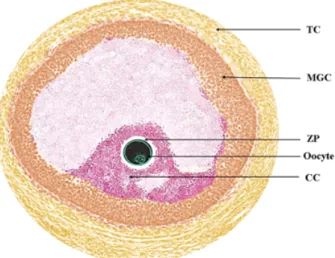
일반적인 체내 환경에서 난포(Fig. 1)는 난자의 성숙에 필요 한 구조적 환경과 기능적 요소를 제공한다(Brower and Schultz, 1982). 이 미세 환경에서 미성숙 난자는 성숙 시 난자 주변의 난구 세포(cumulus cells), 난포 벽면의 과립막 세포(Mural gra- nulosa cells), 그리고 난포막 세포(Theca cells) 등 난소 피질 내 체세포들과의 상호작용을 통하여 발달하게 된다(Brower and Schultz, 1982; Eppig, 2001). 하지만 대부분의 체외 성숙 환경 에서 미성숙 난자는 난포 내 미세 환경으로부터 분리되어 성 숙이 진행되기 때문에, 성숙 시 난포 내 체세포들과의 물질 교 환 및 공간적 상호 작용을 소실하게 된다.
특히 난자의 체외 성숙 과정에서 잃어버리게 되는 난포 내
Fig. 1. Follicular environment during in vivo oocyte maturation.
TC : theca cells, MGC : mural granulosa cells, ZP : zona pellucida, CC : cumulus cells.
미세 환경을 복구하기 위하여 과거 많은 연구자들이 난소 피 질 세포(ovarian cortex cells), 난관 상피 세포(oviductal epithe- lial cells), 과립막 세포, 난포막 세포, 섬유아 세포(fibroblast) 및 난포벽 조각(piece of follicle wall) 등 다양한 유래의 체세 포를 체외 성숙에 적용한 미세 환경의 복원 연구가 다양한 포 유 동물에서 시도되었다(Vannucchi et al., 2006; Chen et al., 2007b; Lin et al., 2009; Shi et al., 2009; Kyasari et al., 2012;
Feng et al., 2013; Karimpour et al., 2014). 일례로 미성숙 돼 지 난자의 체외 성숙 시 난관 상피 세포와 함께 공동 배양을 통해 획득된 성숙 난자에 체외 수정을 실시한 결과, 공동 배 양된 성숙 난자는 대조군보다 유의적으로 높은 단일 정자 수 정률(monospermiy)을 보였고, 이후 배아 발달율 평가에서 유 의적으로 높은 배반포 형성률 및 총 세포수를 나타내는 것이 보고된바 있다(Kidson et al., 2003). 또한 동종에서 난소 피질 세포와 함께 공동 배양을 통해 획득된 성숙 난자는 대조군에 비해 유의적으로 높은 핵 성숙률(metaphase II)이 관찰되었고, 세포질 성숙의 지표로 활용되는 세포질 내 글루타치온(Glu- tathion) 수준이 높게 나타내는 것으로 보고되었다. 이후 체외 수정을 통해 획득된 배아에서 유의적으로 높은 전핵 형성 및 배반포 형성율이 나타났다(Chen et al., 2007).
돼지 외에도 생쥐, 개, 버팔로, 양 등 다른 포유 동물 종에 서 공동 배양 체계를 통해 획득된 체외 성숙 난자에서 높은 핵 성숙율 및 세포질 성숙율을 보이고 있으며, 차후 체외 수정 및 단위 발생을 통해 획득된 배아의 발달능 평가에서 높은 배반포 형성율이 나타나는 것으로 보고되어 왔다(Martino et al., 1995;
Bogliolo et al., 2002; Lin et al., 2009; Karimpour et al., 2014). 지금껏 많은 보고들을 통하여 공동 배양 체계가 체외
성숙 시 적용되었을 때 성숙 난자의 핵성숙율, 세포질 성숙율 및 배아 발달능이 유의적으로 향상되었다. 하지만 공동 배양 체계를 활용하여 체외 성숙 시 부분적인 미세 환경을 복원시 켜 주었을 때 어떠한 요인으로 인하여 성숙효율을 증가되는지 에 대한 이해가 부족한 것이 현실이다. 따라서 생식 세포의 체 외 성숙 간 공동 배양 체계를 적용하는데 있어 체외 성숙 과정 에 발생되는 공동 배양 세포주의 역할 및 그 기전에 대한 연구 의 필요성이 요구된다.
난자와 공동 배양 세포와의 상호 작용
난자와 공동 배양 세포와의 상호 작용을 이해하기 위하여 최근 공동 배양 시 TGF-beta(transforming growth factor-beta) 군의 연관성에 대한 이해가 시도되고 있다. 체내에서 난포의 성공적인 발달을 위해서 난자와 난포벽 과립막 세포 또는 난 포막 세포와 같은 난포 체세포 간의 양 방향적인 소통이 요구 된다(Eppig, 2001; van den Hurk and Zhao, 2005). 특히 난포 의 성장 과정 동안 BMP15(bone morphogenetic protein)과 GDF9(growth differentiation factor)와 같은 TGF-beta 군의 발 현은 설치류, 반추류 및 인간을 포함한 많은 다양한 종에서 양성 조절을 하는 측분비인자(paracrine factor) 중 하나로 제 시된바 있다(Elvin et al., 1999; Elvin et al., 2000; Erickson and Shimasaki, 2003; Shimasaki et al., 2004; Juengel and Mc- Natty, 2005). 특히 난자의 성숙 간 난포 내 GDF9과 BMP15 의 증가는 과립막 세포의 증식을 촉진시키고, 해당 과정 활성 및 여포 자극 호르몬(follicle stimulating hormone, FSH) 의존 적 난포 기능을 조절하는 등 난포의 발달 간 미세 환경을 조 절하는 결정적인 역할을 한다(Gilchrist et al., 2004; Sugiura et al., 2007; Su et al., 2008). 최근 공동 배양 체계에 대한 보고 에 따르면, 공동 배양 시 BMP15, GDF9 그리고 그 해당 수용 체에 대한 mRNA 발현 양상이 변화됨이 보고된 바 있으며 (Kyasari et al., 2012), TGF를 포함하는 인자들과의 연관성의 이해가 시도되고 있다(Lin et al., 2009).
다른 한편으로 난관 상피 세포를 포함하는 체세포와 공동 배양 시 ampiregulin, epiregulin 및 betacellulin과 같은 EGF (epidermal growth factor) 유사 성장 인자의 발현이 증가되는 것이 보고된 바 있다(Rief et al., 2002; Gilchrist et al., 2004).
공동 배양 체계에서 TGF-beta와 EGF 유사 성장 인자의 증가 는 미성숙 난자와 체세포 간의 동반 상승 효과를 가져오게 되 고, 이러한 상호작용의 증가는 공동 배양 시 난자 내의 Mn- SOD, ZnSOD와 같은 항산화 관련 유전자를 증가시킴으로써 성숙 난자의 활성 산소 수준 및 세포 자멸사 현상을 감소시키 며, 세포질 성숙에 영향을 미칠 것이다(Jang et al., 2008a; Jang et al., 2008b). 특히 공동 배양 체계가 적용된 양에서의 보고에
따르면, 공동 배양 시 체세포와 생식 세포 간의 상호 작용의 증가로 체외 수정 후 수정란의 접합자 유전자 활성화(zygotic genome activation)에 관여함으로써 배아의 정상 발달 과정으 로 이끄는 Zar1(Zygote Arrest 1)의 농도가 대조군에 비하여 유의적으로 높게 관찰되었다(Shirazi and Motaghi, 2013).
생식 세포 공동 배양 체계에서의 공간적 환경 영향
생식 세포의 공동 배양 체계는 대사생리학적 측면 외에도 공간적 측면에서도 이해될 수 있는데, 체세포와의 공동 배양을 통해 제공되는 물리적 환경 요소 또한 난자의 성숙 및 향후 배 아의 발달에 영향을 미치는 것으로 보고된 바 있다(Torre et al., 2006; Ma et al., 2007). 실제로 포유동물의 샘 세포(grand cells) 를 이용한 연구에서 세포외기질(extra-cellular matrix)은 세포 의 성장 및 분화에 영향을 주는 것으로 알려졌는데(Abbott, 2003), 이후 공동 배양 시 체세포로부터 제공되는 세포외기질 이 영향을 미치는 것에 기인한 많은 논문이 발표되었다.
특히, 버팔로 연구에서 일차 단계에서 난구 세포를 제거한 미성숙 난자를 삼차원 구조의 난구 세포 구조물 안에서 공동 배양을 실시한 결과, 성숙 후 대조군에 비하여 난자의 성숙률 및 단위 발생 배아의 배반포 형성율이 유의적으로 증가하였 고, 특히 소실된 간극 연접(gap junction)이 재구성되었다고 (Fig. 2) 보고된 바 있다(Feng et al., 2013). 최근 체세포와 생식 세포 간의 상호 작용을 제외하고, 공간적인 영향만을 연구하 기 위하여 체세포가 아닌 젤라틴 또는 콜라겐을 활용한 논문 들이 보고되고 있다(Combelles et al., 2005; Ma et al., 2007).
향후 전략 및 전망
생명공학기술의 발달로 체외 성숙을 포함하는 보조 번식 기 법에 대한 개선이 이루어져 왔으나, 체내 환경의 재현에는 많 은 제한이 있는 것이 사실이다. 무엇보다도 일반적인 체외 성 숙 기술은 미성숙 난자의 성숙 시 난포 내 미세 환경으로부터 분리가 된다는 문제점이 발생되었다. 이러한 문제를 해결하기 위한 대안으로 많은 연구자들이 과립막 세포, 난포막 세포, 섬 유아 세포 등과 같은 체세포를 이용한 부분적인 복원에 관한 시도 및 연구들이 보고되어왔고, 그 결과 핵 성숙율, 세포질 성숙도, 체외 수정 또는 단위 발생을 통해 획득된 배아의 배 반포 형성율과 총 세포수가 증가되는 성숙효율의 향상을 가 져왔다. 이러한 결과는 미성숙 난자의 성숙 시 세포 간 상호 작용 및 세포외기질이 가져오는 물리적 영향에 따른 이해가 가능하게 만들었다. 이처럼 체내 환경과 체외 환경의 정확한 차이와 해결점을 찾고 이해하는데 공동 배양 체계가 그 정보 를 제공할 수 있을 것으로 사료된다.
(A)
(B)
Fig. 2. Illustration of co-culture systems (A) general co-culture system with feeder cells, (B) 3D imensionally restored co- culture system embedding within artificially rolled feeder cells.
참 고 문 헌
Abbott A. 2003. Cell culture: biology's newdimension. Nature 424: 870-2.
Bogliolo L, Zedda MT, Ledda S, Leoni G, Naitana S and Pau S. 2002. Influence of co-culture with oviductal epithelial cells on in vitro maturation of canine oocytes. Reprod. Nutr.
Dev. 42: 265-73.
Brandacher G, Grahammer J, Sucher R and Lee WP. 2012.
Animal models for basic and translational research in re- constructive transplantation. Birth Defects Res. C Embryo Today 96: 39-50.
Brower PT and Schultz RM. 1982. Intercellular communica- tion between granulosa ells and mouse oocytes: existence and possible nutritional role during oocyte growth. Dev.
Biol. 90: 144-53.
Chen XY, Li QW, Zhang SS, Han ZS, Zhao R, Wu SY and Huang J. 2007. Effects of ovarian cortex cell co-culture during in vitro maturation on porcine oocytes maturation, fertilization and embryo development. Anim. Reprod. Sci.
99: 306-316.
Combelles CM, Fissore RA, Albertini DF and Racowsky C.
2005. In vitro maturation of human oocytes and cumulus
cells using a co-culture three-dimensional collagen gel system. Hum. Reprod. 20: 1349-58.
Coticchio G, Dal-Canto M, Guglielmo MC, Mignini-Renzini M and Fadini R. 2012. Human oocyte maturation in vitro.
Int. J. Dev. Biol. 56: 909-18.
Edwards RG. 1965a. Maturation in vitro of human ovarian oocytes. Lancet 2: 926-9.
Edwards RG. 1965b. Maturation in vitro of mouse, sheep, cow, pig, rhesus monkey and human ovarian oocytes.
Nature 208: 349-51.
Elvin JA, Yan C and Matzuk MM. 2000. Oocyte-expressed TGF-beta superfamily members in female fertility. Mol.
Cell. Endocrinol. 159: 1-5.
Elvin JA, Yan C, Wang P, Nishimori K and Matzuk MM.
1999. Molecular characterization of the follicle defects in the growth differentiation factor 9-deficient ovary. Mol.
Endocrinol. 13: 1018-34.
Eppig JJ. 2001. Oocyte control of ovarian follicular develop- ment and function in mammals. Reproduction 122: 829-38.
Erickson GF and Shimasaki S. 2003. The spatiotemporal ex- pression pattern of the bone morphogenetic protein family in rat ovary cell types during the estrous cycle. Reprod.
Biol. Endocrinol. 1: 9.
Feng G, Shi D, Yang S and Wang X. 2013. Co-culture embe- dded in cumulus clumps promotes maturation of denuded oocytes and reconstructs gap junctions between oocytes and cumulus cells. Zygote 21: 231-7.
Gilchrist RB, Ritter LJ and Armstrong DT. 2004. Oocyte- somatic cell interactions during follicle development in mammals. Anim. Reprod. Sci. 82-83: 431-46.
Jang HY, Jung YS, Li ZY, Yoon HJ, Cheong HT, Kim JT, Park CK and Yang BK. 2008a. Protective effect of BOEC co-culture system against nitric oxide on development of bovine IVM/IVF embryos. Reprod. Dev. Biol. 32: 167-173.
Jang H, Jung Y, Cheong H, Kim J, Park C, Kong, H, Lee, H and Yang, B. 2008b. Effects of cell status of bovine ovi- duct epithelial cell (BOEC) on the development of bovine IVM/IVF embryos and gene expression in the BOEC used or not used for the embryo culture. Asian-Aust. J. Anim.
Sci. 21: 980-987.
Jin YX, Jeon Y, Lee SH, Kwon MS, Kim T, Cui XS, Hyun SH and Kim NH. 2014. Production of pigs expressing a transgene under the control of a tetracycline-inducible system. PloS one 9: e86146.
Juengel JL and McNatty KP. 2005. The role of proteins of the transforming growth factor-beta superfamily in the intrao- varian regulation of follicular development. Hum. Reprod.
Update 11: 143-60.
Karimpour MA, Heidari M, Parivar K and Azami NS. 2014.
The effects of fibroblast co-culture and activin A on in vitro growth of mouse preantral follicles. Iran. Biomed. J. 18:
49-54.
Kidson A, Schoevers E, Langendijk P, Verheijden J, Colen- brander B and Bevers M. 2003. The effect of oviductal epithelial cell co-culture during in vitro maturation on sow oocyte morphology, fertilization and embryo development.
Theriogenology 59: 1889-903.
Kyasari OR, Valojerdi MR, Farrokhi A and Ebrahimi B. 2012.
Expression of maturation genes and their receptors during in vitro maturation of sheep COCs in the presence and absence of somatic cells of cumulus origin. Theriogenology 77: 12-20.
Lin YH, Hwang JL, Seow KM, Huang LW, Chen HJ and Tzeng CR. 2009. Effects of growth factors and granulosa cell co-culture on in-vitro maturation of oocytes. Reprod.
Biomed. online 19: 165-70.
Ma S, Lin H, Miao Y, Liu X, Wang B and Dai J. 2007. The effect of three-dimensional demineralized bone matrix on in vitro cumulus-free oocyte maturation. Biomaterials 28:
3198-207.
Martino A, Mogas T, Palomo MJ and Paramio MT. 1995. In vitro maturation and fertilization of prepubertal goat oocytes.
Theriogenology 43: 473-85.
Pincus G and Enzmann EV. 1935. The comparative behavior of mammalian eggs in vivo and in vitro : I. the activation of ovarian eggs. J. Exp. Med. 62: 665-75.
Rief S, Sinowatz F, Stojkovic M, Einspanier R, Wolf E and Prelle K. 2002. Effects of a novel co-culture system on development, metabolism and gene expression of bovine em- bryos produced in vitro. Reproduction (Cambridge, England) 124: 543-56.
Shi L, Yue W, Zhang J, Lv L, Ren Y and Yan P. 2009. Effect of ovarian cortex cells on nuclear maturation of sheep oocy- tes during in vitro maturation. Anim. Reprod. Sci. 113:
299-304.
Shimasaki S, Moore RK, Otsuka F and Erickson GF. 2004.
The bone morphogenetic protein system in mammalian rep- roduction. Endocr. Rev. 25: 72-101.
Shirazi A and Motaghi E. 2013. The in vitro fertilization of ovine oocytes in the presence of oviductal cells and its effect on the expression of zygote arrest 1 (Zar1) and subsequent embryonic development. J. Repro. Infertil. 14: 8-16.
Steptoe PC and Edwards RG. 1978. Birth after the reimplan- tation of a human embryo. Lancet 2: 366.
Su YQ, Sugiura K, Wigglesworth K, O'Brien MJ, Affourtit JP, Pangas SA, Matzuk, MM and Eppig JJ. Oocyte regulation of metabolic cooperativity between mouse cumulus cells and oocytes: BMP15 and GDF9 control cholesterol biosyn- thesis in cumulus cells. Development (Cambridge, England) 135: 111-21.
Sugiura K, Su YQ, Diaz FJ, Pangas SA, Sharma S, Wiggles- worth K, O'Brien MJ, Matzuk MM, Shimasaki S and Eppig JJ. 2007. Oocyte-derived BMP15 and FGFs cooperate to promote glycolysis in cumulus cells. Development (Cam- bridge, England) 134: 2593-603.
Torre ML, Munari E, Albani E, Levi-Setti PE, Villani S, Faustini M, Conte U and Vigo D. 2006. In vitro maturation of human oocytes in a follicle-mimicking three-dimensional coculture. Fertil. Steril. 86: 572-6.
van HR and Zhao J. 2005. Formation of mammalian oocytes and their growth, differentiation and maturation within ova- rian follicles. Theriogenology 63: 1717-51.
Vannucchi CI, Oliveira CM, Marques MG, Assumpcao ME and Visintin JA. 2006. In vitro canine oocyte nuclear matura- tion in homologous oviductal cell co-culture with hormone- supplemented media. Theriogenology 66: 1677-81.
Zhang P, Liu P, Dou H, Chen L, Lin L, Tan P, Vajta G, Gao J, Du Y and Ma RZ. 2013. Handmade cloned transgenic sheep rich in omega-3 fatty acids. PloS one 8: e55941.
(접수: 2014. 3. 17/ 심사: 2014. 3. 17/ 채택: 2014. 3. 24)