Short-term of Reverse Total Shoulder Arthroplasty for the Treatment of Irreparable Massive Rotator Cuff Tear
Jong-Hyuk Park, Seong-Il Wang , Byung-Chang Lee1
Department of Orthopedic Surgery, Chonbuk National University Hospital, Research Institute of Clinical Medicine, Chonbuk National University Medical School, Jeonju, 1Naeun Hospital, Iksan, Korea
Background: To investigate the effectiveness of reverse total shoulder arthroplasty (RTSA) in treating irreparable massive rotator cuff tears (RCTs).
Methods: Twenty-nine patients who underwent RTSA for the treatment of irreparable massive RCTs and completed follow-up for at least 1 year were selected. Their mean age was 69.7 years (range, 59-80 years). The mean follow-up was 17.7 months (range, 12-42 months).
The shoulder range of motion was measured preoperatively and at final follow-up. The functional result was evaluated using visual ana- log scale (VAS) for pain, American Shoulder and Elbow Surgeon (ASES) score, and Korean Shoulder Society (KSS) score. Additionally, the shoulders were categorized into two groups depending on prior history of surgery and the clinical outcomes were analyzed between two groups.
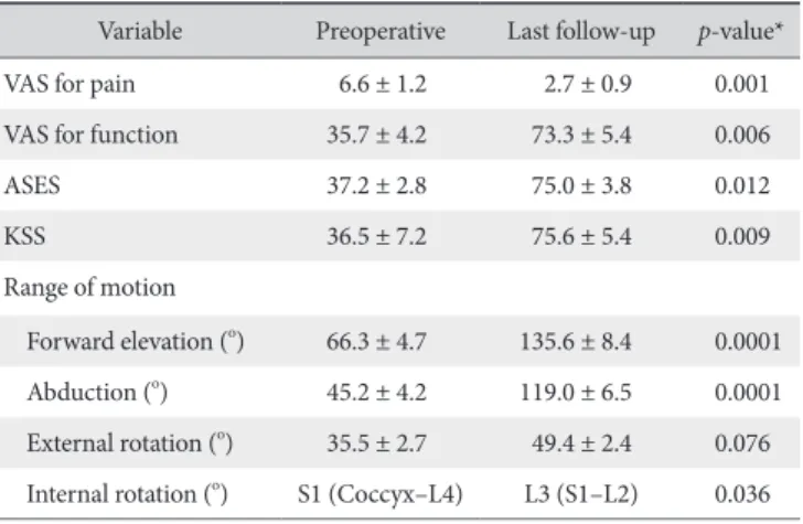
Results: Mean pain VAS improved, from 6.6±1.2 to 2.7±0.9 (p=0.001), and the mean functional VAS from 35.7±4.2 to 73.3±5.4 (p=0.006). The mean ASES score improved from 37.2±2.8 to 75.0±3.8 (p=0.012). The mean KSS improved from 36.5±7.2 to 75.6±5.4 (p=0.009), the mean forward elevation from 66.3±4.7 to 135.6±8.4 (p=0.0001), and the mean abduction from 45.2±4.2 to 119.0±6.5o (p=0.0001). Internal rotation differed significantly from the first sacral to the third lumbar vertebrae (p=0.036). External rotation did not change significantly (p=0.076). There was also no statistically significant difference between groups (no previous opera- tion versus none). Four complications occurred: one superficial infection, one with anterior dislocation, one acromial fracture, and one clavicle fracture.
Conclusions: RTSA provides reliable pain relief and recovery of shoulder function in patients with massive irreparable RCTs in short- term follow-up.
(Clin Shoulder Elbow 2014;17(4):152-158)
Key Words: Rotator cuff; Massive; Arthropathy; Shoulder; Arthroplasty
Introduction
Massive irreparable rotator cuff tears may occur in cases of chronic tears with or without prior surgery. Treatments for ir- reparable rotator cuff tears include conservative treatments,1) arthroscopic debridement, acromial decompression, and partial repair.2,3) Additionally, latissimus dorsi transfer4,5) and arthroplas- ty6-8) have shown variable results.
Although these methods can often provide pain relief, they
are not very reliable and often limited in achieving functional improvement. Hemiarthroplasty provides partial pain relief but poor functional results. Thus, it is typically only useful for elderly people with low motion demands. Total shoulder replacement has been shown to fail due to early glenoid loosening.9)
To overcome these disadvantages, a reverse total shoulder ar- throplasty has been used in place of conventional total shoulder arthroplasty in the treatment of massive rotator cuff tear. Since Grammont and Baulot10) first described the procedure, many Clinics in Shoulder and Elbow Vol. 17, No. 4, December, 2014
http://dx.doi.org/10.5397/cise.2014.17.4.152
Received June 13, 2014. Revised August 26, 2014. Accepted September 14, 2014.
Correspondence to: Seong-Il Wang
Department of Orthopedic Surgery, Chonbuk National University Hospital, 20 Geonji-ro, Deokjin-gu, Jeonju 561-712, Korea Tel: +82-63-250-1760, Fax: +82-63-271-6538, E-mail: [email protected]
Financial support: This pater was (partially) supported by the Chonbuk National University (CBNU) funds for overseas research. Conflict of interests: None.
studies have reported positive results with respect to pain relief and recovery of shoulder function.11,12)
Given this background, we investigated the short-term follow- up outcome of reverse total shoulder arthroplasty for the treat- ment of irreparable massive rotator cuff tear.
Methods
Patients
From October 2008 to February 2011, we selected patients who had undergone reverse total shoulder arthroplasty to treat irreparable massive rotator cuff tear from two institutions.
Among 36 patients who met the requirements, 29 (7 men, 22 women; mean age, 69.7 years; range, 59 to 80 years) who un- derwent at least 1 year of postoperative follow-up (mean, 17.7 months; range, 12 to 42 months) were analyzed. We used the Aequalis reverse type (Tornier, Montbonnot, France) in 20 cases and the Anatomical shoulder (Zimmer Inc., Warsaw, IN, USA) in 9 cases. Glenohumeral arthritis and active forward flexion <90o were observed in 26 cases. There was a case of glenohumeral arthritis with active forward flexion >90o, and two cases without glenohumeral arthritis but a limited active forward flexion (<90o).
The indications for surgery were cuff arthropathy, pseudo- paralysis with a massive irreparable rotator cuff tear that involve two or more rotator cuff tendons with atrophy. Cuff tear ar- thropathy is characterized by rotator cuff dysfunction and end stage glenohumeral arthritis. Painful pseudoparesis is defined as active shoulder elevation of <90o in the presence of free passive anterior elevation. This assessment was based on a combination of findings on physical examination (rotator cuff muscle atropy, dynamic instability, and limited range of motion) and radio- graphs (decreased joint space and abnormal joint position). The classification system described by Hamada et al.13) was used to grade the preoperative radiographs.
The rotator cuff was considered to be irreparable if the rota- tor cuff was chronic massive rotator cuff tears that involve two or more rotator cuff tendons, severe pain for at least 6 months with weakness of rotator cuff muscle, an acromiohumeral interval of less than 6 mm on the true antero-posterior radiograph, fatty infiltration of the supraspinatus and infraspinatus muscles was greater than stage three according to the Goutallier classifica- tion,14) and arthroscopic finding as retraction to glenoid of teared massive rotator cuff with poor tissue quality and non-mobiliza- tion.
The exclusion criteria were insufficient follow-up period (at least 1 years), severe deltoid impairment, preoperative infection history, revision surgery.
Clinical Assessment
In the clinical evaluation, shoulder range of motion was eval- uated in terms of forward elevation, abduction, external rotation
at the side, and internal rotation preoperatively and at the final follow-up. The functional result was evaluated using a Visual Analog Scale (VAS) for pain and function, American Shoulder and Elbow Surgeon (ASES) score, and Korean Shoulder Society (KSS) score. Postoperative complications were also investigated.
Additionally, the shoulders were categorized in two groups depending on whether they had undergone surgery previously:
18 shoulders (Group A) had not undergone previous surgery, whereas 11 shoulders (Group B) had undergone at least one previous procedure.
In Group B, the mean duration to reverse total shoulder arthroplasty after previous surgery was 38.5 ± 20.5 months (range, 12 to 72 months). Five patients were performed with arthroscopic cuff repair, 2 with arthroscopic partial repair and 1 with mini-open repair. They all later complained of re-rupture with aggravating pain. For the other 3 patients, arthroscopic de- bridment was performed as rotator cuff suture was impossible;
however, as they showed no clinical improvement, reverse total shoulder arthroplasty was performed.
The clinical outcomes in Groups A and B were analyzed and compared.
Surgical Technique and Rehabilitation
All procedures were performed in the beach-chair position using general anesthesia. The deltopectoral approach was used, and a portion of the pectoralis major insertion to aid in exposure was released. The subscapularis tendon was detached from the subscapularis footprint with tagging sutures. The joint capsule was released as much as possible towards the anatomical neck of the humerus. The glenohumeral joint was subjected to exten- sion and external rotation to dislocate the head of the humerus.
The humeral head resection was made with a retroversion angle between 0o and 20o according to the individual anatomy using a rotation guide with the forearm axis set to be neutral rotation.
We made the osteotomy in 0o of retroversion for patients with teres minor muscle tears and a preoperative external rotation lag sign. After the head cutting, the glenoid baseplate fixation was followed according to the company instrumentation manual.
We placed the guide for glenoid preparation inferiorly on the glenoid so that the base plate was fully supported by bone and flush with the inferior glenoid margin. We placed the guide wire in approximately 10o inferior tilt, because this position mini- mized the risk of scapular notching.11) Two anterior and posterior screws (4.5 mm) are positioned first to optimize compression of the baseplate with adequate cortical bone fixation. We placed two superior and inferior locking screws (4.5 mm) through the holes in the baseplate with bone purchase into the base of the coracoid and inferior scapular border. Then, we placed the central screw through the glenosphere and into the central hole of the baseplate. The polyethylene insert thickness was chosen based on soft-tissue tension during a trial reduction. In judging
the proper soft-tissue tension, our goal was minimal shucking (1 to 2 mm) with tension of the conjoined tendon, which was tight, but not enough to cause bowstringing of the tendon. Using bone cement, an actual humeral stem was inserted and combined with a polyethylene insert. The subscapularis tendon and joint capsule were reattached to the lesser tuberosity of humerus with No. 2 ethibond that was temporarily hooked.
Patients were placed in an abduction brace for 3 weeks, dur- ing which time only pendulum-type exercises were allowed.
Passive range of motion exercises were initiated carefully 3 weeks after the surgery, beginning with forward flexion exercises.
At 6 weeks after the surgery, active assisted exercises were per- formed. Active range of motion exercises and strength training exercises were started 12 weeks after the surgery.
Statistical Analysis
The Wilcoxon signed rank test was used to compare the preoperative and final follow-up results of VAS, active range of motion (forward flexion, abduction, external rotation, internal rotation), and shoulder function scores (ASES, KSS). The Mann- Whitney U test was used to analyze differences among the subgroups. The significance level was set at p<0.05. All statisti- cal analyses were conducted using the PASW Software ver. 18.0 (IBM Co., Armonk, NY, USA).
Results
Clinical Outcomes of the Entire Population (n=29) The mean VAS score for pain improved, from 6.6 ± 1.2 pre- operatively to 2.7 ± 0.9 at the final follow-up (p=0.001), while the mean VAS score for function improved, from 35.7 ± 4.2 to 73.3 ± 5.4 (p=0.006). The mean total ASES score for the entire population (n=29) improved, from 37.2 ± 2.8 to 75.0 ± 3.8 (p=0.012). The mean KSS improved, from 36.5 ± 7.2 to 75.6
± 5.4 (p=0.009).
Shoulder motion also improved for all patients at the final follow-up. The mean forward elevation improved from 66.3 ± 4.7o to 135.6 ± 8.4o (p=0.0001) and the mean abduction im- proved from 45.2 ± 4.2o to 119.0 ± 6.5o (p=0.0001). Preop- erative internal rotation was noted in one case at the lumbar 4 (L4) level, three cases at the L5 level, 18 cases at the sacrum 1 (S1) level, and seven cases at the coccyx level. At the final follow- up, internal rotations was noted in three cases at the L2 level, 16 cases at the L3 level, six cases at the L4 level, three cases at the L5 level, and one case at the S1 level. These preoperative and final follow-up differences in internal rotation were statistically significant (p=0.036). The mean external rotation improved from 35.5 ± 2.7o to 49.4 ± 2.4o, but the difference was not sta- tistically significant (p=0.076; Table 1).
Clinical Outcomes in the No Previous Surgery (Group A;
n=18) versus Failed Rotator Cuff Repair Group (Group B;
n=11)
In Group A, the mean VAS for pain improved, from 6.3 ± 0.8 preoperatively to 2.1 ± 1.6 at the final follow-up, while the mean VAS score for function improved, from 36.4 ± 5.7 to 75.2
± 3.6. The mean total ASES score improved from 38.6 ± 3.8 to 76.4 ± 2.6, while the mean KSS improved from 37.8 ± 6.8 to 77.1 ± 1.9. In Group B, the mean VAS score for pain improved, from 7.1 ± 1.4 preoperatively to 3.8 ± 2.1 at final follow-up, whereas the mean VAS score for function improved from 34.8
± 7.2 to 70.2 ± 4.6. The mean total ASES score improved, from 34.8 ± 7.6 to 72.8 ± 1.0, while the mean KSS improved, from 34.6 ± 8.2 to 73.1 ± 2.6. Although Group A showed better overall outcomes than Group B, the difference was not statisti- cally significant (Table 2).
With respect to the shoulder range of motion, the mean for- ward elevation improved, from 64.2 ± 5.8o to 138.7 ± 10.8o, while the mean abduction improved, from 44.7 ± 4.7o to 121.4
± 7.3o. Preoperative internal rotation was noted in one case at the L4 level, two cases at the L5 level, 11 cases at the S1 level, and four cases at the coccyx level. At the final followup, the internal rotation had improved to two cases at the L2 level, 10 cases at the L3 level, four cases at the L4 level, one case at the L5 level, and one case at the S1 level. Additionally, the mean external rotation improved from 36.4 ± 3.5o to 50.3 ± 1.7o.
In Group B, the mean forward flexion improved, from 69.7
± 7.2o to 130.4 ± 8.1o, whereas the mean abduction improved from 46.1 ± 5.9o to 115.1 ± 10.3o. Preoperative internal rota- tion was noted in one case at the L5 level, seven cases at the S1 level, and three cases at the coccyx level. At the final follow- up, the internal rotation had improved to one case at the L2 level, six cases at the L3 level, two cases at the L4 level, and two
Table 1. Clinical Outcomes in Total Population (n=29)
Variable Preoperative Last follow-up p-value*
VAS for pain 6.6 ± 1.2 2.7 ± 0.9 0.001
VAS for function 35.7 ± 4.2 73.3 ± 5.4 0.006
ASES 37.2 ± 2.8 75.0 ± 3.8 0.012
KSS 36.5 ± 7.2 75.6 ± 5.4 0.009
Range of motion
Forward elevation (o) 66.3 ± 4.7 135.6 ± 8.4 0.0001 Abduction (o) 45.2 ± 4.2 119.0 ± 6.5 0.0001 External rotation (o) 35.5 ± 2.7 49.4 ± 2.4 0.076 Internal rotation (o) S1 (Coccyx–L4) L3 (S1–L2) 0.036 Values are presented as mean ± standard deviation.
VAS: visual analog scale, ASES: American Shoulder and Elbow Surgeons score, KSS: Korean Shoulder Society score, S: sacrum, L: lumbar.
*Wilcoxon signed rank test.
cases at the L5 level. Additionally, the mean external rotation improved, from 34.1 ± 2.3o to 47.8 ± 3.4o. While the shoulder range of motion in Group A was better than that in Group B, the difference was not statistically significantly (Table 2).
Complications
There were four cases of complications: one case was com- plicated with superficial infection, 1 with anterior dislocation, 1
with acromial fracture, and 1 with clavicle fracture. An anterior dislocation that was caused by a fall 3 months after the surgery did not recur after closed reduction. With respect to an acromial fracture that occurred 7 months after the surgery, K-wires and tension-band wiring were performed to achieve bone union (Fig.
1). Furthermore, conservative treatment of a clavicle fracture found 6 weeks after the surgery by radiography achieved bone union (Fig. 2).
Fig. 1. (A) Preoperative antero-posterior radiograph of a 71-year-old female patient, showing superior migration of the humeral head, combined with glenohu- meral arthritis due to an irreparable rotator cuff tear (Hamada type IV). (B) An antero-posterior radiograph at 7 months after surgery showing an acromial frac- ture. (C) An antero-posterior radiograph at the last follow-up showing union of a previous acromial fracture after K-wire and tension-band wiring.
A B C
Table 2. Clinical Outcomes in Patient with No Previous Surgery (Group A) versus Those with Failed Rotator Cuff Repair (Group B)
Group A Group B
p-value*
Preoperative Last follow-up Preoperative Last follow-up
VAS for pain 6.3 ± 0.8 2.1 ± 1.6 7.1 ± 1.4 3.8 ± 2.1 0.087
VAS for function 36.4 ± 5.7 75.2 ± 3.6 34.8 ± 7.2 70.2 ± 4.6 0.748
ASES 38.6 ± 3.8 76.4 ± 2.6 34.8 ± 7.6 72.8 ± 1.0 0.869
KSS 37.8 ± 6.8 77.1 ± 1.9 34.6 ± 8.2 73.1 ± 2.6 0.628
Range of motion
Forward elevation (o) 64.2 ± 5.8 138.7 ± 10.8 69.7 ± 7.2 130.4 ± 8.1 0.081
Abduction (o) 44.7 ± 4.7 121.4 ± 7.3 46.1 ± 5.9 115.1 ± 10.3 0.162
External rotation (o) 36.4 ± 3.5 50.3 ± 1.7 34.1 ± 2.3 47.8 ± 3.4 0.764
Internal rotation S1 (Coccyx–L4) L3 (S1–L2) S1 (Coccyx–L5) L3 (L5–L3) 0.564
Values are presented as mean ± standard deviation.p-value: no previous surgery (Group A) versus failed rotator cuff repair (Group B).
VAS: visual analog scale, ASES: American Shoulder and Elbow Surgeons score, KSS: Korean Shoulder Society score, S: sacrum, L: lumbar.
*Mann-Whitney U test.
Discussion
The reverse shoulder prosthesis was developed by Gram- mont and Baulot10) to restore shoulder function in case of an glenohumeral osteoarthritis associated with irreparable rotator cuff tear. The implant’s design brings the center of rotation of the glenohumeral joint more medial and the insertion of the deltoid muscle more distally, which then increases the lever arm and the tension of the deltoid muscle. The reverse shoulder prosthesis can, thus, address both degenerative changes of the glenohu- meral joint and a missing rotator cuff.11,12,15) Sirveaux et al.11) reported 80 patients with a mean follow-up period of 3.6 years.
The procedure was associated with good pain relief in 96% of the patients, and their Constant scores improved from 22 to 65 points. Frankle et al.12) reported a minimum 2-year follow-up study of 60 shoulders with cuff-tear arthropathy. Reverse arthro- plasty was associated with statistically significant improvements in pain and function, with a mean active elevation of approxi- mately 105o. However, there was a 17% complication rate and a 12% rate of revision for implant failure.
In the present study, the short-term outcomes (mean, 17.7 months) after reverse total shoulder arthroplasty for the treat- ment of irreparable massive rotator cuff tear demonstrated a significant improvement in the VAS score, ASES score, and KSS at the final follow-up compared with the preoperative results.
Furthermore, forward elevation, abduction, and internal rotation
also improved significantly at the final follow-up compared with the preoperative results. The reverse total shoulder arthroplasty is considered an effective treatment method that produces good clinical outcomes for irreparable massive rotator cuff tears ac- companied by osteoarthritis and pseudoparalysis. However, while the mean external rotation improved, from 35o to 49o in our patients, the results did not show a statistically significant difference from the preoperative results. Simovitch et al.16) sug- gested that improvement in external rotation is uncommon after reverse shoulder arthroplasty, especially when there is fatty infiltration of the teres minor muscle. Werner et al.17) suggested that reverse total shoulder arthroplasty with a Grammont-type prosthesis can restore forward elevation and abduction but not external rotation, which should be explained as a limitation to patients prior to surgery. We believe that performing an osteoto- my in zero retroversion to improve external rotation in patients with teres minor muscle tears and a preoperative external rota- tion lag sign has limitations. Because not all patients underwent magnetic resonance imaging, we believe that the preoperative severity of teres minor muscle tear and fatty infiltration were not fully evaluated.
In the absence of a functional teres minor muscle, some authors have proposed combining implantation of a reverse implant with transfer of the latissimus dorsi around the humeral shaft, through the same deltopectoral approach.18) Biomechani- cal studies suggest that latissimus dorsi muscle transfer improves Fig. 2. (A) Preoperative antero-posterior radiograph of a 64-year-old woman, showing osteoarthritis and the superior migration of the left humeral head accom- panied by the acetabularization of the acromion (Hamada type IV). (B) A radiograph at postoperative week 6 revealing displaced distal clavicle fracture (arrow). (C) A radiograph at postoperative month 8 showing fracture union.
A B C
external rotation torque when combined with a reverse pros- thesis, and that an insertion site on the posterior aspect of the greater tuberosity close to the teres minor muscle insertion pro- duces a greater moment arm.19) For patients with a severe ex- ternal rotation lag and a torn teres minor muscle, this combined procedure may improve final external rotation, which seems to be associated with higher patient satisfication.20,21)
Some researchers have reported that the no previous shoul- der surgery group exhibited significantly higher postoperative mean pain and function scores, according to the ASES sys- tem and Constant scores, than the previous shoulder surgery group.12,17) However, in the present study, an analysis of clinical outcomes between the groups according to the presence or ab- sence of a surgical history as cuff repair or debridement showed no statistically significant difference. However, in the present study, because 11 cases with a surgical history had undergone attempted mini-open, arthroscopic, or partial repair to treat a massive rotator cuff tear rather than a failed total shoulder ar- throplasty or a revision replacement of hemiarthroplasty, a future comparative analysis of more cases is needed.
The reverse total shoulder arthroplasty is a complex proce- dure that changes joint physiology and biomechanics.22) As a result, the surgery may increase the potential for complications.
Typical complications include scapular notching, baseplate fail- ure, periprosthetic fractures, scapular fractures, infections, hema- toma, instability, and nerve lesions.23)
In the present study, there were four cases of complications:
one case was complicated with superficial infection, 1 with an- terior dislocation, 1 with acromial fracture, and 1 with clavicle fracture. We performed closed reduction for the anterior dis- location caused by a fall 3 months after surgery and required the patient to use an arm sling for 3 weeks; no recurrence was noted.
It is known that arm length is increased by approximately 2.5 cm in patients who undergo reverse total shoulder arthroplasty.
This is also accompanied by increased tension of the deltoid muscle and increased loading on the origin of the deltoid muscle with the action of a substantially longer lever arm of the deltoid muscle because of the medial movement of the center of rota- tion. This explains the possible occurrence of fracture at the origin of the deltoid muscle. It has been reported that acromial fractures occur in 1% to 7% of patients who undergo reverse shoulder arthroplasty.24) Crosby et al.25) reported that ipsilateral scapular fracture occurred during follow-up in 5.5% (22/400) of patients undergoing reverse total shoulder arthroplasty.
We diagnosed an acromial fracture that was sustained when the patient lifted a baggage in 7 months after a reverse total shoulder arthroplasty to treat a massive rotator cuff tear accom- panied by arthritis. Bone union was achieved by performing K-wire and tension-band wiring. A 64-year-old woman with a painful massive left rotator cuff tear and glenohumeral arthritis
began to experience pain spontaneously at the anterior aspect of the shoulder at the distal clavicle when performing passive range of motion exercises after removing the abduction sling 3 weeks after the surgery. On radiography 6 weeks after surgery, a distal clavicle fracture was found. She was prescribed 3 additional weeks with the abduction sling and pendulum exercises, after which passive range of motion exercises were restarted care- fully as conservative treatment. The patient achieved a complete bone union, as demonstrated on a radiograph at 8 months.
Our study has several limitations. First, it had a retrospective design and involved two institutions where different surgeons performed the same surgery. As such, patient selection and sur- gical techniques may have been biased.
In the present study, 2 different types of implant material were used. Reverse angle was not aligned during humeral head cutting. Furthermore, as the pre- and postoperative radiographic imaging techniques may have differed between the two insti- tutions, radiographic analysis of events such as notching was excluded. Second, the number of subjects included was small and the minimum follow-up period of 12 months (mean, 17.7 months) was too short to determine implant longevity or evalu- ate the long-term implications of the radiographic findings.
Conclusion
Reverse shoulder arthroplasty provides reliable pain relief and recovery of shoulder function in patients with massive irrepa- rable rotator cuff tear in a short follow-up period. Further long- term studies are needed to fully understand implant limitations and survivorship.
References
1. Rockwood CA Jr, Williams GR Jr, Burkhead WZ Jr. Débride- ment of degenerative, irreparable lesions of the rotator cuff. J Bone Joint Surg Am. 1995;77(6):857-66.
2. Burkhart SS. Arthroscopic treatment of massive rotator cuff tears. Clinical results and biomechanical rationale. Clin Orthop Relat Res. 1991;(267):45-56.
3. Burkhart SS. Arthroscopic treatment of massive rotator cuff tears. Clin Orthop Relat Res. 2001;(390):107-18.
4. Weening AA, Willems WJ. Latissimus dorsi transfer for treatment of irreparable rotator cuff tears. Int Orthop.
2010;34(8):1239-44.
5. Longo UG, Franceschetti E, Petrillo S, Maffulli N, Denaro V. Latissimus dorsi tendon transfer for massive irreparable rotator cuff tears: a systematic review. Sports Med Arthrosc.
2011;19(4):428-37.
6. Pollock RG, Deliz ED, McIlveen SJ, Flatow EL, Bigliani LU.
Prosthetic replacement in rotator cuff-deficient shoulders. J Shoulder Elbow Surg. 1992;1(4):173-86.
7. Williams GR Jr, Rockwood CA Jr. Hemiarthroplasty in rotator cuff-deficient shoulders. J Shoulder Elbow Surg. 1996;5(5):
362-7.
8. Zuckerman JD, Scott AJ, Gallagher MA. Hemiarthroplasty for cuff tear arthropathy. J Shoulder Elbow Surg. 2000;9(3):169- 72.
9. Franklin JL, Barrett WP, Jackins SE, Matsen FA 3rd. Glenoid loosening in total shoulder arthroplasty. Association with rota- tor cuff deficiency. J Arthroplasty. 1988;3(1):39-46.
10. Grammont PM, Baulot E. Delta shoulder prosthesis for rotator cuff rupture. Orthopedics. 1993;16(1):65-8.
11. Sirveaux F, Favard L, Oudet D, Huquet D, Walch G, Molé D.
Grammont inverted total shoulder arthroplasty in the treat- ment of glenohumeral osteoarthritis with massive rupture of the cuff. Results of a multicentre study of 80 shoulders. J Bone Joint Surg Br. 2004;86(3):388-95.
12. Frankle M, Siegal S, Pupello D, Saleem A, Mighell M, Vasey M.
The reverse shoulder prosthesis for glenohumeral arthritis as- sociated with severe rotator cuff deficiency. A minimum two- year follow-up study of sixty patients. J Bone Joint Surg Am.
2005;87(8):1697-705.
13. Hamada K, Fukuda H, Mikasa M, Kobayashi Y. Roentgeno- graphic findings in massive rotator cuff tears. A long-term ob- servation. Clin Orthop Relat Res. 1990;(254):92-6.
14. Goutallier D, Postel JM, Bernageau J, Lavau L, Voisin MC. Fatty muscle degeneration in cuff ruptures. Pre- and postoperative evaluation by CT scan. Clin Orthop Relat Res. 1994;(304):78- 83.
15. Rittmeister M, Kerschbaumer F. Grammont reverse total shoulder arthroplasty in patients with rheumatoid arthritis and nonreconstructible rotator cuff lesions. J Shoulder Elbow Surg.
2001;10(1):17-22.
16. Simovitch RW, Helmy N, Zumstein MA, Gerber C. Impact of fatty infiltration of the teres minor muscle on the outcome of reverse total shoulder arthroplasty. J Bone Joint Surg Am.
2007;89(5):934-9.
17. Werner CM, Steinmann PA, Gilbart M, Gerber C. Treatment of painful pseudoparesis due to irreparable rotator cuff dysfunc- tion with the Delta III reverse-ball-and-socket total shoulder prosthesis. J Bone Joint Surg Am. 2005;87(7):1476-86.
18. Boileau P, Chuinard C, Roussanne Y, Neyton L, Trojani C.
Modified latissimus dorsi and teres major transfer through a single delto-pectoral approach for external rotation deficit of the shoulder: as an isolated procedure or with a reverse ar- throplasty. J Shoulder Elbow Surg. 2007;16(6):671-82.
19. Favre P, Loeb MD, Helmy N, Gerber C. Latissimus dorsi trans- fer to restore external rotation with reverse shoulder arthro- plasty: a biomechanical study. J Shoulder Elbow Surg. 2008;
17(4):650-8.
20. Gerber C, Pennington SD, Lingenfelter EJ, Sukthankar A.
Reverse Delta-III total shoulder replacement combined with latissimus dorsi transfer. A preliminary report. J Bone Joint Surg Am. 2007;89(5):940-7.
21. Boileau P, Chuinard C, Roussanne Y, Bicknell RT, Rochet N, Trojani C. Reverse shoulder arthroplasty combined with a modified latissimus dorsi and teres major tendon transfer for shoulder pseudoparalysis associated with dropping arm. Clin Orthop Relat Res. 2008;466(3):584-93.
22. Kontaxis A, Johnson GR. The biomechanics of reverse anato- my shoulder replacement: a modelling study. Clin Biomech (Bristol, Avon). 2009;24(3):254-60.
23. Farshad M, Gerber C. Reverse total shoulder arthroplasty- from the most to the least common complication. Int Orthop.
2010;34(8):1075-82.
24. Levy JC, Anderson C, Samson A. Classification of postopera- tive acromial fractures following reverse shoulder arthroplasty.
J Bone Joint Surg Am. 2013;95(15):e104.
25. Crosby LA, Hamilton A, Twiss T. Scapula fractures after reverse total shoulder arthroplasty: classification and treatment. Clin Orthop Relat Res. 2011;469(9):2544-9.