Is Extracorporeal Shock Wave Therapy Effective in the Treatment of Myofascial Pain Syndrome?
Jong-Ick Kim, Hyo-Jin Lee, Hyung-Youl Park, Won-Hee Lee1, Yang-Soo Kim
Department of Orthopedic Surgery, Seoul St. Mary’s Hospital, College of Medicine, The Catholic University of Korea, Seoul, 1Department of Orthopedic Surgery, Barunsesang Hospital, Seongnam, Korea
Background: Extracorporeal shock wave therapy (ESWT) is one of the treatment options used for patients with myofascial pain syn- drome (MPS), although its effectiveness is controversial. The purpose of this study was to evaluate the effectiveness of ESWT in the treat- ment of MPS in terms of pain relief and functional improvements.
Methods: We assessed 93 patients with MPS who underwent ESWT from March 2009 to July 2014. After exclusion of 25 patients with shoulder diseases, 68 patients were enrolled in the study. The mean follow-up period was 7.5 months (± 4.2 weeks), and the average duration of symptoms was 5 months (range, 2–16 months). ESWT was applied to intramuscular taut bands and referred pain areas once a week for 3 weeks. Visual analog scale (VAS) pain scores and American Shoulder and Elbow Surgeons (ASES) scores were obtained at an initial assessment and at the 6-week, 3-month, and 6-month follow-up assessments.
Results: VAS pain scores and ASES scores improved significantly after 3 sessions of ESWT (p<0.05). Both scores were improved, al- though not significantly, after 6 weeks (p>0.05).
Conclusions: ESWT is an effective treatment option for patients with MPS.
(Clin Shoulder Elbow 2016;19(1):20-24)
Key Words: Myofascial pain syndromes; Extracorporeal shock wave lithotripsy; Visual analog scale
Copyright © 2016 Korean Shoulder and Elbow Society. All Rights Reserved. pISSN 2383-8337
Clinics in Shoulder and Elbow Vol. 19, No. 1, March, 2016 http://dx.doi.org/10.5397/cise.2016.19.1.20
Received May 28, 2015. Revised September 10, 2015. Accepted September 17, 2015.
Correspondence to: Yang-Soo Kim
Department of Orthopedic Surgery, The Catholic University of Korea, Seoul St. Mary’s Hospital, 222 Banpo-daero, Seocho-gu, Seoul 06591, Korea Tel: +82-2-2258-6117, Fax: +82-2-535-9834, E-mail: [email protected]
Financial support: None. Conflict of interests: None.
Introduction
Myofascial pain syndrome (MPS) is a painful condition arising from skeletal muscle trigger points.1) Application of manual pres- sure to these trigger points can induce local and referred pain consistent with the patient’s symptoms.2) The diagnosis of MPS is based on several clinical manifestations,3) including tender points around the levator scapulae, trapezius, and infraspinatus muscle belly, referred pain to the occipital area and periscapular area, and palpable intramuscular taut bands.4)
The treatment options for MPS include pharmacological and non-pharmacological interventions.5) Nonsteroidal anti-inflam- matory drugs (COX-II inhibitors) and muscle-relaxant agents (afloqualone, GABAergic drugs) are commonly used for treat- ment of MPS. Because an intramuscular taut band remaining
after pharmacological treatment may produce continuous pain, dry needling is occasionally performed to eliminate the band.6) However, dry needling is an invasive technique. Less invasive treatments such as extracorporeal shock wave therapy (ESWT) have recently introduced. Effectiveness of ESWT has been dem- onstrated in treatment of lateral epicondylitis, calcific tendinitis, plantar fasciitis, and tendinitis surrounding various joints.7-9)
The purpose of this study was to evaluate the effectiveness of ESWT in the treatment of MPS in terms of pain relief and func- tional improvements.
Methods
Ninety-three patients diagnosed with MPS in Seoul St. Mary’s Hospital from March 2009 to July 2014 were reviewed. Shoul-
der and cervical spine radiographs were examined, and we also determined whether there were any tender points or palpable taut bands on the muscles around the scapula and neck. The presence of paresthesia or local twitch response during ap- plication of pressure to certain tender points was included in the diagnostic criteria. Of these 93 patients, 25 patients were excluded from the study because of shoulder stiffness, rotator cuff tears, or calcific tendinitis. Thus, 68 patients (17 men and 51 women) were enrolled in the study. The mean age of the enrolled patients was 51 years (range, 29–67 years). The mean follow-up period was 7.5 months (± 4.2 weeks), and the aver- age duration of symptoms was 5 months (range, 2–16 months).
Some patients had previously undergone several treatments (e.g., physical therapy, manual massage, medication, or dry needling);
however none had received treatment within 3 months prior to presenting to our hospital, and none of the patients had previ- ously undergone ESWT. Patients had other associated diseases, such as cervical spine spondylosis. All patients underwent 3 ses-
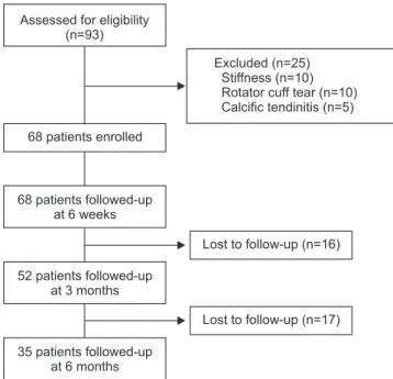
sions of ESWT during a 3-week period. Follow-up assessments were performed at 3 months for 52 patients (76.5%) and at 6 months for 35 patients (51.5%) (Fig. 1).
The above-mentioned pharmacological agents were admin- istered to all patients in conjunction with ESWT treatment. Both nonsteroidal anti-inflammatory drugs and muscle relaxants were used, but the type and dosage varied according to the degree of patient compliance. ESWT was administered as 4,000 impulses of 0.25 mJ/mm2 at a frequency of 7 Hz using the Swiss Dolor- Clast (Electro Medical Systems, Nyon, Switzerland). Pressure was set at 3 bar, and a radial type shock wave was used. After receiving a diagnosis of MPS, all patients underwent ESWT once a week for 3 weeks (for a total of 12,000 impulses). One mem- ber of our shoulder team applied ESWT to the intramuscular taut band and referred pain area, which mainly included the trapezius or levator scapula muscles (1–2 tender points). An ad- ditional session or two was performed in patients who wanted more than 3 ESWT treatments.
A 10-point visual analog scale (VAS) for pain intensity (where 0=no pain and 10=worst possible pain) was used to measure each patient’s response to ESWT. VAS pain scores and American Shoulder and Elbow Surgeons (ASES) scores were checked at the initial visit and at the 6-week, 3-month, and 6-month follow- up visits. These scores are widely used at orthopedic clinics for evaluation of subjective clinical outcomes.
Statistical analyses were performed using SPSS software ver.
12.0 (SPSS Inc., Chicago, IL, USA). The paired t-test was used for comparison of differences in the functional evaluation scores before and after treatment. A p-value <0.05 was considered significant.
Results
Six weeks after ESWT treatment, VAS pain scores had im- proved in 47 patients, deteriorated in 8 patients, and were similar in 13 patients. Three months after ESWT treatment, pain scores had improved in 30 patients, deteriorated in 12 patients, and were similar in 10 patients. Six months after ESWT treat- ment, pain scores had improved in 16 patients, deteriorated in 10 patients, and were similar in 9 patients.
Fig. 1. Flowchart showing the study protocol according to Consolidated Stan- dards of Reporting Trials criteria.
Assessed for eligibility (n=93)
Excluded (n=25) Stiffness (n=10) Rotator cuff tear (n=10) Calcific tendinitis (n=5) 68 patients enrolled
68 patients followed-up at 6 weeks
52 patients followed-up at 3 months
Lost to follow-up (n=16)
Lost to follow-up (n=17) 35 patients followed-up
at 6 months
Table 1. VAS Pain Scores and ADL Scores and ASES Scores Improved aft er 3 Sessions of ESWT Variable
Initial 6 weeks 3 months 6 months
Mean ± SD Mean ± SD p-value Mean ± SD p-value Mean ± SD p-value
VAS 4.82 ± 1.17 3.28 ± 0.72 <0.001 3.17 ± 0.92 <0.001 3.02 ± 0.94 <0.001
ADL 22.80 ± 2.69 26.40 ± 1.83 <0.001 26.20 ± 2.08 <0.001 26.25 ± 2.26 <0.001
ASES 63.85 ± 8.75 77.57 ± 6.38 <0.001 77.80 ± 7.32 <0.001 78.61 ± 6.86 <0.001
Comparison with scores at initial visit and the other visits showedstatistically signifi cant diff erence (p<0.05).
VAS: visual analog scale, ADL: activities of daily life, ASES: American Shoulder and Elbow Surgeons, ESWT: extracorporeal shock wave therapy, SD: standard deviation.
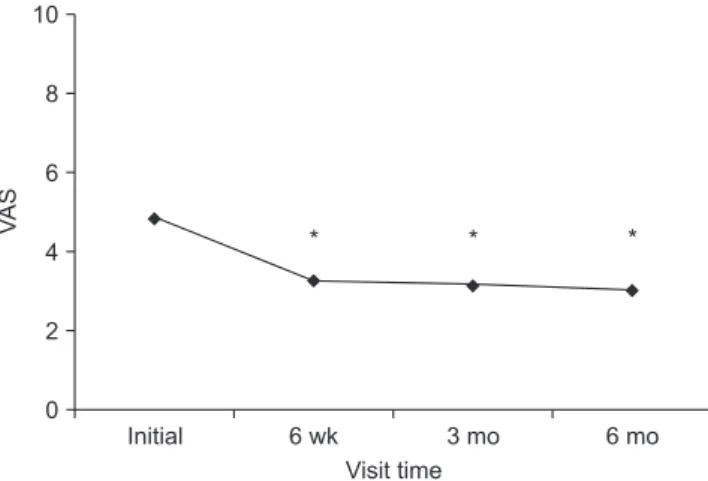
A significant improvement in VAS pain scores was observed after 3 sessions of ESWT (p<0.05) (Table 1). Six weeks after ESWT, the mean VAS pain score had improved from 4.82 points to 3.28 points. At 3 and 6 months, the mean pain score had improved from 4.82 points to 3.17 points and from 4.82 points to 3.02 points, respectively. No significant differences were found between pain scores measured at 6 weeks and 3 months, 6 weeks and 6 months, and 3 months and 6 months.
After 6 weeks follow-up assessment, mean VAS pain scores had improved, but not significantly (p>0.05) (Fig. 2).
Activities of daily life (ADL) scores also improved significantly after 3 sessions of ESWT (p<0.05) (Table 1). Six weeks after treatment, the mean ADL score had improved from 22.80 points to 26.40. At 3 and 6 months, mean ADL scores had improved
from 22.80 points to 26.20 points and from 22.80 points to 26.25 points, respectively. No significant differences were found between scores measured at 6 weeks and 3 months, 6 weeks and 6 months, and 3 months and 6 months. After 6 weeks fol- low-up assessment, the mean ADL score had improved, but not significantly (p>0.05) (Fig. 3).
ASES scores also showed significant improvement after 3 ses- sions of ESWT (p<0.05) (Table 1). Six weeks after treatment, the mean ASES score had improved from 63.85 points to 77.57. At 3 and 6 months, mean ASES scores had improved from 63.85 points to 77.80 points and from 63.85 points to 78.61 points, re- spectively. No significant differences were found between scores measured at 6 weeks and 3 months, 6 weeks and 6 months, 3 months and 6 months. After 6 weeks follow-up assessment, the ASES score had improved, but not significantly (p>0.05) (Fig. 4).
VAS pain scores and ADL scores both improved after 3 ses- sions of ESWT. We conclude that both improvements contrib- uted to the improvements in the ASES score.
Discussion
The mechanisms of ESWT are unclear, however several hypotheses have been proposed based on the cellular and molecular effects of this treatment.10-13) ESWT improves blood circulation in capillaries, and it reduces the tension and stiff- ness of muscles, which can interfere with blood flow and cause excessive stimulation of nociceptors and nerves.14) According to De Sanctis et al.,15) ESWT improves capillary blood circulation in chronic ischemic zones. Referred pain in patients with MPS is due to the ease of inducing central sensitization, because the pe- ripheral muscle nociceptor threshold is lower than that in other systems.16) ESWT may interrupt the cascade of referred pain by Fig. 2. Visual analog scale (VAS) pain scores improved significantly after
extracorporeal shock wave therapy, particularly when initial scores were com- pared with those for the other visits (6-week, 3-month, and 6-month). *Th ese marks mean statistically signifi cant diff erence compared with initial VAS pain score (p<0.05).
*
Initial 6 wk 3 mo 6 mo
10
8
6
4
2
0
*
*
Visit time
VAS
Fig. 3. Activities of daily life (ADL) scores improved signifi cantly aft er extra- corporeal shock wave therapy, particularly when compared with initial scores with the other visits (6-week, 3-month, and 6-month). *Th ease marks mean statistically signifi cant diff erence compared with initial ADL score (p<0.05).
*
Initial 6 wk 3 mo 6 mo
30
25
20
15
10
5
0
* *
ADLscore
Visit time
Fig. 4. American Shoulder and Elbow Surgeon (ASES) scores improved signifi cantly aft er extracorporeal shock wave therapy, particularly when com- paring initial scores with the other visits (6-week, 3-month, and 6-month).
*Th ese marks mean statistically signifi cant diff erence compared with initial ASES score (p<0.05).
Initial 6 wk 3 mo 6 mo
100
80
60
40
20
0
* *
*
ASESscore
Visit time
inhibiting peripheral muscle nociceptors and reducing levels of substance P.13) According to Hausdorf et al.,17,18) ESWT reduces musculoskeletal tissue pain by selectively destroying non-myelin- ated fibers, and it reduced substance P levels in the dorsal root ganglia in an animal study.
The prevalence of MPS is 21% to 85% among individu- als with regional pain.19) Despite its high prevalence rate, the pathophysiology of MPS remains unclear. Travell and Simons3) proposed that damaged muscle fibers become shortened by calcium reflux into the fibers or by acetylcholine secretion at the motor endplate. Ji et al.13) hypothesized that MPS originates from an abnormal increase in the production and release of acetylcholine, which induces sustained depolarization of the post-junctional muscle fiber membrane. Released acetylcholine may cause a continuous release and uptake of calcium ions and produce muscle ischemia as a result of sustained sarcomere shortening and the release of sensitizing substances, such as substance P, bradykinin, calcitonin gene-related peptide, tumor necrosis factor-, interleukin (IL)-1B, IL-6, and IL-8. ESWT may reduce the pain associated with MPS by promoting angiogenesis and increasing perfusion in ischemic tissues and by altering pain signaling at the ischemic tissues caused by calcium influx.
Multiple factors can cause muscle pain around the shoulder and neck. MPS can be caused by poor posture, emotional stress, obsessive-compulsive disorders, or cervical spine disc disease, and these problems influence one another.20,21) Therefore, treat- ing only one of these causes cannot guarantee good results, and achievement of complete remission can be difficult for patients with MPS. ESWT softens taut muscular bands, however other factors such as poor posture or emotional stress cause symptom recurrence.3) The rapport between the doctor and patient is important so that treatment can be continued and patients can be advised about recurrence. A sufficient treatment period and good patient compliance are also important.
In this study, we investigated the effects of ESWT in the treat- ment of MPS of the shoulder by evaluating clinical scores. Few studies have reported a correlation between MPS and shoul- der scores. In this study, ESWT was applied once a week for 3 weeks. After the treatment period, clinical and pain scores showed significant improvements. Symptoms and scores were slightly better at 3 months and 6 months after treatment than at 6 weeks, although this difference was not statistically signifi- cant. It is currently unknown how many sessions of ESWT are required for treatment of MPS, and more studies are required to establish these guidelines.
The limitations of this study include the small patient group, the short-term follow-up period, and the absence of a control group. An additional case-control study using other treatment options will be necessary, as this was the weakest point of our study. Because no diagnostic tools have been confirmed for MPS, our diagnosis mainly depends on the physical examina-
tion. Another limitation of this study is that our patient group was heterogeneous in nature, but these differences were not ad- dressed.
Conclusion
VAS pain scores and ASES scores improved after ESWT treat- ment. ESWT currently represents one of the most effective treat- ment options for patients with MPS.
References
1. Simons DG, Travell J, Simons L. Myofascial pain and dysfunc- tion: the trigger point manual. 2nd ed. Baltimore: Williams &
Wilkins; 1999.
2. Borg-Stein J, Simons DG. Focused review: myofascial pain.
Arch Phys Med Rehabil. 2002;83(3 Suppl 1):S40-7, S48-9.
3. Travell JG, Simons DG. Apropos of all muscles. In: Travell JG, Simons DG, eds. Myofascial pain and dysfunction: the trigger point manual. 1st ed. Baltimore: Williams & Wilkins; 1992.
4. Ge HY. Prevalence of myofascial trigger points in fibromyalgia:
the overlap of two common problems. Curr Pain Headache Rep. 2010;14(5):339-45.
5. Gerber NL, Sikdar S, Hammond J, Shah J. A brief overview and update of myofascial pain syndrome and myofascial trig- ger points. J Spinal Res Found. 2011;6(1):55-64.
6. Tekin L, Akarsu S, Durmuş O, Cakar E, Dinçer U, Kıralp MZ.
The effect of dry needling in the treatment of myofascial pain syndrome: a randomized double-blinded placebo-controlled trial. Clin Rheumatol. 2013;32(3):309-15.
7. Lee SS, Kang S, Park NK, et al. Effectiveness of initial extracor- poreal shock wave therapy on the newly diagnosed lateral or medial epicondylitis. Ann Rehabil Med. 2012;36(5):681-7.
8. Kim YS, Lee HJ, Kim YV, Kong CG. Which method is more effective in treatment of calcific tendinitis in the shoulder? Pro- spective randomized comparison between ultrasound-guided needling and extracorporeal shock wave therapy. J Shoulder Elbow Surg. 2014;23(11):1640-6.
9. Dizon JN, Gonzalez-Suarez C, Zamora MT, Gambito ED. Ef- fectiveness of extracorporeal shock wave therapy in chronic plantar fasciitis: a meta-analysis. Am J Phys Med Rehabil.
2013;92(7):606-20.
10. Wang FS, Yang KD, Chen RF, Wang CJ, Sheen-Chen SM. Ex- tracorporeal shock wave promotes growth and differentiation of bone-marrow stromal cells towards osteoprogenitors associ- ated with induction of TGF-beta1. J Bone Joint Surg Br. 2002;
84(3):457-61.
11. Shah JP, Danoff JV, Desai MJ, et al. Biochemicals associated with pain and inflammation are elevated in sites near to and remote from active myofascial trigger points. Arch Phys Med Rehabil. 2008;89(1):16-23.
12. Jeon JH, Jung YJ, Lee JY, et al. The effect of extracorporeal shock wave therapy on myofascial pain syndrome. Ann Reha- bil Med. 2012;36(5):665-74.
13. Ji HM, Kim HJ, Han SJ. Extracorporeal shock wave therapy in myofascial pain syndrome of upper trapezius. Ann Rehabil Med. 2012;36(5):675-80.
14. Zimmermann R, Cumpanas A, Miclea F, Janetschek G. Extra- corporeal shock wave therapy for the treatment of chronic pelvic pain syndrome in males: a randomised, double-blind, placebo-controlled study. Eur Urol. 2009;56(3):418-24.
15. De Sanctis MT, Belcaro G, Nicolaides AN, et al. Effects of shock waves on the microcirculation in critical limb ischemia (CLI) (8-week study). Angiology. 2000;51(8 Pt 2):S69-78.
16. Giamberardino MA, Costantini R, Affaitati G, et al. Viscero-vis- ceral hyperalgesia: characterization in different clinical models.
Pain. 2010;151(2):307-22.
17. Hausdorf J, Lemmens MA, Heck KD, et al. Selective loss of unmyelinated nerve fibers after extracorporeal shockwave ap- plication to the musculoskeletal system. Neuroscience. 2008;
155(1):138-44.
18. Hausdorf J, Lemmens MA, Kaplan S, et al. Extracorporeal shockwave application to the distal femur of rabbits diminishes the number of neurons immunoreactive for substance P in dorsal root ganglia L5. Brain Res. 2008;1207:96-101.
19. Desai MJ, Saini V, Saini S. Myofascial pain syndrome: a treat- ment review. Pain Ther. 2013;2(1):21-36.
20. Sun A, Yeo HG, Kim TU, Hyun JK, Kim JY. Radiologic assess- ment of forward head posture and its relation to myofascial pain syndrome. Ann Rehabil Med. 2014;38(6):821-6.
21. Adams LM, Turk DC. Psychosocial factors and central sensitiv- ity syndromes. Curr Rheumatol Rev. 2015;11(2):96-108.