Preventing Varus Deformity in Senile Patients with Proximal Humerus Fractures and Poor Medial Support
Young-Kyu Kim, Suk-Woong Kang1 , Jin-Woo Kim
Department of Orthopedic Surgery, Gil Hospital, Gacheon University, Incheon, 1Department of Orthopedic Surgery, Daedong General Hospital, Busan, Korea
Background: We investigated the effectiveness of fibular strut allograft augmentation of proximal humerus fractures to prevent varus de- formity in patients over the age of 65 years with insufficient medial support.
Methods: We analyzed the clinical and radiological outcomes of locking plate fixation with adjunct fibular strut allograft augmentation in 21 patients with proximal humeral fractures. The inclusion criteria were age (65-year-old or older); presence of severe medial comminu- tion; inadequate medial support; and those who could participate in at least a one year follow-up. The average age was 76.4 years. We analyzed each patient’s Constant score, our indicator of clinical outcome. As radiological parameters, we analyzed time-to-bone union;
restoration of the medial hinge; difference between the immediately postoperative and the last follow-up humeral neck-shaft angles;;
and anatomical reduction status, which was assessed using the Paavolainen method.
Results: A successful bone union was achieved in all patients at an average of 11.4 weeks. We found that the average Constant score was 74.2, showing a satisfactory outcome. The average difference in the humeral neck-shaft angles between the immediately postopera- tive time-point and at the final follow-up was 3.09°. According to the Paavolainen method, the anatomical reduction was rated excellent.
The medial hinge was restored in 14 of 21 patients. Although we did not find evidence for osteonecrosis, we found that a single patient had a postoperative complication of screw cut-out.
Conclusions: Fibular strut allografting as an adjunct treatment of proximal humeral fractures may reduce varus deformity in patients with severe medial comminution.
(Clin Shoulder Elbow 2016;19(4):216-222)
Key Words: Proximal humerus fractures; Fibular strut allograft; Open plate fixation Clinics in Shoulder and Elbow Vol. 19, No. 4, December, 2016
https://doi.org/10.5397/cise.2016.19.4.216
Received September 12, 2016. Revised October 26, 2016. Accepted October 30, 2016.
Correspondence to: Suk-Woong Kang
Department of Orthopedic Surgery, Daedong General Hospital, 187 Chungnyeol-daero, Dongnae-gu, Busan 47737, Korea Tel: +82-51-554-8996, Fax: +82-51-553-7575, E-mail: [email protected]
IRB approval (No. GAIRB2016-249).
Financial support: None. Conflict of interests: None.
Introduction
Proximal humeral fractures are one of the most common frac- tures amongst the elderly population. With an increasingly aging population worldwide, the prevalence of proximal humeral frac- tures is likewise increasing.1) Owsley and Gorczyca2) reported that around 36% of elderly patients who undergo plate fixation for proximal humeral fractures are associated with radiologically- determined postoperative complications. They reported that these complications were especially common in combination with 3- or 4-part proximal humeral fractures in over 60-year-old.
Since most elderly patients have poor bone quality, this predis- poses them to combined comminuted fractures of the medial cortex and subsequent loss in medial support, leading them to having a higher risk of postoperative complications such as varus deformity.3,4) In such patients, surgical interventions such as fibu- lar strut allograft has been used.5) In this study, we investigated the effectiveness of fibular strut augmentation as an adjunct to plating in elderly patients with proximal humeral fractures and insufficient medial support.
Methods
Subjects of Study
Between August 2010 and December 2014, we recruited patients aged 65 or over with proximal humeral fractures who for the reason of insufficient medial support had received fibular strut allograft augmentation as an adjunct to locking plate fixa- tion. Of the total of 23 patients who fulfilled these criteria, we enrolled 21 patients who were able to participate in at least a 1-year follow-up. The average age of the patients was 76.4 years (range, 65–90 years) and the study population composed of 2 male and 19 female patients. The average follow-up period was 13.1 months (range, 12–18 months). Using dual energy X-ray absorptiometry, we measured the bone density of the patients’
fractures and the spine, which according to the Lowest T-score showed an average of bone mineral density -2.9. According to the Neer classification system, we found that 16 patients had 2-part fractures and 5 patients with 3-part fractures. According to the AO classification system, we found that 7 patients had A2 fractures; 6 patients had A3 fractures; 7 patients, B2 fractures;
and 1 patient, a B3 fracture.
We measured each patient’s Constant score as our assess- ment of postoperative clinical outcome. And our radiological parameters were measured at five follow-up time-points: 1-, 2-, 4-, 8-, and 12-week postoperatively. We defined a successful bone union as when the fracture site was completely adjoined through callus formation, and humeral neck-shaft angles were measured according to the Paavolainen method6) at the antero- posterior plane. We defined the humeral neck-shaft angle as the angle that forms when the angle between the line that is perpen- dicular to the articular surface of the fracture and the line follow- ing the humeral shaft is bisected. The humeral neck-shaft angle
was measured preoperatively and at the final follow-up (Fig. 1).
We assessed restoration of the medial hinge according to how well the inferiomedial humeral head had been anatomically reduced,7) and we made a comparative analysis of the clinical outcomes according to the restoration status of the medial hinge and to the humeral neck-shaft angle difference. And we also analyzed time-to-bone union and the presence of postoperative complications radiological (Table 1).
We used SPSS Statistics ver. 17.0 (SPSS Inc., Chicago, IL, USA) to perform all statistical analyses. And the nonparametric Mann-Whitney U-test was used to analyze the data. The signifi- cance threshold was set at 0.05.
Surgical Method
Elderly patients aged over 65 years with severe medial comminution and with a medial metaphyseal distance of less than 2 mm were defined as having insufficient medial support (Fig. 2, 3). We used pre-freeze–dried fibular strut allografts 6 to 8 cm in size for the adjunct augmentation. If the strut allograft was too large to be inserted into the intramedullary space, we discarded around a third of the allograft; rather than removing the allograft surface that would form an interface with the proxi- mal humerus, we made an oblique osteotomy of the strut so that it will provide a more stable support once it is impacted into the humeral head (Fig. 4).
In all steps, we attempted to minimize vascular injury in the medial region of the fracture. We pressed the fibular strut al- lograft, which we extracted from a site near the fracture, into the distal intramedullary region, after which we used a hemostat or a small bone clamp to push the allograft towards the proximal fracture site as much as possible. We used a C-arm to assess whether varus deformity was satisfactorily restored and whether the fibular strut allograft made sufficient contact the fracture sur- face; then, the allograft was fixed temporarily using a K-wire. We positioned the plate (proximal humerus internal locking system, PHILOS; Synthes, West Chester, PA, USA) 5 to 8 mm inferior to the tip of the greater tuberosity and at the lateral greater tuberos- ity. In cases where varus adjustment was not enough, we used a bone clamp and mallet to displace the allograft superiorly, re- attempting a varus adjustment. The proximal screw was fixed before the distal screw (Fig. 5).
The patients were administered an immobilizing abduction brace for four postoperative weeks. They were permitted con- tinuous passive motion from the 3rd postoperative day; passive motion within each patient’s capacity from the 3rd postopera- tive week; and active range of motion through the full extent from the 6th postoperative week.
Results
We achieved a successful bone union in all patients, and Fig. 1. Neck-shaft angle measured using the Paavolainen method.
Table 1. Demographic Data of Patients Patient No.SexAge (yr)Cause
F/U (mInl Last naHinge terFoPosrewardl naerAbductioExtt n eer ntBMDta NnsCoUnion AOPre orescrorotion (°)n (°)tiotatao)(°)k)n (°)typeclass(T-sco(wre)NSA (°)tiotiovaNSA (°)neleratoresNSA (°) 12270T1260100100O13513512TA182F12B23-2.5 13060L245100110x1221292TA285F13A22-3.9 9264T755110120120135x16SD382F12A2-2.52 14086T940120160O14014132SD490F12A32-2.8 13077T1260110125O128F115SD878514A22-2.3 115L245100110x125130708776F-3.1SD15A22 127T740115120x130132741276-2.4F7SD12B23 125T940130165O125125841068-2.4M8TA12A32 80L430120140O130130812065-2.4F9TA16B22 86T755150150O130135-3.11306812F10SD12B32 135T940140140O135130811673-2.7M11SD14B23 62L240100100x1221301013075-3.5F12SD15A32 110L455140160O13013088882-3F13SD12A22 70T740125125O1301351211085-2.5F14SD12A22 78T960150150O1351401612583-3.5F15SD12A32 74T1245110130O1351251212383-3.3F16SD12B23 60T950130100110x125TA607212F1713B23-2.5 70T760130130O12013078870-4.6F18SD18A22 74T945100125O1251251211574-2.5F19SD12A32 62T94510511075120125x1068-2.9F20SD15B22 9080T1250130130120125O12SD-2.9F7521212A3 ar tale, M: male, TA: out-cffic accidenrat, SD: slipdow: femw-ule, FeraF/U: follop, BMD: bone minl denngsity, NSA: neck-shaft an.
the average time to bone union was 11.4 weeks (range, 8–16 weeks). Our indicator of clinical outcome, the Constant score was on average 74.2 points (range, 60–88 points) at the 1-year postoperative follow-up, showing a good clinical outcome asso- ciated with the fibular graft augmentation.
Anatomical reduction was carried out according to the Paavolainen method, and in all the final follow-up humeral neck-shaft angle was excellent (range, 120°–140°). The aver- age preoperative humeral neck-shaft angle was 110.14° (range, 60°–135°), the average immediately postoperative humeral neck-shaft angle was 130.80° (range, 125°–140°); and the aver- age final follow-up humeral neck-shaft angle was 127.71° (range, 120°–140°). Thus, we found that the difference between the im- mediately postoperative and the final follow-up values was 3.09°.
The average forward elevation, abduction, external rotation, and internal rotation was 129.04°, 118.33°, 38.33°, and T10–T11, respectively.
After treatment, we found that 14 of 21 patients (66.7%) had a successful medial hinge restoration. Those with medial hinge restoration showed an average Constant score of 79 points, and those without had a significantly lower Constant score of 64.57 points (p=0.000). Preoperatively, we found that the patients in the successful medial hinge restoration group had had a lower average preoperative humeral neck-shaft angle (115.85°), and thus a less severe varus deformity, than patients whose medial hinge did not restore (98.71°). Yet this difference was not statisti- cally significant (p=0.224). However, we observed a statistically significant difference at the final follow-up with respect to the av-
A B C
Fig. 2. Radiographs of an 80-year-old female patient treated with locking plate fixation using an anatomical plate and screws show varus angulation and commi- nution of the fracture fragments preoperatively (A), a successful clinical outcome postoperatively (B), and at the final follow-up (C).
A B C
Fig. 3. Radiographs of a 76-year-old female patient after plate fixation using an anatomical plate and screw show angulation of the fractured fragments preopera- tively (A), successful clinical outcomes postoperatively (B), and at the final follow-up (C).
erage humeral neck-shaft angle: the patients in the former group had a significantly higher average humeral neck-shaft angle than those of the latter group (129.86° vs. 123.43°; p=0.025). For the range of motion, patients with successful medial hinge res- toration showed postoperative abduction, external rotation, and internal rotation of 137.86°, 125.35°, and 49.64°, respectively.
The respective values for patients without successful medial res- toration were 111.42°, 104.28°, and 45.71°, respectively; the two groups significantly differed in terms of only abduction and external rotation (Table 2). We found that the type of fracture did not influence the success and failure of medial hinge restora- tion.
In terms the postoperative complications, we did not observe any medial support loss, fibular fractures, or severe varus defor- mity, resulting from unsuccessful plating. Nor did we observe any incidents of osteonecrosis. Even though one patient pre-
sented with a screw cutout because a successful bone union was apparent we kept a vigilant eye throughout the follow-up: the patient later presented with a satisfactory Constant score of 60 points.
Discussion
Proximal humeral fractures compose 10% of all fractures amongst elderly patients over 60 years, and with an aging popu- lation its prevalence is increasing.1) Various treatment methods exist for proximal humeral fractures such as suture fixation, per- cutaneous K-wire fixation, plate fixation, intramedullary nailing, and arthroplasty. But for elderly patients who commonly have concomitant osteoporosis it is difficult to attain a satisfactory fixation on account of their poor bone quality, meaning that the
Table 2. Results of the Medial Hinge Restoration*
Variable Medial hinge
restoration (+) Medial hinge
restoration (-) p-value
Age (yr) 76.85 76.42 0.971
BMD (T-score) -2.89 -2.97 0.636
Union (wk) 11.43 11.42 0.971
Pre NSA (°) 115.85 98.71 0.224
Post NSA (°) 131.07 130.29 0.931
Last NSA (°) 129.86 123.43 0.025
Forward elevation (°) 137.86 111.42 0.001
Abduction (°) 125.35 104.28 0.006
External rotation (°) 49.64 45.71 0.535
Internal rotation T10–T11 T10–T11 0.971
Constant score 79 64.57 0.000
BMD: bone mineral density, NSA: neck-shaft angle.
*Mann-Whitney U-test.
Fig. 4. Oblique cut-out of a fibular strut allograft.
A B C
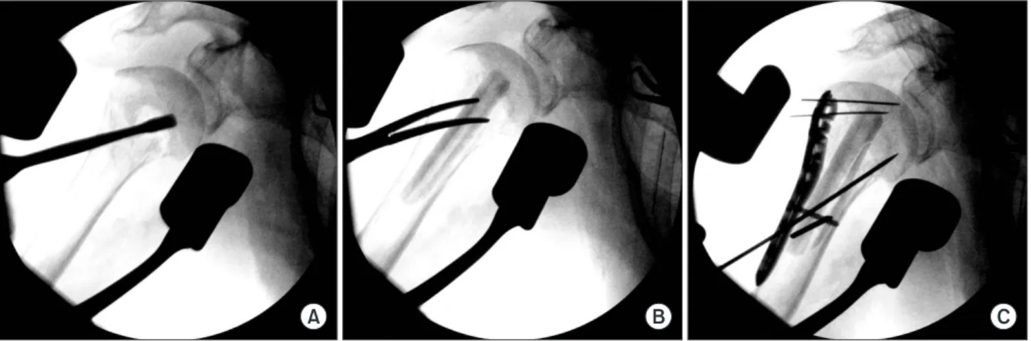
Fig. 5. Image intensification shows the anatomical reduction of the proximal humerus fracture (A); the inserted fibular allograft (B); and the anatomical plate fix- ated using a screw (C).
gold standard of treatment is yet to exist for these subset of pa- tients.8)
Solberg et al.9) suggested that in patients older than 55 years the presence or absence of early varus deformity and the length of the fracture at the medial metaphyseal region are important factors concerning treatment prognosis. Thus, we followed the method of intramedullary fibular allograft augmentation pro- posed by Gardner et al.10) in elderly patients with a distance be- tween the medial comminution fracture and the medial interos- seous fragment of less than 2 mm, because we thought that this may prevent re-establishment of medial support.
In a human cadaver study where the outcomes after locking plates and intramedullary fibular allografting were compared to outcomes after locking plates alone, it was found that employing an additional adjunct treatment provided stronger fixation.11,12) Hettrich et al.13) looked at the clinical outcomes after fibular allograft and locking plate in patients over 70 year olds with proximal humeral and concomitant medial comminution frac- tures. They found that the difference between the intraopera- tive and the final follow-up humeral neck-shaft angles was only 2.2° (135.4° vs. 133.2°), showing good clinical outcomes. This humeral neck-shaft angle difference in our study was 3.09°. This comes to show that even with the addition of fibular strut grafts a stable fracture reduction that is maintained through to the final follow-up via medial support can be attained whilst preserving the proximal humerus length.
In their comparative study, Gardner et al.4) found that, after treatment of proximal humeral fractures, patients with medial support showed an average 1.2-mm loss in humeral length, whereas those without medial support, a 5.8-mm loss. We found that the 14 patients who showed successful medial hinge restoration showed satisfactory clinical outcomes, and their hu- meral neck-shaft angles at the final follow-up were significantly better than the other 7 patients whose medial hinge did not re- store. In the latter cases, we observed that varus deformity began within 2 and 4 weeks of the treatment. It seems that those who have severe varus deformity preoperatively have less change of anticipating a successful medial hinge restoration. However, further studies are required to confirm this association between preoperative varus deformity and treatment outcome. Still, our results suggest that fibular allograft augmentation does not al- ways guarantee a successful medial hinge restoration and other steps must be carried out to ascertain as much medial support as possible, thereby ensuring a more favorable prognosis postop- eratively.
Likewise, Neviaser et al.14) have reported good clinical out- comes after fibular grafting, generally using 6- to 8-cm fibula allografts. They suggested that with fibular graft augmentation it was vital to form a natural medial arch and that a sturdy fixa- tion can be attained by tailoring the placement of graft to each fracture. They also used temporary K-wire fixation to adjust
the fibular allograft if necessary. Using cadaveric specimens, Chow et al.15) used 8-cm long allografts from the fibular shaft cut into around 5-cm constructs, which were fixated onto the distal fracture site. We also chose to use 8-cm fibular allografts, which were inserted in a manner that provides the most me- dial support and then were temporarily fixed. Then, the plate was permanently fixed on the lateral humerus, the distal end of which was also temporarily stabilized beforehand, with screws on the proximal and distal ends. Saltzman et al.16) used a saw and burr to shape the fibular allograft so it fits the intramedullary area, whereas Matassi et al.17) stabilized the graft with screws after medially impacting the graft first during fibular insertion.
For the authors, in incidences where the intramedullary area of the distal humerus could not accommodate the fibular graft, we ostetomized the bone obliquely to the width; the oblique cut was made so that the fibular graft and proximal surface area of contact could be maximized.
The prevalence of osteonecrosis in patients with proximal humeral fractures has been reported to range from 2.2% to 8%.18,19) In their study, Neviaser et al.14) reported that none of the 36 patients who were treated with fibular grafts presented with complete osteonecrosis. They proposed that by carrying out a fibular graft transfer, they were able to prevent reduction loss and, thus, osteonecrosis, notwithstanding the poor bone quality and the medial comminution by structurally enhancing the bio- mechanics of the humerus. Although our study requires greater statistical power to conclude for certain that the prevalence of osteonecrosis was decreased as a result the treatment, several pre-existing reports have already shown that the prevalence of osteonecrosis after fibular graft augmentation is low.16)
One of the study’s limitations is that, as a retrospective study, the findings may have been influenced by selection bias. Al- though we did not see a difference with respect to the distribu- tion of Neer classification of the medial comminution fractures, since we did not make an analysis of the severity of fractures among patients, we may have included in our fibular allograft augmentation group those whose fractures are not severe enough to justify them having received this treatment. Thus, a prospective analysis may be required to assess the long-term outcomes of the intervention. Second, the total number of cases numbered only 21; therefore, it is difficult to anticipate a statis- tically significant difference based on whether a medial hinge restoration has been achieved. Third, because the study design consisted of a relatively short follow-up, the manifestation of long-term complications, such as osteonecrosis, later on cannot be out-ruled. Lastly, the study requires a more detailed analysis of why the medial hinge could not be restored in some patients.
Still, in spite of the small study population and these limitations, compared to other studies that have reported the outcomes of fibular strut allografts, ours is notable in that we were able to achieve good clinical outcomes, especially given that our study
population consisted of only elderly patients.
Conclusion
In sum, in case of insufficient medial support amongst elderly patients with proximal humeral fractures, fibular strut graft aug- mentation as an adjunct to plate fixation may facilitate an ana- tomical reduction and reduce varus deformity.
References
1. Palvanen M, Kannus P, Niemi S, Parkkari J. Update in the epi- demiology of proximal humeral fractures. Clin Orthop Relat Res. 2006;442:87-92.
2. Owsley KC, Gorczyca JT. Fracture displacement and screw cutout after open reduction and locked plate fixation of proximal humeral fractures [corrected]. J Bone Joint Surg Am.
2008;90(2):233-40.
3. Thanasas C, Kontakis G, Angoules A, Limb D, Giannoudis P.
Treatment of proximal humerus fractures with locking plates: a systematic review. J Shoulder Elbow Surg. 2009;18(6):837-44.
4. Gardner MJ, Weil Y, Barker JU, Kelly BT, Helfet DL, Lorich DG.
The importance of medial support in locked plating of proxi- mal humerus fractures. J Orthop Trauma. 2007;21(3):185-91.
5. Gardner MJ, Lorich DG, Werner CM, Helfet DL. Second- generation concepts for locked plating of proximal humerus fractures. Am J Orthop (Belle Mead NJ). 2007;36(9):460-5.
6. Paavolainen P, Björkenheim JM, Slätis P, Paukku P. Operative treatment of severe proximal humeral fractures. Acta Orthop Scand. 1983;54(3):374-9.
7. Nho SJ, Brophy RH, Barker JU, Cornell CN, MacGillivray JD.
Management of proximal humeral fractures based on current literature. J Bone Joint Surg Am. 2007;89 Suppl 3:44-58.
8. Ring D. Current concepts in plate and screw fixation of os- teoporotic proximal humerus fractures. Injury. 2007;38 Suppl 3:S59-S68.
9. Solberg BD, Moon CN, Franco DP, Paiement GD. Locked plating of 3- and 4-part proximal humerus fractures in older patients: the effect of initial fracture pattern on outcome. J Or- thop Trauma. 2009;23(2):113-9.
10. Gardner MJ, Boraiah S, Helfet DL, Lorich DG. Indirect medial reduction and strut support of proximal humerus fractures us- ing an endosteal implant. J Orthop Trauma. 2008;22(3):195- 200.
11. Bae JH, Oh JK, Chon CS, Oh CW, Hwang JH, Yoon YC. The biomechanical performance of locking plate fixation with in- tramedullary fibular strut graft augmentation in the treatment of unstable fractures of the proximal humerus. J Bone Joint Surg Br. 2011;93(7):937-41.
12. Mathison C, Chaudhary R, Beaupre L, Reynolds M, Adeeb S, Bouliane M. Biomechanical analysis of proximal humeral fixa- tion using locking plate fixation with an intramedullary fibular allograft. Clin Biomech (Bristol, Avon). 2010;25(7):642-6.
13. Hettrich CM, Neviaser A, Beamer BS, Paul O, Helfet DL, Lorich DG. Locked plating of the proximal humerus using an endosteal implant. J Orthop Trauma. 2012;26(4):212-5.
14. Neviaser AS, Hettrich CM, Beamer BS, Dines JS, Lorich DG.
Endosteal strut augment reduces complications associated with proximal humeral locking plates. Clin Orthop Relat Res.
2011;469(12):3300-6.
15. Chow RM, Begum F, Beaupre LA, Carey JP, Adeeb S, Bouliane MJ. Proximal humeral fracture fixation: locking plate construct
± intramedullary fibular allograft. J Shoulder Elbow Surg.
2012;21(7):894-901.
16. Saltzman BM, Erickson BJ, Harris JD, Gupta AK, Mighell M, Romeo AA. Fibular strut graft augmentation for open reduc- tion and internal fixation of proximal humerus fractures: a sys- tematic review and the authors’ preferred surgical technique.
Orthop J Sports Med. 2016;4(7):2325967116656829.
17. Matassi F, Angeloni R, Carulli C, et al. Locking plate and fibular allograft augmentation in unstable fractures of proximal hu- merus. Injury. 2012;43(11):1939-42.
18. Lee CW, Shin SJ. Prognostic factors for unstable proximal hu- meral fractures treated with locking-plate fixation. J Shoulder Elbow Surg. 2009;18(1):83-8.
19. Brunner F, Sommer C, Bahrs C, et al. Open reduction and in- ternal fixation of proximal humerus fractures using a proximal humeral locked plate: a prospective multicenter analysis. J Orthop Trauma. 2009;23(3):163-72.