A Randomized Comparative Study of a Standard Anterior Capsular Release versus Inferior Extended Release for the Treatment of
Shoulder Stiffness
Ahmed Abdullah Alzeyadi, Yang-Soo Kim, Hyo-Jin Lee, Sung-Ryeoll Park, Gwang Young Sung, Dong-Jin Kim, Ji-Hwan Jung, Jong-Ho Kim
Department of Orthopedic Surgery, Seoul St. Mary’s Hospital, College of Medicine, The Catholic University of Korea, Seoul, Korea
Background: To compare the clinical outcomes of arthroscopic capsular release in patients with and without inferior capsular release for shoulder stiffness.
Methods: Between January 2010 and December 2015, 39 patients who underwent arthroscopic capsular release for shoulder stiffness were enrolled and randomized into two groups. In group I, 19 patients underwent arthroscopic capsular release of the rotator interval and anterior capsule. In group II, 20 patients underwent arthroscopic capsular release of the anterior to inferior capsule, including the ro- tator interval. The American Shoulder and Elbow Surgeons score, Constant scoring system, Simple Shoulder Test, visual analogue scale for pain, and range of motion (ROM) were used for evaluation before surgery, at 3, 6, and 12 months after surgery and on the last follow-up.
Results: Preoperative demographic data revealed no significant differences (p>0.05). The average follow-up was 16.07 months. Both groups showed significantly increased ROM at the last follow-up compared with preoperative (p<0.05). At the last follow-up, no sta- tistical differences were found (p>0.05) between groups I and II in functional scores and ROM (forward flexion, p=0.91; side external rotation, p=0.17; abduction external rotation, p=0.72; internal rotation, p=0.61). But we found that group II gained more flexion com- pared to group I at 3 months and 6 months (p<0.05) after the surgery.
Conclusions: Both techniques of capsular release are effective for stiffness shoulder. However, the extended inferior capsular release shows superiority in forward flexion over anterior capsular release alone during 6 months of follows-up (level of evidence: Level I, thera- peutic randomized controlled trial).
(Clin Shoulder Elbow 2017;20(3):117-125)
Key Words: Shoulder; Stiffness; Frozen shoulder; Arthroscopic capsular release Clinics in Shoulder and Elbow Vol. 20, No. 3, September, 2017
https://doi.org/10.5397/cise.2017.20.3.117
Received December 26, 2016. Revised June 30, 2017. Accepted July 3, 2017.
Correspondence to: Jong-Ho Kim
Department of Orthopedic Surgery, Seoul St. Mary’s Hospital, College of Medicine, The Catholic University of Korea, 222 Banpo-daero, Seocho- gu, Seoul 06591, Korea
Tel: +82-2-2258-6359, Fax: +82-2-535-9834, E-mail: [email protected] IRB approval (No. KC12OISI0532).
Financial support: None. Conflict of interests: None.
Introduction
Shoulder stiffness is a common problem that is traditionally considered to be self-limiting.1,2) Conservative treatment has usu- ally been effective, with satisfying results reported in 60% to 80%
of cases.3) Many patients with rotator cuff tears not only have pain, but also have decreased range of motion (ROM) of the shoulder, which inhibits their function in daily life. Many factors
can lead to such shoulder stiffness in patients with rotator cuff tears. The exact prevalence of shoulder stiffness combined with rotator cuff tears is unknown, but it is thought that mild shoulder stiffness, defined as at least a 20° of restriction in total ROM, ex- ists in more than 40% of patients with rotator cuff tears.4)
Arthroscopic release of the glenohumeral joint has recently been used with success in the treatment of recalcitrant frozen- shoulder syndrome.5-7) Release of the contracture of the Coraco-
humeral ligament (CHL) and thickening capsule is believed to be essential to treatment of frozen shoulder.7,8) However, the extent of release is still the subject of debate. Besides the CHL and ro- tator interval, some authors have recommended release of the subscapularis tendon,9) inferior capsule,10) posterior capsule,11,12) or global capsule13) to improve elevation and internal rotation (IR), as well as external rotation (ER). Pearsall et al.14) reported that information regarding preoperative motion loss would precisely direct selective capsular release.
A number of authors have also recommended posterior cap- sular release to improve IR.11,12) However, according to Snow et al.15) additional posterior capsular release was not associated with any significant difference in outcome when compared with anterior release. Chen16) derived a similar result with extended posterior capsular release, showing that there was no advantage in function or ROM. Kim et al.17) performed a level I study to compare anterior and inferior capsular release with anterior, inferior and posterior capsular release. In a one-year follow-up, there were no significant differences in ROM and functional scores between groups. Ranalletta et al.18) suggested that isolated anteroinferior capsular release provides reliable improvement in pain and ROM for primary adhesive capsulitis.
The goal of this study was to determine the extent of capsular release needed in arthroscopic surgery for shoulder stiffness. To accomplish this, we compared the clinical outcomes of capsular release between those with and without inferior capsular release for shoulder stiffness. The hypothesis of our study was that the addition of inferior capsular release would contribute to im- proved outcomes.
Methods
Between January 2010 and December 2015, 152 consecu- tive patients with rotator cuff tear with concomitant stiffness were enrolled. Inclusion criteria were as follows: patients whose main symptom was shoulder stiffness, patients with small- to medium-sized full-thickness rotator cuff tear, and pain and functional limitation of the shoulder for at least 3 months. All patients underwent operation after the first visit to an outpatient clinic and had no conservative treatment period. There are no universally accepted criteria for the diagnosis of stiffness, but we determined stiffness by passive range of shoulder motion to be forward flexion of less than 100° (maximal 150°; forward flexion is glenohumeral motion without scapulohumeral rhythm), ER of less than 45°, or IR of the back at a level lower than the first lumbar spine junction.17,19) Plain radiographs of the shoulder (true anteroposterior, supraspinatus outlet, and axial view) and shoul- der magnetic resonance imaging were checked in all patients.
Patients with concomitant glenohumeral pathologies (Bankart lesion, superior labrum anterior to posterior lesion), symptomatic cervical spinal lesion and large or massive cuff tears (tear size larger than 3 cm or more than 2 concomitant tendon tears) were excluded, as were patients with a previous history of operation or fracture. Overall, 55 patients were finally excluded. The base- line characteristics of the enrolled patients are shown in Fig. 1.
Patients were randomized into two groups through com- puter-generated blocked-randomization numbers (http://www.
randomizer.org). After arthroscopic confirmation of inclusion and exclusion criteria, the surgical procedure was determined by a random number taken from a sealed envelope at the time of
Group I allocated to a standard anterior release (n=49)
Excluded (n=55)
Not meeting the including criteria
Received allocated surgery (n=49)
Lost follow-up (n=30)
Analyzed (n=19)
Allocation
Follow-up
Analyzed
Group II allocated to anterior to inferior capsule release (n=48)
Received allocated surgery (n=48)
Lost follow-up (n=28)
Analyzed (n=20) Assessed for eligibility (n=152)
Enrollment
Randomized (n=97)
Fig. 1. Flow chart shows the conduct of the study according to Consolidated Standards of Reporting Trials (CONSORT) criteria.
surgery. All patients were blinded to this treatment and informed about the advantages and disadvantages of both treatments.
In group I, 19 patients underwent arthroscopic capsular re- lease of the rotator interval, anterior (a standard anterior release).
In group II, 20 patients underwent arthroscopic capsular release of the anterior to inferior capsule, including the rotator interval (a standard anteroinferior release). All patients gave informed consent before surgery and underwent the same rehabilitation protocol after the surgery. This study was approved by the insti- tutional review board.
Biceps long head
Subscapularis
CA ligament
Glenoid
Fig. 2. Right shoulder glenohumeral joint viewing from the posterior portal.
The rotator interval is done in the right shoulder glenohumeral joint until coracoacromial (CA) ligament and coracoid process base are exposed.
Humeral head
Anterior capsule
Glenoid
Fig. 3. Right shoulder glenohumeral joint viewing from posterior portal. Ar- throscopic rotator interval release and anterior capsular release up to the 5:30 position in the right shoulder.
Humeral head
Inferior capsule
Glenoid
Fig. 4. Right shoulder glenohumeral joint viewing from trans-cuff portal.
Arthroscopic rotator interval release, anterior capsular release and extended release of the inferior capsule up to 7:00 in the right shoulder.
Table 1. Preoperative Patient Demographic Data
Variable Group*
p-value Group I Group II
Patient 19 20
Age (yr) 62.32 60.05 0.41
Sex (men/women) 6/13 7/13
Follow-up period (mo) 16.83 15.30
Initial ROM (°)
FF 130.00 121.5 0.61
ERa 78.42 76.00 0.35
ERs 73.16 68.50 0.30
IR 4.53 4.05 0.65
VAS for pain 4.75 5.35 0.41
ASES score 52.89 44.95 0.26
Constant scoring system 73.44 67.40 0.12
KSS 66.78 62.60 0.27
Initial tear size (cm)
AP 1.26 ± 6.1 1.34 ± 5.7 0.65
ML 1.69 ± 5.2 1.66 ± 4.7 0.83
Values are presented as number only, mean only, or mean ± standard deviation.
ROM: range of motion, FF: forward flexion, ERa: external rotation (ER) at 90°
of abduction, ERs: ER at side, IR: internal rotation, VAS: visual analogue scale, ASES: American Shoulder and Elbow Surgeons, KSS: Korean shoulder score, AP: anteroposterior, ML: mediolateral.
*Patients who underwent arthroscopic capsular release for shoulder stiffness were enrolled and randomized into two groups: group I, patients who under- went arthroscopic capsular release of the rotator interval, anterior (a standard anterior release); group II, patients who underwent arthroscopic capsular release of the anterior to inferior capsule, including the rotator interval (a standard anteroinferior release).
Surgical Technique
A senior shoulder specialist conducted all surgical procedures with the patient under general anesthesia. The patients under- went standard glenohumeral arthroscopy in the lateral decubitus position. In group I, capsular release began with the rotator interval via 3.0 mm 90° electrocautery (Arthrocare, Sunnyvale, CA, USA) through the anterior portal (Fig. 2). Anterior capsular release began below the biceps origin by preserving the glenoid labrum. The process began superiorly with resection of the supe-
rior glenohumeral ligament and CHL. Release continued as far medial as the coracoid process until visualization of the vertical- oriented fibers of the coracoacroimal ligament and conjoint tendon. Next, the middle glenohumeral ligament was released without damaging the subscapularis tendon, and then the ante- rior band of the inferior glenohumeral ligament was released up to the 5:30 position in the right shoulder (Fig. 3).
In group II, the same procedures were conducted, but there was extended release of the inferior capsule up to the 7:00 position until the posterior border of the inferior glenohumeral ligament (Fig. 4). The posterior capsule was not addressed. To avoid axillary nerve damage, capsular release was conducted just off the glenoid rim without violating the glenoid labrum.
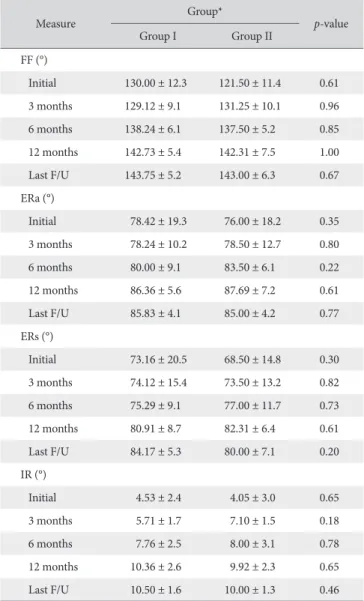
We believe that axillary nerve damage can be prevented if the Table 2. Comparison of Range of Motion
Measure Group*
p-value
Group I Group II
FF (°)
Initial 130.00 ± 12.3 121.50 ± 11.4 0.61
3 months 129.12 ± 9.1 131.25 ± 10.1 0.96
6 months 138.24 ± 6.1 137.50 ± 5.2 0.85
12 months 142.73 ± 5.4 142.31 ± 7.5 1.00
Last F/U 143.75 ± 5.2 143.00 ± 6.3 0.67
ERa (°)
Initial 78.42 ± 19.3 76.00 ± 18.2 0.35
3 months 78.24 ± 10.2 78.50 ± 12.7 0.80
6 months 80.00 ± 9.1 83.50 ± 6.1 0.22
12 months 86.36 ± 5.6 87.69 ± 7.2 0.61
Last F/U 85.83 ± 4.1 85.00 ± 4.2 0.77
ERs (°)
Initial 73.16 ± 20.5 68.50 ± 14.8 0.30
3 months 74.12 ± 15.4 73.50 ± 13.2 0.82
6 months 75.29 ± 9.1 77.00 ± 11.7 0.73
12 months 80.91 ± 8.7 82.31 ± 6.4 0.61
Last F/U 84.17 ± 5.3 80.00 ± 7.1 0.20
IR (°)
Initial 4.53 ± 2.4 4.05 ± 3.0 0.65
3 months 5.71 ± 1.7 7.10 ± 1.5 0.18
6 months 7.76 ± 2.5 8.00 ± 3.1 0.78
12 months 10.36 ± 2.6 9.92 ± 2.3 0.65
Last F/U 10.50 ± 1.6 10.00 ± 1.3 0.46
Values are presented as mean ± standard deviation.
FF: forward flexion, F/U: follow-up, ERa: external rotation (ER) at 90° of ab- duction, ERs: ER at side, IR: internal rotation.
*Patients who underwent arthroscopic capsular release for shoulder stiffness were enrolled and randomized into two groups: group I, patients who under- went arthroscopic capsular release of the rotator interval, anterior (a standard anterior release); group II, patients who underwent arthroscopic capsular release of the anterior to inferior capsule, including the rotator interval (a standard anteroinferior release).
Table 3. Comparisons of Range of Motion Improvement by Period
Measure Group*
p-value
Group I Group II
FF (°)
Initial to 3 months -0.88 9.75 0.03
Initial to 6 months 8.24 16.00 0.03
Initial to 12 months 12.73 20.81 0.17
Initial to last F/U 13.75 21.50 0.06
ERa (°)
Initial to 3 months -0.96 2.00 0.43
Initial to 6 months 1.58 6.5 0.08
Initial to 12 months 7.94 11.69 0.41
Initial to last F/U 8.41 9.00 0.67
ERs (°)
Initial to 3 months 0.96 5.00 0.28
Initial to 6 months 2.13 8.50 0.17
Initial to 12 months 7.75 13.81 0.23
Initial to last F/U 11.01 11.50 0.66
IR (°)
Initial to 3 months 1.18 3.05 0.14
Initial to 6 months 3.24 3.95 0.50
Initial to 12 months 5.83 5.97 0.96
Initial to last F/U 5.97 5.95 0.98
Values are presented as mean only.
FF: forward flexion, F/U: follow-up, ERa: external rotation (ER) at 90° of ab- duction, ERs: ER at side, IR: internal rotation.
*Patients who underwent arthroscopic capsular release for shoulder stiffness were enrolled and randomized into two groups: group I, patients who under- went arthroscopic capsular release of the rotator interval, anterior (a standard anterior release); group II, patients who underwent arthroscopic capsular release of the anterior to inferior capsule, including the rotator interval (a standard anteroinferior release).
electrocautery stays just off the glenoid rim. Moreover, electrical stimulation of electrocautery was helpful in detecting proximity to the axillary nerve before direct injury.
For small-sized (<1 cm) rotator cuff tears, a single-row repair was conducted, and a trans osseous equivalent repair (suture bridge technique) was performed for medium-sized (1–3 cm) tears. The selection of the surgical method was based on the location and shape of the torn tendon. Acromioplasty was per- formed for all type II and III acromions, along with the removal of sub-acromial spurs. For type I acromions, the acromial under- surface was smothered.
Rehabilitation
The same standardized rehabilitation protocols were applied to both groups postoperatively. Starting on the first day after the operation, pendulum circumduction was conducted, including gentle passive ROM exercise. An abduction brace was applied for 1 month after the operation. Pulley exercises were prescribed to increase the flexion after 1 month. When passive shoulder ROM was restored to 90%, isometric exercises in all planes were
recommended. Thera-Band exercises, strengthening exercises for the muscles stabilizing the scapula, and advanced muscle strengthening exercises with dumbbells were taught 12 weeks after the operation. All listed procedures were recommended until the last visit after 12 months. No limit was imposed on use of the shoulder within a tolerable extent.
Outcome Measurement
Each patient was assessed before surgery and at 3, 6, and 12 months after surgery and at the last follow-up via the American Shoulder and Elbow Surgeons score20) and Constant scoring system. ROM including forward flexion, ER at side, ER at 90° of abduction, and IR of the treated shoulder was measured with a goniometer, and a visual analogue scale (VAS) for pain (0, no pain; 10, the most severe pain) was used to evaluate all patients at each visit. IR, measured in the sitting position, was evaluated by the tip of the thumb reaching the vertebral level. For analysis, the vertebral level was numbered serially: 0 for any level below the sacral region and 1 point added for each level above the sacrum. The postoperative cuff tendon integrity was assessed at
Initial 3 months 150
140
130
120
110
Forwardflexion()o
Group I Group II
6 months 12 months Last Initial 3 months
90
80
70
60
50
Externalrotationatside()o
Group I Group II 6 months 12 months Last
Initial 3 months
Externalrotationatabduction()o
Group I Group II
6 months 12 months Last Initial 3 months
12
10
8
6
4
2
Internalrotation(numberofspinelevel) 0
Group I Group II 6 months 12 months Last 90
80
70
60
50
Fig. 5. Range of motion (forward flexion, external rotation at abduction and side, internal rotation) had improved significantly in both groups at last follow-up.
There were no significant differences between the two groups at each time point. Patients who underwent arthroscopic capsular release for shoulder stiffness were enrolled and randomized into two groups: group I, patients who underwent arthroscopic capsular release of the rotator interval, anterior (a standard ante- rior release); group II, patients who underwent arthroscopic capsular release of the anterior to inferior capsule, including the rotator interval (a standard antero- inferior release).
8 weeks and 12 month after the operation using magnetic reso- nance imaging (MRI). All assessment data were prospectively collected by a clinical researcher who was blinded to the current study. Patients were also blinded during the assessment.
Statistical Methods
Statistical analysis was performed with IBM SPSS ver. 21.0 (IBM Co., Armonk, NY, USA). The Mann-Whitney test was used to compare differences between the outcomes of the 2 groups, and the Wilcoxon signed rank test was used to compare the dif- ferences in functional evaluation scores before and after surgery for each group. A p<0.05 was considered significant.
Results
There were no significant differences in age or sex between groups (p>0.05) (Table 1). Initial clinical scores before surgi- cal release in both groups revealed no significant differences (p>0.05) (Table 1). The mean follow-up period was 16.83 months for the standard anterior capsular release group (group
I) and 15.30 months for the extended inferior capsular release group (group II). In group I, there were 6 men and 13 women, with an average age of 62.32 years. In group II, there were 7 men and 13 women, with an average age of 60.05 years. On postoperative 12 months follow-up MRI, there was no signifi- cant difference in re-tear rate between groups. Three patients in group I (15.8%) and four patients in group II (20.0%) had type 4 and 5 re-tears according to the Sugaya classification (p>0.05).
The all ROM and functional scores improved significantly in both groups (p<0.05) at final follow-up. In group I, the mean improvement of ROM was 13.75° in forward flexion, 11.01° in ER at side, 8.41° in ER at 90° abduction and 5.97 spinal levels in IR on the last follow-up. In group II, the mean improvement of ROM was 21.50° in forward flexion, 11.5° in ER at side, 9° in ER at 90° abduction and 5.95 spinal levels in IR at the last follow-up (Table 2).
We found that group II gained more flexion compared to group I from 0 to 3 months and from 0 to 6 months (p<0.05) (Table 3). However, no significant differences were found in other ROM, pain, and functional scores between groups at
Initial 3 months 10
8
6
4
2
0
VASforpain
Group I Group II
6 months 12 months Last Initial 3 months
100
80
60
40
20
0
ASESscore
Group I Group II 6 months 12 months Last
Initial 3 months
Constantscoringsystem
Group I Group II
6 months 12 months Last Initial 3 months
0
KSS
Group I Group II 6 months 12 months Last 100
80
60
40
20
0
100
80
60
40
20
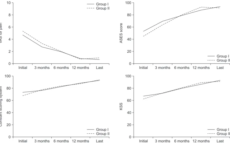
Fig. 6. Functional scores including visual analogue scale (VAS) score for pain, American Shoulder and Elbow Surgeons (ASES) score, Constant score, and Ko- rean shoulder score (KSS) had improved significantly in both groups at last follow-up. There were no significant differences between the two groups at each time point. Patients who underwent arthroscopic capsular release for shoulder stiffness were enrolled and randomized into two groups: group I, patients who under- went arthroscopic capsular release of the rotator interval, anterior (a standard anterior release); group II, patients who underwent arthroscopic capsular release of the anterior to inferior capsule, including the rotator interval (a standard anteroinferior release).
each time point (p>0.05) (Fig. 5, 6, Table 3, 4). No postopera- tive complications of infection, instability, or axillary nerve injury were observed in these patients.
Discussion
Shoulders with rotator cuff tears that were stiff before sur- gery were more likely to be stiff after surgery. A stiffer shoulder postoperatively correlated with better rotator cuff integrity post-
operatively.21) Therefore, concomitant rotator cuff repair with capsular release should be recommended to the patients who have rotator cuff repair with stiffness to achieve overall improved clinical outcomes.
Arthroscopic capsular release has become a reliable method for restoring ROM in patients with idiopathic frozen shoulder for which physical therapy and conservative care have failed.5) A nearly normal ROM and good outcome scores can be obtained with this procedure. In the current study, ROM significantly improved compared with preoperative values in both groups;
however, it showed superiority for group II in the forward flexion until 6 months after surgery.
Histologic studies, open exploration, and arthroscopic obser- vations have revealed that the CHL and rotator interval are the major affected areas in frozen shoulder and should be released to restore passive ER.7,22-24) Therefore, it was believed that the an- terior capsular structures were required to be released to restore ER and abduction.14,25) However, none of the studies conducted to date discussed the extent to which the anterior structures need to be released. In addition to the rotator interval, CHL, and middle glenohumeral ligament, Pearsall et al.9) recommended releasing the intra-articular subscapularis tendon without signifi- cant morbidity.
A release of the superior and middle glenohumeral ligaments, the rotator interval, the CHL, and the intra-articular portion of the subscapularis increased the range of ER, whereas release of the anteroinferior capsule, including the anterior band of the inferior glenohumeral ligament, improved the elevation range.
However, arthroscopic posterior capsular release is controversial.
Selective posterior capsular release or circumferential release around the joint capsule was advocated in some studies for re- covery of IR.6,20) Kim et al.17) found that the range of ER tended to be better in the posterior extended capsular release group until 6 months after surgery, but that it did not shown any signifi- cant differences and that observed differences did not last be- yond one year after surgery. Moreover, Codding et al.26) reported that selective arthroscopic posterior-inferior capsular release may be a reasonable solution in overhead athletes with symptomatic glenohumeral IR deficit.
We believe that treatment should be directed at the rotator interval and the contracted CHL, as this is the site of primary pathology in idiopathic adhesive capsulitis. We found that the range of elevation tended to be better in group II (standard an- terior inferior capsular release) until 6 months after surgery. We cautiously state that there was an apparent benefit in terms of flexion with additional inferior capsular release. Miyazaki et al.27) found that there were better improvements in pain, IR and ER in patients who underwent anteroinferior capsulotomy.
It should be noted that there were several limitations to this study. First, the size of the enrolled patients is quite small.
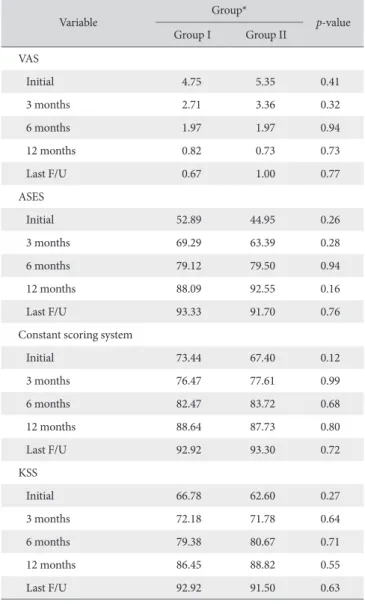
Table 4. Comparisons of Functional Scores
Variable Group*
p-value Group I Group II
VAS
Initial 4.75 5.35 0.41
3 months 2.71 3.36 0.32
6 months 1.97 1.97 0.94
12 months 0.82 0.73 0.73
Last F/U 0.67 1.00 0.77
ASES
Initial 52.89 44.95 0.26
3 months 69.29 63.39 0.28
6 months 79.12 79.50 0.94
12 months 88.09 92.55 0.16
Last F/U 93.33 91.70 0.76
Constant scoring system
Initial 73.44 67.40 0.12
3 months 76.47 77.61 0.99
6 months 82.47 83.72 0.68
12 months 88.64 87.73 0.80
Last F/U 92.92 93.30 0.72
KSS
Initial 66.78 62.60 0.27
3 months 72.18 71.78 0.64
6 months 79.38 80.67 0.71
12 months 86.45 88.82 0.55
Last F/U 92.92 91.50 0.63
Values are presented as mean only.
VAS: visual analogue scale, F/U: follow-up, ASES: American Shoulder and Elbow Surgeons, KSS: Korean shoulder score.
*Patients who underwent arthroscopic capsular release for shoulder stiffness were enrolled and randomized into two groups: group I, patients who under- went arthroscopic capsular release of the rotator interval, anterior (a standard anterior release); group II, patients who underwent arthroscopic capsular release of the anterior to inferior capsule, including the rotator interval (a standard anteroinferior release).
Moreover, there was no control group. For these reasons, it was difficult to determine what percentage of patients would have improved without rotator interval release and capsulectomy.
Second, the mean follow-up period was relatively short. For both groups, it was 16.1 months. However, shoulder stiffness does not recur once it has healed, and it is unlikely that the re- sults may get worse with time. Finally, there were no long-term results. Overall, the results might have been more reliable with more enrolled patients and additional long-term data.
Conclusion
Arthroscopic capsular release is a reliable method for reliev- ing pain and improving clinical function in patients with shoul- der stiffness. However, the addition of inferior capsular release shows superiority in forward flexion over anterior capsular re- lease alone at 6 months follows-up.
References
1. Codman EA. The shoulder: rupture of the supraspinatus ten- don and other lesions in or about the subacromial bursa. Bos- ton: T. Todd Company; 1934. 216-24.
2. Watson L, Dalziel R, Story I. Frozen shoulder: a 12-month clinical outcome trial. J Shoulder Elbow Surg. 2000;9(1):16- 22.
3. Shaffer B, Tibone JE, Kerlan RK. Frozen shoulder. A long-term follow-up. J Bone Joint Surg Am. 1992;74(5):738-46.
4. Tauro JC. Stiffness and rotator cuff tears: incidence, ar- throscopic findings, and treatment results. Arthroscopy.
2006;22(6):581-6.
5. Warner JJ, Allen A, Marks PH, Wong P. Arthroscopic release for chronic, refractory adhesive capsulitis of the shoulder. J Bone Joint Surg Am. 1996;78(12):1808-16.
6. Harryman DT 2nd, Matsen FA 3rd, Sidles JA. Arthroscopic management of refractory shoulder stiffness. Arthroscopy.
1997;13(2):133-47.
7. Harryman DT 2nd, Sidles JA, Harris SL, Matsen FA 3rd. The role of the rotator interval capsule in passive motion and stabil- ity of the shoulder. J Bone Joint Surg Am. 1992;74(1):53-66.
8. Ozaki J, Nakagawa Y, Sakurai G, Tamai S. Recalcitrant chronic adhesive capsulitis of the shoulder. Role of contracture of the coracohumeral ligament and rotator interval in pathogenesis and treatment. J Bone Joint Surg Am. 1989;71(10):1511-5.
9. Pearsall AW 4th, Holovacs TF, Speer KP. The intra-articular component of the subscapularis tendon: anatomic and histo- logical correlation in reference to surgical release in patients with frozen-shoulder syndrome. Arthroscopy. 2000;16(3):236- 42.
10. Massoud SN, Pearse EO, Levy O, Copeland SA. Operative
management of the frozen shoulder in patients with diabetes.
J Shoulder Elbow Surg. 2002;11(6):609-13.
11. Nicholson GP. Arthroscopic capsular release for stiff shoulders:
effect of etiology on outcomes. Arthroscopy. 2003;19(1):40-9.
12. Ide J, Takagi K. Early and long-term results of arthroscopic treatment for shoulder stiffness. J Shoulder Elbow Surg.
2004;13(2):174-9.
13. Jerosch J. 360 degrees arthroscopic capsular release in patients with adhesive capsulitis of the glenohumeral joint: indication, surgical technique, results. Knee Surg Sports Traumatol Ar- throsc. 2001;9(3):178-86.
14. Pearsall AW 4th, Osbahr DC, Speer KP. An arthroscopic tech- nique for treating patients with frozen shoulder. Arthroscopy.
1999;15(1):2-11.
15. Snow M, Boutros I, Funk L. Posterior arthroscopic capsular re- lease in frozen shoulder. Arthroscopy. 2009;25(1):19-23.
16. Chen SK. Arthroscopic and histologiccal findings in the idio- pathic frozen shoulder. J Shoulder Elbow Surg. 1996;5(2):S26.
17. Kim YS, Lee HJ, Park IJ. Clinical outcomes do not support arthroscopic posterior capsular release in addition to anterior release for shoulder stiffness: a randomized controlled study.
Am J Sports Med. 2014;42(5):1143-9.
18. Ranalletta M, Rossi LA, Zaidenberg EE, et al. Midterm out- comes after arthroscopic anteroinferior capsular release for the treatment of idiophatic adhesive capsulitis. Arthroscopy. 2017;
33(3):503-8.
19. Kim YS, Lee HJ, Park I, Im JH, Park KS, Lee SB. Are delayed operations effective for patients with rotator cuff tears and concomitant stiffness? An analysis of immediate versus delayed surgery on outcomes. Arthroscopy. 2015;31(2):197-204.
20. Richards RR, An KN, Bigliani LU, et al. A standardized method for the assessment of shoulder function. J Shoulder Elbow Surg. 1994;3(6):347-52.
21. McNamara WJ, Lam PH, Murrell GA. The relationship be- tween shoulder stiffness and rotator cuff healing: a study of 1,533 consecutive arthroscopic rotator cuff repairs. J Bone Joint Surg Am. 2016;98(22):1879-89.
22. Neer CS 2nd, Satterlee CC, Dalsey RM, Flatow EL. The anato- my and potential effects of contracture of the coracohumeral ligament. Clin Orthop Relat Res. 1992;(280):182-5.
23. Omari A, Bunker TD. Open surgical release for frozen shoul- der: surgical findings and results of the release. J Shoulder Elbow Surg. 2001;10(4):353-7.
24. Yamaguchi K, Sethi N, Bauer GS. Postoperative pain control following arthroscopic release of adhesive capsulitis: a short- term retrospective review study of the use of an intra-articular pain catheter. Arthroscopy. 2002;18(4):359-65.
25. Ogilvie-Harris DJ, Biggs DJ, Fitsialos DP, MacKay M. The resis- tant frozen shoulder. Manipulation versus arthroscopic release.
Clin Orthop Relat Res. 1995;(319):238-48.
26. Codding J, Dahm DL, McCarty LP 3rd, May JH, Tucker LH, Buss DD. Arthroscopic posterior-inferior capsular release in the treatment of overhead athletes. Am J Orthop (Belle Mead NJ).
2015;44(5):223-7.
27. Miyazaki AN, Santos PD, Silva LA, Sella GD, Carrenho L, Checchia SL. Clinical evaluation of arthroscopic treatment of shoulder adhesive capsulitis. Rev Bras Ortop. 2016;52(1):61- 8.