Is Pancapsular Release More Effective than Selective Capsular Release for the Treatment of Adhesive Capsulitis?
Nam Hoon Moon*, Seung-Jun Lee , Won Chul Shin, Sang Min Lee, Kuen Tak Suh
Department of Orthopaedic Surgery, Pusan National University Yangsan Hospital, Yangsan, Korea
Background: We assessed the effectiveness of arthroscopic capsular release for the treatment of adhesive capsulitis. Further, we tried to ascertain the clinical benefits, if any, of pancapsular release over selective capsular release, where the two differ by performing or not performing a posterior capsular release, respectively.
Methods: Thirty-five consecutive patients with either primary or secondary adhesive capsulitis who failed conservative treatment for more than 6 months were enrolled in the study. A total of 16 patients allocated in group 1 received a pancapsular release that comprises the release of the rotator interval, anteroinferior capsular, and the posterior capsular release, whereas 19 patients in group 2 received a selective capsular release that comprises only the release of the rotator interval release and anteroinferior capsular release. The clinical outcomes, visual analogue scale (VAS) score, Constant score, and range of motion, were assessed preoperative and postoperatively.
Results: In both groups, the preoperative VAS score, Constant score, and ROM showed a significant improvement by the 6-month fol- low-up. We found that the immediate postoperative internal rotation was significantly higher in group 1 than group 2. Despite significant differences seen between the two groups at the initial postoperative period, there were no significant differences in Constant score, VAS score, and the ROM at all the subsequent follow-ups between the two groups.
Conclusions: Arthroscopic capsular release for the treatment of adhesive capsulitis is very effective. However, pancapsular release did not show any advantage over selective capsular release in terms of overall clinical outcome.
(Clin Shoulder Elbow 2015;18(1):28-35)
Key Words: Adhesive capsulitis; Pancapsular release; Selective capsular release Clinics in Shoulder and Elbow Vol. 18, No. 1, March, 2015
http://dx.doi.org/10.5397/cise.2015.18.1.28
Received August 18, 2014. Revised January 22, 2015. Accepted February 5, 2015.
*Current affiliation: Department of Orthopaedic Surgery, Pusan National University Hospital, Busan, Korea Correspondence to: Seung-Jun Lee
Department of Orthopaedic Surgery, Pusan National University Yangsan Hospital, 20 Geumo-ro, Mulgeum-eup, Yangsan 626-770, Korea Tel: +82-55-360-2125, Fax: +82-55-360-2155, E-mail: [email protected]
Financial support: None. Conflict of interests: None.
Introduction
In general, adhesive capsulitis of the shoulder is conservative- ly treated. Conservative treatments such as drug therapy, physio- therapy, and injection-based therapy have been shown to lead to remission of the condition by 60% to 80%. However, when these methods fail surgical treatments using arthroscopy may be considered as a second-line option.1,2) Of the arthroscopic in- terventions, the most common and effective approach reported to treat adhesive capsulitis is arthroscopic capsular release. And despite the existence of several studies that support its effective- ness in treating this condition, none have attempted to define the guidelines on the extent of the capsular release required
for a successful outcome.3,4) Further, some discrepancies in pre- existing findings exist between studies. A few authors have shown that to achieve enhanced internal rotation and cross body adduction in adhesive capsulitis patients, pancapsular re- lease, which is a type of a selective capsular release that includes the release of the rotator interval, anteroinferior capsule, and additionally, the posterior capsule, should be implemented. In stark contrast, others have shown that selective capsular release alone, i.e. the release of the rotator interval and the anteroinfe- rior capsule, gives results that are comparable to the results after posterior capsular release.5-9)
To rule out one of the possiblility that selective capsular re- lease alone could give clinical outcomes that are comparable to
those of posterior capsular release for the treatment of adhesive capsulitis, we investigated whether selective capsular release and pancapsular release have any relative advantage over one another for the treatment of adhesive capsulitis in terms of the clinical outcome and range of motion (ROM).
Methods
Subjects of Study
In our study, we enrolled patients suffering from adhesive capsulitis who despite having had conservative treatments such as drug therapy, physiotherapy, and intra-articular steroid therapy for at least 3 to 6 months between May 2010 to January 2011 had showed persistent, non-resolution of shoulder pain and joint stiffness. Patients able to undergo capsular release and to attend up to at least the 6-month follow-up session were included in the study. We took preoperative x-rays and magnetic resonance imaging (MRI) scans of all the patients, and those revealed through MRI to have a rotator cuff tear, a Bankart’s lesion, or a superior labrum anterior and posterior lesion were excluded from the study. In total, 35 patients were enrolled in the study.
The average age of the patients was 56 years of age (range, 38 to 82 years), and the ratio of sex was 9 males to 26 females. Adhe- sive capsulitis was found on the right shoulder in 24 patients and on the left side in 11 patients. Of the 35 patients, 27 patients had suffered the condition on their dominant shoulder. We cat- egorized the patients in terms of etiology of the frozen shoulder;
as either primary or post-traumatic. Between May 2010 to May 2012, we carried out 16 operations of pancapsular release, and between June 2012 to January 2014, we carried out 19 opera- tions of selective capsular release. For our subsequent analyses, we sub-grouped the patients into the following two groups;
group 1 who received pancapsular release and group 2 who re- ceived selective capsular release.
Surgical Methods
The surgery proceeded with the patient in a beach chair posi- tion and under general anesthesia. Preoperatively, we measured each patient’s ROM (forward flexion, external rotation, and in- ternal rotation at neutrality, etc.) before making the surgical prep- arations. We created a posterior portal to feed the arthroscopy through the portal and to examine the intra-articular space and assess the extent and position of the synovitis and capsular con- tracture. We also created an anterior portal, through which we inserted the planer and molder machine and used it to remove the hyper-proliferated synovium surrounding the rotator interval and the long head of biceps tendon. After cleaning the region out, we made an incision in the superior glenohumeral ligament using an electrocautery (ArthroCare, Sunyvale, CA, USA) all the while taking care not to induce labral injury. After the release of the rotator interval, we unveiled the coracohumeral ligament up
to the base of the coracoid process and revealed the conjoined tendon. Then, we continued with the release by following the glenoid rim and sticking to the glenoid surface as much as pos- sible thereby releasing the anterior capsule and the middle gle- nohumeral ligament. Finally, once we observed the muscle fiber of the subscapularis, which we arbitrarily decided as a marker of sufficient release, the electrocautery was veered posteriorally to perform the release of the inferior glenohumeral ligament and the anterior capsule in the 6’oclock direction. After the ar- throscopic anterior capsular release was finished, we monitored the patients’ ability to carry out forward flexion, internal rotation, and external rotation.
For the posterior capsular release, the arthroscopy was in- serted from the anterior portal and the electrocautery was in- serted through the posterior portal. We performed the release in the posterior-superior direction to the glenoid rim and in the 6 o’clock direction. Once again, the release was stopped when the muscle fiber of the subscapularis was observed. When work- ing in the 6 o’clock direction, we made sure to be aware of pos- sible axillary nerve injury (Fig. 1, 2).
Lastly, we inserted the arthroscopic tube into the subacromial space and examined this region along with the subacromial surface and the rotator cuffs. If we found a coracoacromial liga- ment tear, subacromial impingement syndrome, or excessive adhesion due to the subacromial bursitis, we implemented an arthroscopic decompression.
Rehabilitation
From the next day of surgery, the patients, under patient con- trolled analgesia, began passive joint exercises using a rod with pulley and contineous passive movement. The same rehabilita- tion protocol was issued to both patient groups irrespective of the mode of surgery they had received. The aim of the rehabili- tation was to achieve as much passive ROM of the joints as per- mitted by pain. Active exercise was commenced from the 6th week of surgery, and muscle-strengthening exercises were begun from 3 months of surgery when the patient was able to achieve all ROM of the joints under no pain.
We measured the visual analogue scale (VAS) score, Constant score, and the ROM (forward elevation, internal rotation, and external rotation) in all the patients at the preoperative, 1-day postoperative, 1-month postoperative, 3-month postoperative, and at the 6-month postoperative follow-up. We analyzed the differences in pain alleviation and ROM between the two pa- tient groups.
Statistical Analysis
All statistical analyses were performed using IBM SPSS Statis- tics ver. 21.0 (IBM Co., Armonk, NY, USA). Initially, a normality test was implemented for our comparative analysis in terms of age, follow-up duration, and prepoerative and postoperative
ROM and pain intensity. If samples were shown to have normal distribution, a Student t-test was carried out, and if not, a non- parametric Mann-Whitney test was carried out. To compare the sex ratio, shoulder dominance, and diabetic status between the two groups, we used the chi-square test and the Fisher’s exact
test. Preoperative and postoperative changes in ROM and pain were compared between the two groups using a paired t-test for data with normal distribution and the Wilcoxon signed rank test for data without a normal distribution.
A B
C D
Fig. 1. (A) Arthroscopic electrocautery re- leased thickened, contracted anteroinferior capsule (black arrow). (B) After complete release of anteroinferior capsule, the interval was wide open and the muscle belly of sub- scapularis (black arrow) could be seen. (C) Arthroscopic electrocautery released postero- superior recess (black arrow). (D) Posteriorly muscle fibers of the infraspinatus could be seen as the thickened capsule (black arrow) was released.
A B
Fig. 2. Schematic drawings of the capsular re- lease for the adhesive capsulitis. (A) Selective capsular release. (B) Pancapsular release.
Results
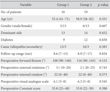
The etiologies of the 35 frozen shoulders were primary in 29 cases and post-traumatic in 9 cases. The mean follow-up period was 8.7 months. We did not find a significant difference be- tween the pancapsular release group and the selective capsular release group in terms of age, sex, co-diabetic complications, nor preoperative ROM, VAS score, and Constant score (Table 1). In the 4 patients shown to have a coracoacromial ligament injury, subacromial impingement syndrome, or subacromial bursitis, we carried out a subacromial decompression. Further, we per- formed tenotomies in 11 patients who had either severe de- generative changes or fractures of the long head biceps brachii muscle.
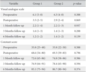
In both patient groups, the postoperative values for VAS score, Constant score, forward flexion, internal rotation, and external rotation at 6-month follow-up improved from their re- spective preoperative values in a statistically significantly manner (Table 2, Fig. 3). We found that group 1 showed a statistically enhanced postoperative value in only one parameter, internal rotation, than that of group 2. Even so, this significant difference disappeared at later follow-ups (Table 3). Lastly, we did not find any significant difference between neither the VAS score nor the Constant score between the groups at all follow-up periods (Table 4).
Discussion
In this study, the authors aimed to see the efficacy of two types of arthroscopic capsular release, pancapsular release and
selective capsular release, for adhesive capsulitis of the shoulder and to elucidate if one type of release had a relative advantage over the other. In our study on 35 adhesive capsulitis patients, we could not see a significant benefit of pancapsular release over selective capsular release or vice versa for the treatment of adhesive capsulitis. But, nonetheless, we were able to show that arthroscopic capsular release for adhesive capsulitis is highly effective even when performed in patients who had already re- ceived an unsuccessful conservative management.
Adhesive capsulitis of the shoulder results in capsular con- tracture induced by progressive fibrosis and in severe pain with debilitating effects on the shoulder such as limited motion. The prevalence of adhesive capsulitis is around 2% to 5%.10,11) Cod- man,12) who was the first to described adhesive capsulitis, coined the term ‘frozen shoulder’ based on his findings of bursitis in and around the shoulder joint and of an adhered rotator cuff, which in turn induced tendinitis that spread to the subacromial space.
Like Codman,12) Neviaser13) agreed with the pathophysiology of adhesive capsulitis being fibrosis, infection, and joint contracture, but argued that adhesive capsulitis is a more accurate term that describes the condition and pathophysiology than ‘frozen shoul- der’. Currently, these two terms are used interchangeably, but in light of typical symptoms of adhesive capsulitis such as joint stiffness and pain, the authors also believe that the latter term is more appropriate than the earlier alias.
Table 1. Preoperative Demographic Data of Patients
Variable Group 1 Group 2 p-value
No. of patients 16 19
Age (yr) 55.4 (41–71) 58.9 (38–82) 0.351
Gender (male/female) 3/13 6/13 0.667
Dominant side 13 14 0.452
Diabetes 9 12 0.830
Cause (idiopathic/secondary) 13/3 16/3 0.383
Follow-up range (mo) 8.6 (7–15) 8.9 (7–17) 0.834 Preoperative forward flexion (o) 108 (90–140) 116 (90–145) 0.152 Preoperative external rotationn (o) 3 (-10–20) 2 (-20–25) 0.743 Preoperative internal rotation (o) 22 (0–40) 22 (0–40) 0.573 Preoperative visual analogue scale 6.1 (5–8) 6.3 (5–8) 0.545 Preoperative Constant score 35.8 (25–48) 33.8 (22–50) 0.388 Values arepresented as number only or median (range).
Group 1: who received pancapsular release, Group 2: who received selective capsular release.
Table 2. Comparison between Preoperative and Postoperative Results in Both Groups
Variable Preoperative 6 Months follow-up Forward flexion (o)
Group 1 108 (90–140) 166 (160–180)
Group 2 116 (90–145) 163 (150–170)
External rotation (o)
Group 1 2.8 (-10–20) 40.0 (30–60)
Group 2 1.6 (-20–25) 42.6 (20–70)
Internal rotation (o)
Group 1 20.0 (0–40) 51.9 (30–70)
Group 2 22.1 (0–40) 50.5 (30–70)
Visual analogue scale score
Group 1 6.1 (5–8) 1.2 (1–3)
Group 2 6.3 (5–8) 1.4 (1–2)
Constant score
Group 1 35.8 (25–48) 85.1 (75–94)
Group 2 33.8 (22–50) 86.7 (80–94)
Values arepresented as median (range). p<0.001.
Group 1: who received pancapsular release, Group 2: who received selective capsular release.
The underlying mechanism for the pathogenesis of adhe- sive capsulitis is unknown, but synovitis-induced expression of growth factors such as transforming growth factor-beta is known to leads to fibrosis. Further, 20% to 30% of patients with adhe- sive capsulitis are associated with minor injuries but whether or not they are in any way related to the pathophysiology of adhe- sive capsulitis is not understood.14) Some regard this condition as a autoimmunity problem that causes degenerative lesions, but again there is no conclusive evidence to support this statement.
Although a study by Bulgen et al.15) and Rizk and Pinals16) on HLA-B27 showed that HLA-B27 is increased in adhesive cap- sulitis patients more than normal patients, this increase was not statistically significant.
In general, the first line of management is a conservative one, which shows a success rate of around 60% to 80%.1,2) If conser- vative management using drug therapy, physiotherapy, or injec- tion therapy of at least 3 to 6 months shows no signs of improve- ment of ROM and pain, surgical methods can be reconsidered.
Typical surgical methods for adhesive capsulitis are manipulation under general anesthesia or capsular release. However, manipu- lation may cause complications such as humeral fractures, rota-
tor cuff tear, long head biceps brachii muscle tears, and glenoid fractures. Arthroscopic capsular release is known to be effective for adhesive capsulitis. Many papers show that pathological structures implicated in adhesive capsulitis may be the capsules surrounding the articular joints. Other implicated structures are coracohumeral ligament, axillary recess, and the subacromial space.17-19)
Through cadaveric studies, Bowen and Warren,20) Harryman et al.,21) and Ovesen and Nielsen22) found that the release of the superior and middle glenohumeral ligaments, rotator intervals, and the intra-articular subscarpularis can improve the external rotation of the shoulder, whereas release of the anteroposterior capsule or the inferior glenohumeral ligament can improve the forward flexion. Further, they also found that the release of the posterior capsule can improve the internal rotation. These results provide a basis for using arthroscopy-based capsular release as the treatment modality for adhesive capsulitis. Still, clinical stud- ies that define or give guidelines as to the amount of capsular release is effective or required are rare, and results of which are inconsistent.
Authors who state that a pancapsular release is effective sug-
Preop. Postop. 1 mo 3 mo 6 mo 80
60
40
20
0
Internalrotation()
Preop. Postop. 1 mo 3 mo 6 mo 60
40
20
0
Externalrotation()
Preop.Postop. 1 mo 3 mo 6 mo 180
160
140
100
80
Forwardflexion()
120
Preop.Postop. 1 mo 3 mo 6 mo 100
80
60
40
20
Constantscore
Preop. Postop. 1 mo 3 mo 6 mo 8
6
4
2
VASscore
Group 1 Group 2
Group 1 Group 2
Group 1 Group 2 Group 1
Group 2
Group 1 Group 2
Fig. 3. There was highly significant improvement of range of motion (forwad flexion, internal rotation, external rotation) and functional scores (visual analogue scale [VAS] score, Constant score) 6 months follow-up in both groups. In group 1, there was significant improvement in internal rotation posteratively. However postoperative (postop.) VAS score was higher compared with group 2. There was no significant difference in Constant score, VAS score and range of motion 1 month, 3 months, 6 months follow-up between the two groups. Group 1: who received pancapsular release, Group 2: who received selective capsular release, Preop.: preoperative.
gest that because posterior-superior capsule contracture limits cross body adduction and internal rotation at abduction, a pos- terior capsular releases leads to an enhanced internal rotation postopertively.4-7) Nicholson5) showed good clinical outcomes in 68 patients with adhesive capsulitis after arthroscopic pancap- sular release, and proposed that for an enhanced postoperative internal rotation, performing a posterior capsular release is rec- ommended. Another study carried out Ide and Takagi6) on 44 patients with adhesive capsulitis showed a successful outcome after pancapsular release.
Conversely, when Snow et al.8) and Chen et al.23) compared the results of pancapsular release that includes a posterior cap- sular release with those of selective capsular release for adhesive capsulitis, they did not find a significant difference in terms of internal rotation. Similarly, the study by Kim et al.9) on 75 pa- tients with adhesive capsulitis found that the mode of surgery, posterior capsular release or anterior capsular and rotator inter- val release, did not affect the postoperative pain or ROM. Thus, these authors concluded that when performing an arthroscopic capsular release a release of the posterior region is unnecessary.
In this study, we found that the extent of the arthroscopic capsular release did not affect the clinical success of the treat- ment. All the showed a satisfactory outcome of surgery allowing
us to conclude that in case where conservative methods are ineffective surgery methods are effective second-line of options.
At the final follow-up, in agreement with the studies by Snow et al.8) and Kim et al.,9) we found that the postoperative ROM and pain between patients of different treatment groups did not show a significant difference. Interestingly, at a day postopera- tion, we found that the angle of internal rotation was significantly higher in patients who received pancapsular release than those who received selective capsular release. However, the signifi- cance disappeared at later follow-ups. The explanation for the significant increase in internal rotation at 1 day postoperation in patients may be as according to Jerosch’s hypothesis3) that poste- rior capsular contracture may be the most important pathologi- cal factor in adhesive capsulitis and inhibits internal rotation of the shoulders, and thus releasing this would contribute to the enhancement of ROM. However, further confirmative studies are needed as this significant difference is no longer seen at later follow-ups.
At the final follow-up, we did not find a significant differ- ence in neither VAS score nor Constant score between the two groups. However, a day postoperation, we found that the VAS score was higher in the pancapsular release group than the se- lective capsular release group. We believe the reason for this elevated score is that with greatercapsular release there may be greater tissue damage, such as unintentional damage of the sub- scapularis fibrotic tissue underneath the thin posterior capsule that may lead to surgery-related pain.
Through this study, we found that although a temporary ben- efit of pancapsular release over selective capsular release was seen in terms of the internal rotation, this benefit was no longer Table 3. Postoperative Improvement of Range of Motion
Variable Group 1 Group 2 p-value
Forward flexion (o)
Preoperative 108 (90–140) 116 (90–145) 0.152 Postoperative 148 (100–165) 143 (110–165) 0.331 1 Month follow-up 157 (145–170) 152 (125–170) 0.165 3 Months follow-up 163 (150–180) 157 (130–170) 0.112 6 Months follow-up 166 (160–180) 163 (150–170) 0.162 External rotation (o)
Preoperative 3 (-10–20) 2 (-20–25) 0.743
Postoperative 10 (0–20) 15 (0–30) 0.125
1 Month follow-up 17 (10–30) 21 (10–40) 0.116 3 Month follow-up 21 (10–40) 24 ( 10–45) 0.280 6 Month follow-up 40 (20–60) 43 (30–50) 0.448 Internal rotation (o)
Preoperative 20 (0–40) 22 (0–40) 0.573
Postoperative 43 (20–60) 34 (20–60) 0.037
1 Month follow-up 44 (30–70) 47 (30–80) 0.510 3 Month follow-up 43 (20–60) 49 (30–70) 0.209 6 Month follow-up 52 (30–70) 51 (30–60) 0.747 Values arepresented as median (range).
Group 1: who received pancapsular release, Group 2: who received selective capsular release.
Table 4. Pain Relief of Patients Who Had Arthroscopic Capsular Release
Variable Group 1 Group 2 p-value
Visual analogue scale
Preoperative 6.1 (5–8) 6.3 (4–8) 0.388
Postoperative 3.5 (2–5) 2.9 (2–4) 0.069
1 Month follow-up 2.2 (1–4) 2.2 (1–3) 0.937 3 Months follow-up 1.6 (1–3) 1.4 (1–3) 0.288 6 Months follow-up 1.2 (1–2) 1.4 (1–2) 0.139 Constant score
Preoperative 35.8 (25–48) 33.8 (22–50) 0.388 Postoperative 68.6 (54–80) 69.3 (59–83) 0.796 1 Month follow-up 72.6 (65–86) 74.8 (58–86) 0.386 3 Months follow-up 76.9 (64–91) 78.4 (65–90) 0.594 6 Months follow-up 85.1 (75–94) 86.7 (80–94) 0.374 Values arepresented as median (range).
Group 1: who received pancapsular release, Group 2: who received selective capsular release.
applicable at later follow-ups. Further, we found that the pan- capsular release was associated with an increase in postopera- tive pain. Our finding supports the notion of many researchers that the main pathological region in adhesive capsulitis lies in the anterior and anterior-inferior capsular regions such as the cora- cohumeral ligament, the rotator interval, and the axillary recess rather than the posterior capsular region. Especially, since we found that at least by the final follow-up the extent of capsular release does not have an effect on the clinical outcome, there is no benefit in carrying out a pancapsular release.24,25)
There are limitations to this study. First, we carried out the surgeries in two bulks in terms of the type of surgery, thus allocat- ing the patients into the treatment-type according to the order they were hospitalized rather than taking into consideration the extent of hypertrophy in either of the anterior or the posterior capsule. Even though the patients in each group received the same diagnoses of adhesive capsulitis, the extent of hypertrophy which may vary within group was not segmented and the same surgery was applied disregarding this. In this study, some patients had severe shoulder stiffness due to hypertrophy of both their anterior and the posterior capsules. These patients received a posterior articular capsule release. It may be interesting to see if a similar clinical outcome can be achieved when such patients do not receive a posterior capsular release despite the presence of hypertrophy in that region. Secondly, since our study encom- passed only a small sample, potential false negative data or type II error may mean a clinically significant finding may have gone unnoticed. Thus, prospective studies including a greater num- ber of cases are needed, and those differentiating the extent of hypertrophy in the anterior and posterior capsules and assessing their effect on the outcome of capsular release are required.
Conclusion
Arthroscopic capsular release for adhesive capsulitis of the shoulder is an effective treatment method that gives satisfac- tory clinical outcome. In this study, we found that pancapsular release had no relative advantage over selective capsular release to achieve good clinical outcomes for the treatment of adhesive capsulitis.
References
1. Hannafin JA, Chiaia TA. Adhesive capsulitis. A treatment ap- proach. Clin Orthop Relat Res. 2000;(372):95-109.
2. Shaffer B, Tibone JE, Kerlan RK. Frozen shoulder. A long-term follow-up. J Bone Joint Surg Am. 1992;74(5):738-46.
3. Harryman DT 2nd. Shoulders: frozen and stiff. In: The Ameri- can Academy of Orthopedic Surgeons, eds. Instructional course lectures. Vol. 42. Rosemont, IL: The American Academy of Orthopedic Surgeons; 1993. 247-57.
4. Warner JJ, Allen A, Marks PH, Wong P. Arthroscopic release for chronic, refractory adhesive capsulitis of the shoulder. J Bone Joint Surg Am. 1996;78(12):1808-16.
5. Nicholson GP. Arthroscopic capsular release for stiff shoulders:
effect of etiology on outcomes. Arthroscopy. 2003;19(1):40-9.
6. Ide J, Takagi K. Early and long-term results of arthroscopic treatment for shoulder stiffness. J Shoulder Elbow Surg. 2004;
13(2):174-9.
7. Jerosch J. 360 degrees arthroscopic capsular release in patients with adhesive capsulitis of the glenohumeral joint--indication, surgical technique, results. Knee Surg Sports Traumatol Ar- throsc. 2001;9(3):178-86.
8. Snow M, Boutros I, Funk L. Posterior arthroscopic capsular re- lease in frozen shoulder. Arthroscopy. 2009;25(1):19-23.
9. Kim YS, Lee HJ, Park IJ. Clinical outcomes do not support arthroscopic posterior capsular release in addition to anterior release for shoulder stiffness: a randomized controlled study.
Am J Sports Med. 2014;42(5):1143-9.
10. Pearsall AW, Speer KP. Frozen shoulder syndrome: diagnostic and treatment strategies in the primary care setting. Med Sci Sports Exerc. 1998;30(4 Suppl):S33-9.
11. Noël E, Thomas T, Schaeverbeke T, Thomas P, Bonjean M, Revel M. Frozen shoulder. Joint Bone Spine. 2000;67(5):393- 400.
12. Codman EA. The shoulder: rupture of the supraspinatus ten- don and other lesions in or about the subacromial bursa. Bos- ton, MA: T Todd Company; 1934.
13. Neviaser JS. Adhesive capsulitis of the shoulder. J Bone Joint Surg Am. 1945;27(2):211-22.
14. Rodeo SA, Hannafin JA, Tom J, Warren RF, Wickiewicz TL. Im- munolocalization of cytokines and their receptors in adhesive capsulitis of the shoulder. J Orthop Res. 1997;15(3):427-36.
15. Bulgen DY, Binder A, Hazleman BL, Park JR. Immunological studies in frozen shoulder. J Rheumatol. 1982;9(6):893-8.
16. Rizk TE, Pinals RS. Histocompatibility type and racial incidence in frozen shoulder. Arch Phys Med Rehabil. 1984;65(1):33-4.
17. Ozaki J, Nakagawa Y, Sakurai G, Tamai S. Recalcitrant chronic adhesive capsulitis of the shoulder. Role of contracture of the coracohumeral ligament and rotator interval in pathogenesis and treatment. J Bone Joint Surg Am. 1989;71(10):1511-5.
18. Pollock RG, Duralde XA, Flatow EL, Bigliani LU. The use of arthroscopy in the treatment of resistant frozen shoulder. Clin Orthop Relat Res. 1994;(304):30-6.
19. Watson L, Dalziel R, Story I. Frozen shoulder: a 12-month clinical outcome trial. J Shoulder Elbow Surg. 2000;9(1):16-22.
20. Bowen MK, Warren RF. Ligamentous control of shoulder sta- bility based on selective cutting and static translation experi- ments. Clin Sports Med. 1991;10(4):757-82.
21. Harryman DT 2nd, Sidles JA, Harris SL, Matsen FA 3rd. The role of the rotator interval capsule in passive motion and stabil- ity of the shoulder. J Bone Joint Surg Am. 1992;74(1):53-66.
22. Ovesen J, Nielsen S. Anterior and posterior shoulder instability.
A cadaver study. Acta Orthop Scand. 1986;57(4):324-7.
23. Chen J, Chen S, Li Y, Hua Y, Li H. Is the extended release of the inferior glenohumeral ligament necessary for frozen shoul- der? Arthroscopy. 2010;26(4):529-35.
24. Uitvlugt G, Detrisac DA, Johnson LL, Austin MD, Johnson C.
Arthroscopic observations before and after manipulation of frozen shoulder. Arthroscopy. 1993;9(2):181-5.
25. Wiley AM. Arthroscopic appearance of frozen shoulder. Ar- throscopy. 1991;7(2):138-43.

![Fig. 3. There was highly significant improvement of range of motion (forwad flexion, internal rotation, external rotation) and functional scores (visual analogue scale [VAS] score, Constant score) 6 months follow-up in both groups](https://thumb-ap.123doks.com/thumbv2/123dokinfo/5333762.175453/5.892.75.810.116.582/significant-improvement-internal-rotation-external-rotation-functional-constant.webp)