Evaluating Scapular Notching after Reverse Total Shoulder Arthroplasty
Young-Kyu Kim, Jun-Sung Won , Chang-Kyu Park, Jong-Geun Kim
Department of Orthopedic Surgery, Gachon University Gil Medical Center, Incheon, Korea
Background: Scapular notching can happen at diverse location depending on implant design or operative technique, therefore, it is eas- ily misdiagnosed. Thus, this study purposed to suggest a method helpful to assess scapular notching.
Methods: The subjects were 73 cases of reverse shoulder arthroplasty (RSA) for cuff tear arthropathy during the period from May 2009 to April 2014 and followed-up for over a year. There was medialized RSA in 22 cases, bone increased offset RSA (BIO-RSA) in 36 cases, and metal increased offset RSA (metal-RSA) in 15 cases. Scapular notching was not determined by bone defect at the inferior of gleno- sphere as Sirveaux’s classification, but scapular notching at the site where the rotational route of the polyethylene of humeral implant met the scapular neck were examined. The results were compared with conventional method.
Results: By conventional method, scapular notching was observed in 10 cases (45.5%) in medialized RSA, 12 cases (33.3%) in BIO-RSA, and none in metal-RSA. By new method, it was observed in 9 cases (40.9%) in medialized RSA, 10 cases (27.8%) in BIO-RSA, and none of metal-RSA. The site of scapular notching was apart from glenoshpere in 18 cases, and at inferior of glenosphere in 1 case. Absorption of bone graft was observed in 4 (11.1%) out of 36 cases of BIO-RSA.
Conclusions: It is hard to distinguish scapular notching from absorption of bone graft in BIO-RSA, and bone absorption at the lateral lower end of glenoid in medialized RSA. Thus, it is considered useful to assess scapular notching at the site where the rotational route of the polyethylene insert meets scapular neck.
(Clin Shoulder Elbow 2015;18(4):248-253)
Key Words: Rotator cuff tear arthropathy; Reverse total shoulder arthroplasty; Scapular notching; Bone absorption Clinics in Shoulder and Elbow Vol. 18, No. 4, December, 2015
http://dx.doi.org/10.5397/cise.2015.18.4.248
Received October 4, 2015. Revised December 19, 2015. Accepted December 19, 2015.
Correspondence to: Jun-Sung Won
Department of Orthopedic Surgery, Gachon University Gil Medical Center, 21 Namdong-daero 774beon-gil, Namdong-gu, Incheon 21565, Korea Tel: +82-32-460-3384, Fax: +82-32-460-3388, E-mail: [email protected]
Financial support: None. Conflict of interests: None.
Introduction
Various complications are reported after reverse total shoul- der arthroplasty (RTSA) of rotator cuff arthropathy. Scapular notching is the most common complication observed in 44% to 96% of all the cases.1-8) Studies have not reported a correlation between scapular notching and the clinical results or implant failure,2,9) but it has been shown that superior invasion of scapu- lar notching into the inferior screw that supports the glenoid base plate can adversely affect the glenoid component or shoul- der function.1,10) As the pathological effects of scapular notching exacerbates with time,9,11) it must be treated promptly and its occurrence minimized.
According to Sirveaux’s classification10) of scapular notching, the most widely used classification system, scapular notching is most frequently classified into the group that reflects that scapu- lar notching has developed at the inferior glenoid component.
Because scapular notching can develop along the surfaces of contact between the medial humeral polyethylene implant and the scapular neck, it has the potential to develop anywhere from the areas inferiolateral to the glenoid component to the medial portion of the scapular neck. It is difficult to differentiate scapu- lar notching from partial absorption of the graft bone that occurs in bony increased offset RSTA (BIO-RTSA). In this study, we pro- pose a method to evaluate and to diagnose more accurately the scapular notching that occurs at the sites of contact between the
humeral implant polyethylene and the scapular neck.
Methods
Of the 80 patients with rotator cuff arthropathy who under- went RTSA between May 2009 and April 2014, we enrolled 73 patients who could be followed-up for at least one year. We excluded from our data evaluation 1 patient with inflammation, 1 patient who died from chronic diseases, and 5 patients who were lost during follow-up. We performed medialized RTSA (Tornier, Montbonnot Saint Martin, France) in 22 patients, bony increased-offset RTSA (Tornier) in 36, and metal increased-offset RTSA (DJO, Vista, CA, USA) in 15. The mean age of the patients was 70.9 years (range, 59 to 81 years). They consisted of 18 men and 55 women, and arthropathy was on the dominant- side in 58 patients and on the non-dominant-side in 15 patients.
The mean follow-up period was 18.8 months (range, 13 to 56 months).
Surgery was performed by a single surgeon who specialized in treating pathologies of the shoulder joint. A deltopetoral ap- proach was used with the patient under general anesthesia and lying at a 30o beach-chair position. We used a 155o guide for the Tonier implant cases and a 135o guide for the DJO implant cases to cut the humoral neck at 0o to 20o of humeral retroversion. For the bony increased-offset RTSA, we harvested a 10-mm thick cancellous bone from the humeral head for bone grafting. For the insertion of the glenoid component, we reamed the inferior glenoid surface further than the superior joint surface, inclined the component 10o inferiorally using a guide, and exposed the inferior cancellous bone of the joint surface. For the medialized RTSA, the glenoid plate was aligned to the inferior margin of the glenoid joint surface so that we could expose and fix the glenoid hemisphere. For the bony increased-offset RTSA, the glenoid component was fixed in alignment to the inferior margin of the bone graft and of the glenoid. For both the medialized reverse shoulder arthroplasty (RSA) and BIO-RSA, 36-mm diameter gle- noid hemispheric implants were used. For the metal increased- offset RTSA, 32-mm diameter, 6-mm lateralized glenoid hemi- spheric implants were used to fix the glenoid plate aligned to the glenoid inferior margin. The humeral implants were fixed using cement in all patients. The implants were reduced using a trial polyethylene; then, their range of motion and stability and the surrounding muscle tension were confirmed to determine the thickness of polyethylene. Upon completion of the surgery, the patients were applied an abduction brace up to the sixth week of surgery. The subjects began pendulum and isometric exer- cises the day after the surgery and passive exercises from the 3rd week of surgery. The abduction brace was removed at the 6th postoperative week, after which active exercises were initiated.
From the 10th postoperative week, the patients gradually began muscle strengthening exercises.
Using Sirveaux’s classification10) system, we evaluated and classified postoperative scapular notching on true anteroposte- rior simple radiographs at the following time points: 3rd, 6th, 9th, and 12th postoperative months, and every year thereafter.
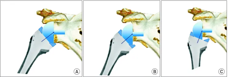
Considering that scapular notching develops at the contact point of the medial side of the humeral implant polyethylene and the scapular neck, we observed the contact point of the rotating platform of the humeral polyethylene and the scapular neck in the true anteroposterior radiologic images. The anticipated area of contact on the medial humeral polyethylene implant follow- ing the glenoid hemisphere were drawn into a hypothetical cir- cle, and the bone loss that developed within the area where the circle met the inferior margin of the scapular neck was defined as scapular notching (Fig. 1). The platform on which the rota- tional path of the humeral implant met the scapula was decided on the basis of the implant design and of the location of the glenoid component. In this study, we proposed, as mentioned, a novel method of defining scapular notching. We compared the frequency and the location of scapular notching determined after using this method and those found using the conventional method.
For statistical analyses, we compared the frequency and the location of scapular notching between the medialized RTSA group and the bony increased-offset RTSA group using the chi- square test. We set statistical significance as p<0.05.
Results
Using the conventional method of evaluating scapular notch-
Fig. 1. The anticipated points on the medial humeral implant polyethylene line according to the glenoid hemisphere were made to become a circle, and the bone loss that developed at the area where the circle met the inferior mar- gin of the scapular neck was defined as scapular notching.
ing, we found 22 patients (30.1%) with scapular notching: 10 patients (45.5%) from the medialized RTSA group, 12 patients (33.3%) from the bony increased-offset RTSA group, and none from the metal increased-offset RTSA group. Using the novel method suggested by us, we found 19 patients (26.0%) with scapular notching: 9 patients (40.9%) from the medialized RTSA group, 10 patients (27.8%) from the bony increased-offset RTSA group, and none from the metal increased-offset RTSA group.
In a patient from the medialized RTSA group, bone loss was observed inferior to the glenoid margin, but it was not on the rotational path of the humeral polyethylene; thus, it was not deemed as scapular notching (Fig. 2). In the bony increased- offset RTSA group, absorption of the graft bone was observed in 4 (11.1%) of 36 patients (Fig. 3). Of the 4 patients, 2 developed scapular notching not on the rotational path of the humeral im- plant, so they were deemed as having “no scapular notching”
according to the evaluation system we proposed. We found that all the bone loss that developed in the scapular neck were er- roneously diagnosed as scapular notching when they were diag- nosed using the conventional classification system. The scapular notching of the 22 patients who underwent medialized RTSA were classified: 2 patients (9.1%) had type 1 scapular notching, 3 patients (13.6%), type 2 scapular notching, 2 patients (9.1%), type 3 scapular notching, and 2 patients (9.1%), type 4 scapular notching. The scapular notching of the 36 patients who under- went bony increased-offset shoulder joint RTSA were classified as follow: 7 patients (19.4%) had type 1 scapular notching, 1 patient (2.8%), type 2 scapular notching, and 2 patients (5.6%),
type 3 scapular notching. Thus, we found that the frequency of types 2 and 3 scapular notching in the increased-offset RTSA group was significantly less than that those of the medialized RTSA group (p=0.032).
Fig. 2. In the one decrease scapular notching in the medialized reverse total shoulder arthroplasty group, bone loss was observed inferiorly to the glenoid margin, but it was not on the rotational path of the humeral polyethylene was thus not deemed a scapular notching.
A B
C D
Fig. 3. (A, B) Two cases developed not on the rotational path of the humeral implant and were thus deemed as ‘no scapular notching’
cases according to the analysis our method.
(C, D) Scapular notching of 2 cases were ob- served with bone absorption.
Discussion
Scapular notching after RTSA is a frequent postoperative complication, and many studies have proposed ways to reduce its frequency. Studies have shown that positioning of the glenoid component toward the inferior glenoid and with an incline of 10° inferiorly reduces the frequency of scapular notching in RTSA.6,12-14) Other studies have shown that a lateral offset of the center of rotation of the glenoid component and a smaller neck- shaft angle of the humeral implant reduces the frequency.5,15-17) Yet despite these efforts to minimize the incidence of postopera- tive scapular notching through improved surgical techniques and through diversification of implant types, the number of studies that investigate the location or the severity of postoperative scapular notching is insufficient.
Because more and more surgical methods place the glenoid component more inferiorally and use an inferiorly protruded im- plant in an effort to reduce scapular notching, scapular notching that do develop do so in a position that is more medial to the inferior of the glenoid component than is described in Sirveaux’s classification (Fig. 4).1,14) Thus, the current conventional method of scapular notching classification would classify erroneously and non-discriminatorily any bone loss, those that are and are not induced by scapular notching, that develop at the inferior mar- gin of the glenoid component or at the scapular neck as scapular notching. To address this, we have developed a novel method of evaluating scapular notching that facilitates an accurate identifi- cation of initial scapular notching location and detection of even mild bone loss induced by scapular notching.
In the study conducted by Boileau et al.,18) 98% of the 42 pa- tients who received bony increased-offset RTSA were shown to have a successful graft bone union, but the extent of graft bone absorption was not investigated. In the current study, of the 36
patients who received bony increased-offset RTSA, we found that 4 had graft bone absorption (11%) through shoulder antero- posterior radiographs. All the 4 cases of graft bone absorption developed at the lateral side of the graft and were observed not at the center of the graft bone but at the superior and inferior sides. When Sirveaux’s classification system of scapular notching is used, the graft bone absorption that developed just inferior to the glenoid component cannot be differentiated from scapular notching that occurs at the same place. As scapular notching develops as a result of the wear of the medial humeral polyeth- ylene, the location of scapular notching can be anticipated to a certain extent as described above. If bone loss develops more laterally than to the anticipated location, it is probably a case of bone absorption rather than of scapular notching. Graft bone absorption frequently develops at the lateral side where blood supply is insufficient, and it has been shown that the level of bone absorption levels at the superolateral and at the inferiolat- eral sides of the graft bone are similar.
In this study, we evaluated graft bone absorption using the shoulder true anteroposterior simple radiographs. Computed to- mography (CT) scans were used for more precise evaluation, but only for the 1 case of bone absorption because of interference from artifact induced by a metal implant. In a cadaveric study, it was found that bone loss near the glenoid component was observed in 46% of cases and that bone loss was observed in 38%.19) CT arthrography can be used to confirm the stability of the glenoid component after RSTA, but a 50% negative predic- tive value of this method limits its usefulness.20)
The diagnostic power of the conventional method is limited in terms of its capacity to differentiate bone absorption from scapular notching. When using the conventional method, all bone loss that develops at the scapular neck are classified as scapular notching; as such, the bone loss at the locations other
A B C
Fig. 4. Due to the surgical method of inferiorly moving the glenoid component, and to the use of the inferiorly protruded implant, scapular notching develops more medially to the inferior of the glenoid component than as described in Sirveaux’ classification. (A) A glenosphere placed on the inferior margin of the gle- noid. (B, C) Placement of the glenosphere bellow the inferior rim of the glenoid.
than those that make contact with the humeral polyethylene are erroneously classified as scapular notching. In other words, the bone loss on the scapular neck and on the rotational path of the humeral implant polyethylene shown on shoulder true antero- posterior simple radiographs may be classified as scapular notch- ing.
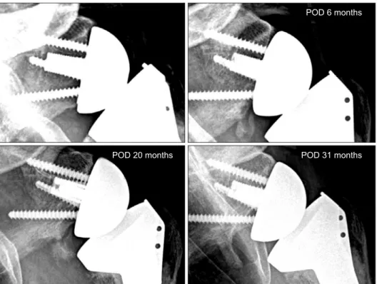
In the medialized RTSA group, we found that one patient whose diagnosis of scapular notching differed when using the two methods of analysis had a scapular notching that developed near the glenoid component and more lateral to the rotational path of the humeral polyethylene implant. This patient was not classified as having scapular notching according to our novel method. But because bone loss is usually observed at the inferio- lateral margin of the glenoid component in patients who receive RTSA, this patient was considered as having scapular notching according to the conventional method of scapular notching eval- uation. According to the proposed method, however, the case is one of scapular notching if the glenoid bone loss developed on the rotational path of the glenoid component; otherwise, the case can be considered one of simple bone loss (Fig. 5).
We found that bone loss usually developed a year after the surgery with no later increases in bone loss. The region of bone loss was the area where the humeral implant polyethylene did not make contact. We thought that the duration was too short for debris uptake induced by polyethylene abrasion to be the cause of bone loss. Rather, we thought that an avascular envi- ronment created as we reamed the glenoid to expose the can- cellous bone and dissected the nearby soft tissues to secure the visual field and surgical space was to blame for the bone loss.
Still, further studies are necessary to clarify this. The Sirveaux’s classification system for scapular notching uses the position of the inferior screw as the guideline for classification, but the ideal screw location during a surgery differs from one patient to an- other.21,22) Therefore, the location of the screw is an inherently inconsistent marker of the size and grade of scapular notching. A scapular notching classification that can overcome this drawback is based on the distance between the glenoid component peg or the central screw and the inferior margin of the scapular neck and on the invasion height and ratio of the scapular notching.17) When the scapular notching proceeds superiorly to the screw that fixes the glenoid plate, the condition is assumed to be not a case of mechanical scapular notching but one of debris uptake caused by polyethylene abrasion. Further studies to delineate the exact cause of superior procession of scapular notching may also be necessary.
In this study, we proposed a method to differentiate scapular notching from simple bone loss. But limitations to the scope of this study include that we did not attempt to clarify the cause of bone absorption or to elucidate a correlation between simple bone absorption and clinical outcomes, which was difficult for the study was a short-term follow-up study.
Conclusion
Absorption of graft bone in bony increased-offset RTSA and of inferiolateral glenoid in medialized RTSA cannot be easily dif- ferentiated from scapular notching. To aid the differentiation, a close examination for scapular notching at the junction of the
POD 6 months
POD 31 months POD 20 months
Fig. 5. The case is scapular notching if the glenoid bone loss developed on the rotational path of the glenoid component. POD: post- operative day.
rotational path of the humeral implant polyethylene and the scapular neck may be useful.
References
1. Simovitch RW, Zumstein MA, Lohri E, Helmy N, Gerber C.
Predictors of scapular notching in patients managed with the Delta III reverse total shoulder replacement. J Bone Joint Surg Am. 2007;89(3):588-600.
2. Werner CM, Steinmann PA, Gilbart M, Gerber C. Treatment of painful pseudoparesis due to irreparable rotator cuff dysfunc- tion with the Delta III reverse-ball-and-socket total shoulder prosthesis. J Bone Joint Surg Am. 2005;87(7):1476-86.
3. Florie EE, Crosby LA. Scapular notching: danger ahead? Semi Arthroplast. 2013;24(1):24-7.
4. Zumstein MA, Pinedo M, Old J, Boileau P. Problems, compli- cations, reoperations, and revisions in reverse total shoulder arthroplasty: a systematic review. J Shoulder Elbow Surg.
2011;20(1):146-57.
5. Valenti P, Sauzières P, Katz D, Kalouche I, Kilinc AS. Do less medialized reverse shoulder prostheses increase motion and reduce notching? Clin Orthop Relat Res. 2011;469(9):2550-7.
6. Lévigne C, Garret J, Boileau P, Alami G, Favard L, Walch G.
Scapular notching in reverse shoulder arthroplasty: is it impor- tant to avoid it and how? Clin Orthop Relat Res. 2011;469(9):
2512-20.
7. Sanchez-Sotelo J. Reverse total shoulder arthroplasty. Clin Anat.
2009;22(2):172-82.
8. Nicholson GP, Strauss EJ, Sherman SL. Scapular notching:
Recognition and strategies to minimize clinical impact. Clin Orthop Relat Res. 2011;469(9):2521-30.
9. Lévigne C, Boileau P, Favard L, et al. Scapular notching in reverse shoulder arthroplasty. J Shoulder Elbow Surg. 2008;
17(6):925-35.
10. Sirveaux F, Favard L, Oudet D, Huquet D, Walch G, Molé D.
Grammont inverted total shoulder arthroplasty in the treat- ment of glenohumeral osteoarthritis with massive rupture of the cuff. Results of a multicentre study of 80 shoulders. J Bone Joint Surg Br. 2004;86(3):388-95.
11. Favard L, Levigne C, Nerot C, Gerber C, De Wilde L, Mole D.
Reverse prostheses in arthropathies with cuff tear: are survivor- ship and function maintained over time? Clin Orthop Relat Res. 2011;469(9):2469-75.
12. Edwards TB, Trappey GJ, Riley C, O’Connor DP, Elkousy HA,
Gartsman GM. Inferior tilt of the glenoid component does not decrease scapular notching in reverse shoulder arthroplasty:
results of a prospective randomized study. J Shoulder Elbow Surg. 2012;21(5):641-6.
13. Bigorre N, Lancigu R, Bizot P, Hubert L. Predictive factors of scapular notching in patients with reverse shoulder arthroplas- ty. Orthop Traumatol Surg Res. 2014;100(7):711-4.
14. de Wilde LF, Poncet D, Middernacht B, Ekelund A. Pros- thetic overhang is the most effective way to prevent scapular conflict in a reverse total shoulder prosthesis. Acta Orthop.
2010;81(6):719-26.
15. Edwards TB, Riley C, Shani RH, O’Connor DP, Elkousy HA, Gartsman GM. Bony increased offset reverse shoulder arthro- plasty: does it make a difference? results of a prospective ran- domized control trial. J Shoulder Elbow Surg. 2013;22(10):e35.
16. Erickson BJ, Frank RM, Harris JD, Mall N, Romeo AA. The influence of humeral head inclination in reverse total shoul- der arthroplasty: a systematic review. J Shoulder Elbow Surg.
2015;24(6):988-93.
17. Kowalsky MS, Galatz LM, Shia DS, Steger-May K, Keener JD. The relationship between scapular notching and reverse shoulder arthroplasty prosthesis design. J Shoulder Elbow Surg.
2012;21(10):1430-41.
18. Boileau P, Moineau G, Roussanne Y, O’Shea K. Bony in- creased-offset reversed shoulder arthroplasty: minimizing scapular impingement while maximizing glenoid fixation. Clin Orthop Relat Res. 2011;469(9):2558-67.
19. Ferreira LM, Knowles NK, Richmond DN, Athwal GS. Effec- tiveness of CT for the detection of glenoid bone graft resorp- tion following reverse shoulder arthroplasty. Orthop Traumatol Surg Res. 2015;101(4):427-30.
20. Mallo GC, Burton L, Coats-Thomas M, Daniels SD, Sinz NJ, Warner JJ. Assessment of painful total shoulder arthroplasty using computed tomography arthrography. J Shoulder Elbow Surg. 2015;24(10):1507-11.
21. Stephens BF, Hebert CT, Azar FM, Mihalko WM, Throckmor- ton TW. Optimal baseplate rotational alignment for locking- screw fixation in reverse total shoulder arthroplasty: a three- dimensional computer-aided design study. J Shoulder Elbow Surg. 2015;24(9):1367-71.
22. Parsons BO, Gruson KI, Accousti KJ, Klug RA, Flatow EL. Op- timal rotation and screw positioning for initial glenosphere baseplate fixation in reverse shoulder arthroplasty. J Shoulder Elbow Surg. 2009;18(6):886-91.