9
<원례보저>
Molecular characterizations of phosphoprotein of rabies virus circulating in Korea
Ha-Hyun Kim, Dong-Kun Yang*, Jeong Kuk Jeon, Soo-Dong Cho, Jae-Young Song
Animal, Plant and Fisheries Quarantine and Inspection Agency, Anyang 430-757, Korea(Received: January 26, 2012; Revised: March 8, 2012; Accepted: March 14, 2012)
Abstract : Rabies is a major zoonotic disease that causes approximately 55,000 human deaths worldwide on an annual basis. The nucleocapsid protein and glycoprotein genes of the Korean rabies virus (RABV) have been subjected to molecular and phylogenetic analyses. Although the phosphoprotein (P) has several important functions in viral infection and pathogenicity, the genetic characterizations of the P of Korean RABV isolates have not yet been established. In the present study, we conducted genetic analyses of P genes of 24 RABV isolates circulating in the Republic of Korea (hereafter, Korea) from 2008 to 2011.
This study revealed that the P genes of Korean RABVs are genetically similar to those of RABV strains of lyssavirus genotype I including V739 (dogs, Korea), NNV-RAB-H (humans, India), NeiMeng925 (raccoon dogs, China), and RU9.RD (raccoon dogs, Russia). Among Korean isolates, the RABV P genes showed low variability in the variable domains among Korean isolates; they had specific consensus sequences and amino acid substitutions capable of identifying geographic characteristics and retained specific sequences thought to be important for viral function. These results provide important genetic characteristics and epidemiological information pertaining to the P gene of the Korean RABV.
Keywords : molecular epidemiology, P gene, rabies virus
Introduction
Rabies is a fatal viral disease in animals and humans that each year causes approximately 55,000 human deaths worldwide [22]. Rabies can be transmitted by various host species such as bats, red foxes, skunks, raccoon dogs, dogs, wolves, and mongooses according to geographic region [11]. Prior to 1993, dogs were considered the main vector for transmission of the rabies virus (RABV) to humans or other animals in the Korea.
Since 1993, the raccoon dog has been the main vector between domestic and wild animals.
The first case of rabies was reported in 1907. Since then, many rabies cases were reported in Korea up to 1945 [7]. The occurrence of rabies decreased dramatically to an average of 32 cases per year by 1984, with no case of rabies reported between 1985 and 1992 due to effective implementation of the RABV control program using live and inactivated vaccines [10]. However, since rabies was identified in a dog in Gyeonggi-do Province
in 1993, a few other rabies cases have been reported in Gyeonggi-do and Gangwon-do provinces each year [23].
Although a renewed national RABV control program that distributed bait vaccine was conducted in early 2000, rabies still occurs in some areas of Korea [23].
According to national data, no case was reported in Gyeonggi-do Province since 2007, whereas 18 rabies cases were reported in Gangwon-do Province (KAHIS program; Animal, Plant and Fisheries Quarantine and Inspection Agency, Korea).
The lyssavirus nucleocapsid protein (N) genes were divided into seven genotypes (GTs) by phylogenetic analysis: 1, rabies virus (RABV); 2. Lagos bat virus (LBV); 3. Mokola virus (MOKV); 4. Duvenhage virus (DUVV); 5. European bat lyssavirus (EBLV) group 1;
6. European bat lyssavirus (EBLV) group 2; and 7.
Australian bat lyssavirus (ABLV) [1, 5].
The RABV contains approximately 12 kb of unsegmented negative-strand RNA for encoding the genes of the nucleocapsid protein (N), phosphoprotein
*Corresponding author
Tel: +82-31-467-1783, Fax: +82-31-467-1797 E-mail: [email protected]
(P), matrix protein (M), glycoprotein (G), and the polymerase (L) [11]. The N protein is involved in encapsidation of the genomic RNA and the formation of an active cytoplasmic ribonucleoprotein complex essential for viral replication [25]. The nucleotide (nt) sequence of the N gene has been used as a molecular marker to explain geographic characteristics of RABV on regional and global levels [19]. The G protein is critically important for attachment of the virus to the cell, pathogenicity, immunogenicity, and neurovirulence [11, 24]. The G gene has been used as another marker to study genetic diversity and antigenic typing because of having several major antigenic sites and relatedness with pathogenicity [24].
Along with the G protein, both the P and the M rabies proteins have been reported to be associated with the pathogenicity of RABV [12, 14]. The P protein forms a ribonucleoprotein (RNP) that wraps around the viral RNA along with the N and L proteins. It plays an important role in transcription and replication in conjunction with the L protein [11, 12, 14]. Moreover, the P protein counteracts the host’s interferon responses in infected cells [2, 3].
The genetic characterization of RABV is considered
important for developing diagnostic and preventive measures, including a more effective vaccine. Most recently, the genetic characterizations of the N and G proteins of Korean RABV were analyzed in comparison with those of other reported RABVs [24]. The N and G genes of 11 Korean RABV isolates collected from animals diagnosed with rabies between 2008 and 2009 were subjected to molecular and phylogenetic analyses [24].
Although the P protein has been shown to have several important functions in viral infection and pathogenicity, the genetic characterization of the P gene of Korean RABV isolates had not been analyzed. In the present study, we analyzed the epidemiological and genetic characteristics of the P gene of 24 RABV isolates circulating in Korea between 2008 and 2011.
Materials and Methods
Samples
Twenty-four isolates of RABV were obtained from brain samples collected from cattle, dogs, and raccoon dogs in Gangwon-do Province, Korea, from 2008 to 2011 (Table 1). These samples had previously been Table 1. The P genes of Korean isolates detected and analyzed in this study
Isolate Species of origin Region Year of isolation Accession Number
KRVR0801 Raccoon dog Sokcho 2008 JN786934
KRVC0802 Dog Inje 2008 JN786930
KRVR0803 Raccoon dog Sokcho 2008 JN786935
KRVR0804 Raccoon dog Sokcho 2008 JN786936
KRVR0901 Raccoon dog Goseong 2009 JN786937
KRVB0902 Cattle Goseong 2009 JN786915
KRVB0903 Cattle Hongcheon 2009 JN786916
KRVB0904 Cattle Goseong 2009 JN786917
KRVB0905 Cattle Inje 2009 JN786918
KRVR0906 Raccoon dog Goseong 2009 JN786938
KRVB0907 Cattle Inje 2009 JN786919
KRVB0908 Cattle Goseong 2009 JN786920
KRVB0909 Cattle Goseong 2009 JN786921
KRVB0910 Cattle Goseong 2009 JN786922
KRVB1001 Cattle Goseong 2010 JN786923
KRVB1002 Cattle Goseong 2010 JN786924
KRVC1003 Dog Goseong 2010 JN786931
KRVB1004 Cattle Yangyang 2010 JN786925
KRVC1005 Dog Goseong 2010 JN786932
KRVB1006 Cattle Goseong 2010 JN786926
KRVC1007 Dog Goseong 2010 JN786933
KRVB1008 Cattle Goseong 2010 JN786927
KRVB1101 Cattle Goseong 2011 JN786928
KRVB1102 Cattle Inje 2011 JN786929
diagnosed as rabies-positive by the indirect fluorescence antibody test (FAT) using a monoclonal antibody against the N protein of RABV (Median Diagnostics, Korea).
RNA extraction and RT-PCR
Total RNA was extracted from the lysates of 24 samples using the Qiagen RNeasy Mini Kit (Qiagen, USA) according to the manufacturer’s instructions. RT- PCRs were performed using a sequencing primer set for the P genes of 24 RABV isolates (Table 2). RT-PCR was conducted in a reaction mixture consisting of 10µL of denatured RNA, 1µL of each primer (50 pmol), 10 µL of 5× buffer (12.5 mM MgCl2), 2µL of dNTP mix, 2µL of enzyme mix (reverse transcriptase and Taq polymerase), and 24µL of distilled water (Qiagen, USA).
The cycling profile consisted of cDNA synthesis at 42oC for 30 min, followed by 35 cycles at 95oC for 45 sec, 50oC for 45 sec, and 72oC for 1 min, with a final extension at 72oC for 5 min. The PCR products were visualized using electrophoresis on 1.8% agarose gel containing ethidium bromide.
Cloning and DNA sequencing
RT-PCR products were purified using a QIAEX II Gel Extraction kit (Qiagen, USA) and were ligated with the pGEM-T easy vector (Promega, USA) according to the manufacturers’ instructions. Plasmid DNA was isolated from amplified Escherichia coli (DH5α), and recombinant plasmids were identified by EcoRI enzyme digestion (Bioneer, Korea). DNA sequencing of the purified plasmids was carried out using an ABI 3730xl automated DNA sequencer (Applied Biosystems, USA).
Molecular characterization
P genes of the 24 Korean isolates were compared with those of the other RABV isolates (Table 3) submitted to NCBI using the DNAsis MAX DNA Basic module (MiraiBio, USA). Phylogenetic analyses were conducted using MEGA 4 [21]. Genetic distances were calculated
using the Kimura 2-parameter correction at the nt level, and the phylogenetic tree was constructed using the neighbor-joining method with 1,000 bootstrap replicates.
The nt and amino acid (aa) sequence similarities of P genes of the 24 Korean isolates were performed using the homology and distance matrices method of DNAMAN program (ver. 6.0, Lynnon BioSoft; Canada).
Results
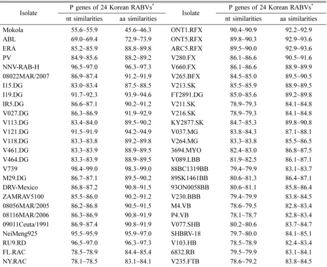
The nts of 24 Korean RABV isolates were sequenced and all data were deposited in GenBank under accession numbers JN786915-JN786938. The P genes of all 24 isolates showed 894 nts encoding 297 aas, which is quite common for RABV isolates. The similarities of nt and aa sequences of the 24 Korean RABV P genes were found to be 98.9~100% and 98.6~100%, respectively, scoring high similarities regardless of the species of origin (Table 4).
The P gene open reading frame sequences of 24 Korean RABV isolates were compared with those of previously reported RABV isolates. Among the reported RABV isolates, the P genes of the Korean RABVs showed the highest nt and aa similarities (98.4~99.0%
and 98.3~99.0%, respectively) with Korean dog RABV isolate V739 (Table 4). In addition, Korean RABVs showed high (> 95%) nt and aa similarities with NNV- RAB-H (humans, India), NeiMeng925 (raccoon dogs, China), and RU9.RD (raccoon dogs, Russia; Table 4).
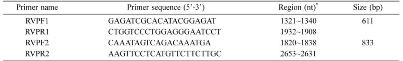
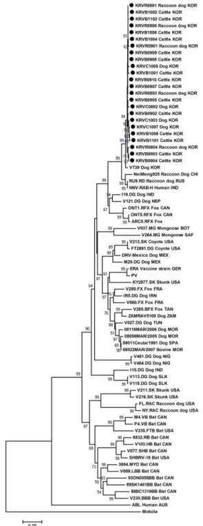
Similar to previous reports [12], the phylogenetic tree of P gene nt sequences in this study showed two large genetic clusters (Chiroptera-related and Carnivora-related RABVs; Fig. 1). All 24 Korean RABVs were included in the Carnivora-related RABV cluster and were closely clustered with one another within the same lineage regardless of species origin. In addition, Korean isolates were most closely related with the V739 isolated from Korean dogs as well as being closely related to NeiMeng925 (raccoon dogs, China), RU9.RD (raccoon
Table 2. Oligonucleotide primers for amplifying the P genes of rabies viruses in this study
Primer name Primer sequence (5’-3’) Region (nt)* Size (bp)
RVPF1 GAGATCGCACATACGGAGAT 1321~1340 611
RVPR1 CTGGTCCCTGGAGGGAATCCT 1932~1908
RVPF2 CAAATAGTCAGACAAATGA 1820~1838 833
RVPR2 AAGTTCCTCATGTTCTTCTTGC 2653~2631
*The position of primers are based on the PV strain (GenBank accession no. M13215).
dogs, Russia), and NNV-RAB-H (humans, India). These findings are consistent with the similarities in nts.
Finally, skunk (V211.SK and V216.SK) and raccoon dog (FL.RAC and NY.RAC) RABV isolates detected in the United States were included in the Chiroptera-related RABV cluster in this study, as previously reported [12]
(Fig. 1).
Two conserved domains (CD1 and CD 2) and two
variable domains (VD1 and VD2) have been identified based on aa sequence multiple alignments of RABV P gene (Fig. 2). Significant differences were not detected between the conserved and variable domains in the 24 Korean RABV P genes in this study (Fig. 2). However, specific consensus sequences of Korean RABVs lineage including V739, NeiMeng925, RU9.RD, and NNV-RAB- H in the phylogenetic tree were detected as KEPS (N/
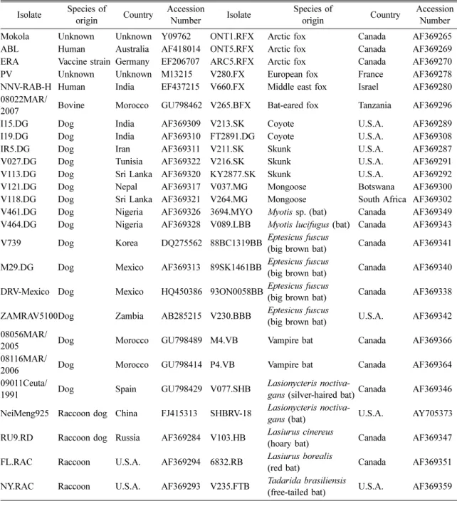
Table 3. Reference rabies virus isolates used in this study Isolate Species of
origin Country Accession
Number Isolate Species of
origin Country Accession Number
Mokola Unknown Unknown Y09762 ONT1.RFX Arctic fox Canada AF369265
ABL Human Australia AF418014 ONT5.RFX Arctic fox Canada AF369269
ERA Vaccine strain Germany EF206707 ARC5.RFX Arctic fox Canada AF369270
PV Unknown Unknown M13215 V280.FX European fox France AF369278
NNV-RAB-H Human India EF437215 V660.FX Middle east fox Israel AF369280 08022MAR/
2007 Bovine Morocco GU798462 V265.BFX Bat-eared fox Tanzania AF369296
I15.DG Dog India AF369309 V213.SK Coyote U.S.A. AF369289
I19.DG Dog India AF369310 FT2891.DG Coyote U.S.A. AF369308
IR5.DG Dog Iran AF369311 V211.SK Skunk U.S.A. AF369287
V027.DG Dog Tunisia AF369322 V216.SK Skunk U.S.A. AF369291
V113.DG Dog Sri Lanka AF369320 KY2877.SK Skunk U.S.A. AF369292
V121.DG Dog Nepal AF369317 V037.MG Mongoose Botswana AF369300
V118.DG Dog Sri Lanka AF369321 V264.MG Mongoose South Africa AF369302
V461.DG Dog Nigeria AF369326 3694.MYO Myotis sp. (bat) Canada AF369349 V464.DG Dog Nigeria AF369328 V089.LBB Myotis lucifugus (bat) Canada AF369343 V739 Dog Korea DQ275562 88BC1319BB Eptesicus fuscus
(big brown bat) Canada AF369341 M29.DG Dog Mexico AF369313 89SK1461BB Eptesicus fuscus
(big brown bat) Canada AF369340 DRV-Mexico Dog Mexico HQ450386 93ON0058BB Eptesicus fuscus
(big brown bat) Canada AF369338 ZAMRAV5100Dog Zambia AB285215 V230.BBB Eptesicus fuscus
(big brown bat) U.S.A. AF369342 08056MAR/
2005 Dog Morocco GU798489 M4.VB Vampire bat Canada AF369366
08116MAR/
2006 Dog Morocco GU798414 P4.VB Vampire bat Canada AF369364
09011Ceuta/
1991 Dog Spain GU798429 V077.SHB Lasionycteris noctiva-
gans (silver-haired bat)Canada AF369346 NeiMeng925 Raccoon dog China FJ415313 SHBRV-18 Lasionycteris noctiva-
gans (bat) U.S.A. AY705373
RU9.RD Raccoon dog Russia AF369284 V103.HB Lasiurus cinereus
(hoary bat) Canada AF369347
FL.RAC Raccoon U.S.A. AF369294 6832.RB Lasiurus borealis
(red bat) Canada AF369351
NY.RAC Raccoon U.S.A. AF369293 V235.FTB Tadarida brasiliensis
(free-tailed bat) U.S.A. AF369359
S) LGGVTTA encoded by aas 61~72 within the VD1 (Fig. 2). In addition, a specific substitution between VDI and VD2 was found in aa 91 of Korean RABV P genes (Fig. 2). This aa was substituted by Val91 in only Korean RABVs and was not found in other isolates such as PV (Leu91) and V739 (Ile91) (Fig. 2).
Phosphoacceptors related to protein kinase C (PKC) or RABV protein kinase in the P protein of the CVS strain were reported as Ser63, Ser64, Ser162, Ser210, and Ser271 [6]. All but phosphoacceptor Ser63 were retained in all Korean RABVs. Instead, Ser63 was substituted with Pro63 in all Korean RABVs (Fig. 2). The four N- terminally truncated P proteins could be synthesized due to translational initiation at internal Met20, Met53, Met69, and Met83 in the CVS strain [4, 14]. Of the four aforementioned methionine residues, Met69 was substituted
by Val69 in all Korean RABVs (Fig. 2).
The binding site for the cellular protein, cytoplasmic dynein light chain (LC8), was recently detected in P protein residues 139~151 (N’-RSSEDKSTQTTGR-C’) of the PV strain of RABV and is thought to be involved in viral RNP axoplasmic transport along the microtubule network with the D143 and Q147 residues being critical in this interaction [9, 17, 18]. Also, the consensus sequence (K/R)XTQT has been reported to be the common target-acceptor of LC8 and is a conserved LC8 binding motif [13]. This consensus sequence was located within the VD2 region and strongly conserved in all Korean RABVs examined in this study (Fig. 2). Finally, the lysine-rich motif (N’-FSKKYKF-C’), an important component of C-terminal N protein-binding, was also conserved in aas 209~215 in all Korean RABVs (Fig. 2).
Table 4. The similarities (%) of nucleotide (nt) and amino acid (aa) sequences Isolate P genes of 24 Korean RABVs*
Isolate P genes of 24 Korean RABVs* nt similarities aa similarities nt similarities aa similarities
Mokola 55.6~55.9 45.6~46.3 ONT1.RFX 90.4~90.9 92.2~92.9
ABL 69.0~69.4 72.9~73.9 ONT5.RFX 89.8~90.3 92.9~93.6
ERA 85.2~85.9 88.8~89.8 ARC5.RFX 89.5~90.0 92.9~93.6
PV 84.9~85.6 88.2~89.2 V280.FX 86.1~86.6 90.5~91.6
NNV-RAB-H 96.5~97.0 96.3~97.3 V660.FX 86.1~86.6 88.9~89.9
08022MAR/2007 86.9~87.4 91.2~91.9 V265.BFX 84.5~85.0 89.5~90.5
I15.DG 83.0~83.4 87.5~88.5 V213.SK 85.5~85.9 88.9~89.5
I19.DG 91.7~92.3 93.9~94.6 FT2891.DG 85.0~85.6 89.2~89.8
IR5.DG 86.6~87.1 90.2~91.2 V211.SK 78.9~79.3 84.1~84.8
V027.DG 86.3~86.9 91.9~92.9 V216.SK 78.9~79.3 84.1~84.8
V113.DG 83.4~84.0 89.5~90.2 KY2877.SK 84.7~85.3 89.8~90.8
V121.DG 91.5~91.9 94.2~94.9 V037.MG 83.8~84.3 87.1~88.1
V118.DG 83.3~83.8 89.2~89.8 V264.MG 83.3~83.8 85.5~86.5
V461.DG 83.3~83.9 88.9~89.5 3694.MYO 82.4~83.0 86.8~87.5
V464.DG 83.3~83.9 88.9~89.5 V089.LBB 81.9~82.5 86.1~87.1
V739 98.4~99.0 98.3~99.0 88BC1319BB 79.4~79.9 83.1~83.7
M29.DG 86.7~87.1 89.5~90.2 89SK1461BB 80.6~81.3 86.4~87.1
DRV-Mexico 86.8~87.2 90.8~91.5 93ON0058BB 80.6~81.1 85.8~86.4
ZAMRAV5100 85.5~86.0 90.2~91.2 V230.BBB 79.4~79.9 83.8~84.5
08056MAR/2005 86.2~86.8 90.5~91.5 M4.VB 78.6~79.5 82.8~83.4
08116MAR/2006 86.3~86.9 90.8~91.9 P4.VB 78.1~78.7 82.8~83.4
09011Ceuta/1991 86.9~87.4 90.8~91.9 V077.SHB 80.2~80.6 83.7~84.7
NeiMeng925 95.5~95.9 95.9~97.0 SHBRV-18 79.7~80.0 84.1~85.1
RU9.RD 96.5~97.0 96.3~97.3 V103.HB 78.5~78.9 82.4~83.4
FL.RAC 78.5~78.9 84.4~85.4 6832.RB 79.5~79.9 83.1~84.1
NY.RAC 78.1~78.5 83.1~84.1 V235.FTB 78.6~79.2 83.8~84.5
*The similarities (%) of nt and aa sequences of 24 Korean RABV P genes were found to be 98.9~100% and 98.3~100%, respectively.
Fig. 1. Phylogenetic analysis based on the ORF nucleotide sequences of P gene of 24 Korean rabies virus (RABV) isolates with other RABV isolates submitted to NCBI GenBank. Mokola virus used as an outgroup. Genetic distances were calculated using the Kimura-2 parameter model at the nucleotide level and the phylogenetic tree was constructed using the neighbor- joining method with 1,000 bootstrap replicates using MEGA 4. Bootstrap values above 50 are shown. The abbreviations of countries are as follows: AUS, Australia; BOT, Botswana; CAN, Canada; CHI, China; FRA, France; GER, Germany;
IND, India; IRN, Iran; ISR, Israel; KOR, Korea; MEX, Mexico; MOR, Morocco; NEP, Nepal; NIG, Nigeria; RUS, Russia;
SAF, South Africa; SLK, Sri Lanka; SPA, Spain; TAN, Tanzania; TUN, Tunisia; USA, United States; ZAM, Zambia.
Discussion
In the P gene, intragenotypic nt and aa similarities of 73.5% and 79.3%, respectively, were indicated while
intergenotypic similarities between RABV and other virus GTs ranged from 65.9% and 69.7% (for ABLVs) down to 56% and 45.5% (for MOKVs) [14]. Among previously reported RABV isolates, the P genes of the Fig. 2. Comparison of P protein amino acid sequences of 24 Korean RABVs with those of PV strain and V739 (Korean dog) isolate. Only different amino acids from the PV sequence are indicated and dots indicate amino acids that are in agreement with the sequence of PV strain. Boxes with CD1 and CD2 indicate conserved domains, while boxes with VD1 and VD2 delimitate variable domains. Black triangles indicate the positions of serine residues identified as phosphoacceptors in the P protein of the CVS strain. White triangles indicate the positions of the four methionines used for internal translation initiation in the CVS strain. Asterisk indicates specific amino acid substitutions of P genes of only 24 Korean RABVs found in amino acid 91 (valine). The lysine-rich motif (FSKKYKF) and LC8 binding motif (RSSEDKSTQTTGR) were shown as continuous and double-continuous underlining, respectively.
Korean RABVs showed the highest nt and aa similarities (95.5~99.0% and 95.9~99.0%, respectively) with V739 (dogs, Korea), NNV-RAB-H (humans, India), NeiMeng 925 (raccoon dogs, China), and RU9.RD (raccoon dogs, Russia) of GT I of lyssavirus.
In the phylogenetic tree, we found all 24 Korean RABV isolates to be categorized in the Carnivora-related RABV cluster. All 24 were closely clustered together within the same lineage regardless of species origin. The Korean RABV isolates of this study were most closely related to the V739 isolated from Korean dogs, as well as being close to NeiMeng925 (raccoon dogs, China), RU9.RD (raccoon dogs, Russia), and NNV-RAB-H (humans, India).
Based on the nt sequences of the N and G genes, the Korean RABV isolates were confirmed as GT I of lyssavirus, were clustered into four distinct subgroups (Gangwon I, II, III, and Gyeonggi) with high similarities. The isolates were most closely related to the NeiMeng1025B (raccoon dogs, Eastern China), and 857r (raccoon dogs, Russia) [24].
Similar to the N and G genes, P gene sequence analyses revealed that all of the Korean isolates were classified into GT I of the lyssavirus. However, unlike the N and G genes, the P gene was not separated into distinct subgroups because the 24 isolates were collected only from Gangwon-do Province. In the phylogenetic tree, P genes of Korean RABV isolates were most closely related with NeiMeng925 (raccoon dogs, China) and RU9.RD (raccoon dogs, Russia). These findings are consistent with the results found for the N and G genes.
Overall, the Korean RABV isolates show genetic closeness with RABV strains from Northeastern Asia (China and Russia) and are pathogenic in several hosts including dogs, raccoon dogs, and cattle.
Previous reports have suggested that two conserved domains (CD1 and CD2) and two variable domains (VD1 and VD2) have been identified based on aa sequence multiple alignments of the RABV P gene [14, 15]. The variable domains located on the surface structure of the P protein are believed to be involved in host/viral interactions and adaptation to the host environment, and are expected to be useful in elucidating the adaptive evolution of the rabies virus [12, 14, 16]. However, the VD and CD of the P genes among the 24 Korean RABV isolates examined in the current study were found to be highly conserved. Prior to the current findings, a commonly held belief was that variants of Korean RABV
in the variable domains occurred at low frequencies. We found that the Korean RABV maintained a conserved sequence of the P gene during transmission.
In addition, specific consensus sequences of Korean RABVs lineage including V739, NeiMeng925, RU9.RD, and NNV-RAB-H in the phylogenetic tree were detected as KEPS(N/S)LGGVTTA encoded by aas 61~72 within VD1. Moreover, a specific substitution was found in aa 91 of Korean RABV P genes between VD1 and VD2.
These findings suggest that the specific consensus sequences and aa substitution using P genes are helpful in identifying epidemiological characteristics like geo- graphic origins of Korean RABVs lineage or Korean isolates.
Of the five serine phosphoacceptors identified for the CVS strain, Ser210 and Ser271 within the PKC phos- phoacceptor were conserved in all lyssavirus GTs (1~7) [12, 14]. The phosphoacceptors of Ser64, Ser162, Ser210, and Ser271 were retained in all Korean RABVs, while Ser63 was substituted with Pro63 in Korean RABVs.
Variation in P phosphorylation patterns, especially within the N-terminal region, is predicted and might form the basis of subtle differences in viral-host interactions [14].
Further analyses of phosphorylation patterns would likely offer explanations for the function of potential variations.
Four N-terminally truncated P proteins could be synthesized due to four methionine residues located in- frame of single P protein sequences in the CVS strain.
In particular, approximately one-half of the RABVs examined, along with the GT 4 lyssaviruses, retained Met69 [4, 14]. In addition, the Met20 is retained in all lyssavirus GTs [14]. In this study, among the four methionine residues, Met69 is substituted with Val69, and Met20 is retained in all Korean RABVs. Further studies are needed to identify the biological characteristics and potential functions of truncated P proteins synthesized by methionine residues.
The binding motif for cytoplasmic dynein light chain (LC8) involving viral RNP axoplasmic transport along the microtubule network was recently found in P proteins of the PV strain [8, 17, 18]. The consensus sequence (K/R)XTQT of the P gene was thought to be a conserved motif and the common binding site of LC8 [13]. Although consensus sequence was located within the VD2 region, it is still strongly conserved in all Korean RABVs examined in the current study. In addition, the lysine-rich motif (N’-FSKKYKF-C’)
identified as an important C-terminal component for N protein binding [9], was also conserved in aas 209~215 in all Korean RABVs. These findings suggest that the functional binding sites of the P gene are conserved in Korean RABV. This conservation of GTs is clearly functionally important for Korean RABV to transport viral RNP with LC8 and to bind the N protein, which are essential for RABV pathogenicity and replication.
In conclusion, this study revealed that the P genes of Korean RABVs are genetically close to those of RABV strains of the lyssavirus GT I from Northeastern Asia, including V739 (dogs, Korea), NNV-RAB-H (humans, India), NeiMeng925 (raccoon dogs, China), and RU9.RD (raccoon dogs, Russia). The P genes of Korean RABVs showed low variability in the variable domains; they had specific consensus sequence and aa substitution capable of identifying geographic characteristics and retained specific sequences thought to be important for viral function in the P genes of RABV. This study will be helpful in understanding the genetic characteristics and viral functions of Korean RABV on the basis of the P gene.
References
1. Bourhy H, Kissi B, Tordo N. Molecular diversity of the Lyssavirus genus. Virology 1993, 194, 70-81.
2. Brzozka K, Finke S, Conzelmann KK. Identification of the rabies virus alpha/beta interferon antagonist:
phosphoprotein P interferes with phosphorylation of interferon regulatory factor 3. J Virol 2005, 79, 7673- 7681.
3. Brzozka K, Finke S, Conzelmann KK. Inhibition of interferon signaling by rabies virus phosphoprotein P:
activation-dependent binding of STAT1 and STAT2. J Virol 2006, 80, 2675-2683.
4. Chenik M, Chebli K, Blondel D. Translation initiation at alternate in-frame AUG codons in the rabies virus phosphoprotein mRNA is mediated by a ribosomal leaky scanning mechanism. J Virol 1995, 69, 707-712.
5. Gould AR, Hyatt AD, Lunt R, Kattenbelt JA, Hengstberger S, Blacksell SD. Characterisation of a novel lyssavirus isolated from Pteropid bats in Australia. Virus Res 1998, 54, 165-187.
6. Gupta AK, Blondel D, Choudhary S, Banerjee AK.
The phosphoprotein of rabies virus is phosphorylated by a unique cellular protein kinase and specific isomers
of protein kinase C. J Virol 2000, 74, 91-98.
7. Hyun BH, Lee KK, Kim IJ, Lee KW, Park HJ, Lee OS, An SH, Lee JB. Molecular epidemiology of rabies virus isolates from South Korea. Virus Res 2005, 114, 113-125.
8. Jacob Y, Badrane H, Ceccaldi PE, Tordo N.
Cytoplasmic dynein LC8 interacts with lyssavirus phosphoprotein. J Virol 2000, 74, 10217-10222.
9. Jacob Y, Real E, Tordo N. Functional interaction map of lyssavirus phosphoprotein: identification of the minimal transcription domains. J Virol 2001, 75, 9613- 9622.
10. Kim CH, Lee CG, Yoon HC, Nam HM, Park CK, Lee JC, Kang MI, Wee SH. Rabies, an emerging disease in Korea. J Vet Med B Infect Dis Vet Public Health 2006, 53, 111-115.
11. Knipe DM, Howley PM, Griffin DE, Lamb RA, Martin MA, Roizman B, Straus SE. Field Virology.
4th ed. pp. 1221-1277, Lippincott Williams & Wilkins, Philadelphia, 2001.
12. Kobayashi Y, Okuda H, Nakamura K, Sato G, Itou T, Carvalho AA, Silva MV, Mota CS, Ito FH, Sakai T. Genetic analysis of phosphoprotein and matrix protein of rabies viruses isolated in Brazil. J Vet Med Sci 2007, 69, 1145-1154.
13. Lo KW, Naisbitt S, Fan JS, Sheng M, Zhang M. The 8-kDa dynein light chain binds to its targets via a conserved (K/R)XTQT motif. J Biol Chem 2001, 276, 14059-14066.
14. Nadin-Davis SA, Abdel-Malik M, Armstrong J, Wandeler AI. Lyssavirus P gene characterisation provides insights into the phylogeny of the genus and identifies structural similarities and diversity within the encoded phosphoprotein. Virology 2002, 298, 286-305.
15. Nadin-Davis SA, Huang W, Wandeler AI.
Polymorphism of rabies viruses within the phospho- protein and matrix protein genes. Arch Virol 1997, 142, 979-992.
16. Nadin-Davis SA, Sheen M, Abdel-Malik M, Elmgren L, Armstrong J, Wandeler AI. A panel of monoclonal antibodies targeting the rabies virus phosphoprotein identifies a highly variable epitope of value for sensitive strain discrimination. J Clin Microbiol 2000, 38, 1397-1403.
17. Poisson N, Real E, Gaudin Y, Vaney MC, King S, Jacob Y, Tordo N, Blondel D. Molecular basis for the interaction between rabies virus phosphoprotein P and
the dynein light chain LC8: dissociation of dynein- binding properties and transcriptional functionality of P.
J Gen Virol 2001, 82, 2691-2696.
18. Raux H, Flamand A, Blondel D. Interaction of the rabies virus P protein with the LC8 dynein light chain.
J Virol 2000, 74, 10212-10216.
19. Smith JS, Orciari LA, Yager PA, Seidel HD, Warner CK. Epidemiologic and historical relationships among 87 rabies virus isolates as determined by limited sequence analysis. J Infect Dis 1992, 166, 296-307.
20. Talbi C, Lemey P, Suchard MA, Abdelatif E, Elharrak M, Nourlil J, Faouzi A, Echevarra JE, Vazquez Morn S, Rambaut A, Campiz N, Tatem AJ, Holmes EC, Bourhy H. Phylodynamics and human- mediated dispersal of a zoonotic virus. PLoS Pathog 2010, 6, e1001166.
21. Tamura K, Dudley J, Nei M, Kumar S. MEGA4:
Molecular Evolutionary Genetics Analysis (MEGA) software version 4.0. Mol Biol Evol 2007, 24, 1596- 1599.
22. World Health Organization (WHO). WHO expert committee on rabies. World Health Organ Tech Rep Ser 1992, 824, 1-84.
23. Yang DK, Oh YI, Cho SD, Kang HK, Lee KW, Kim YH, Song JY. Molecular identification of the vaccine strain from the inactivated rabies vaccine. J Bacteriol Virol 2011, 41, 47-54.
24. Yang DK, Park YN, Hong GS, Kang HK, Oh YI, Cho SD, Song JY. Molecular characterization of Korean rabies virus isolates. J Vet Sci 2011, 12, 57-63.
25. Yang J, Hooper DC, Wunner WH, Koprowski H, Dietzschold B, Fu ZF. The specificity of rabies virus RNA encapsidation by nucleoprotein. Virology 1998, 242, 107-117.