J Vet Sci 2016, 17(3), 369-375ㆍhttp://dx.doi.org/10.4142/jvs.2016.17.3.369
JVS
Received 15 Sep. 2015, Revised 15 Dec. 2015, Accepted 30 Dec. 2015
*Corresponding author: Tel/Fax: +86-20-8528-0283; E-mail: [email protected] Supplementary data is available at http://www.vetsci.org only.
Journal of Veterinary Scienceㆍⓒ 2016 The Korean Society of Veterinary Science. All Rights Reserved.
This is an Open Access article distributed under the terms of the Creative Commons Attribution Non-Commercial License (http://creativecommons.org/licenses/
pISSN 1229-845X eISSN 1976-555X
Molecular characterization and phylogenetic analysis of pseudorabies virus variants isolated from Guangdong province of southern China during 2013–2014
Jindai Fan
1, Xiduo Zeng
2, Guanqun Zhang
2, Qiwen Wu
1, Jianqiang Niu
2, Baoli Sun
1, Qingmei Xie
1, Jingyun Ma
1,*
1College of Animal Science, South China Agricultural University, Guangzhou 510642, China
2Guangdong Wen’s Food Co. Ltd., Xinxing 527400, China
Outbreaks of pseudorabies (PR) have occurred in southern China since late 2011, resulting in significant economic impacts on the swine industry. To identify the cause of PR outbreaks, especially among vaccinated pigs, 11 pseudorabies virus (PRV) field strains were isolated from Guangdong province during 2013–2014. Their major viral genes (gE, TK, gI, PK, gD, 11K, and 28K) were analyzed in this study.
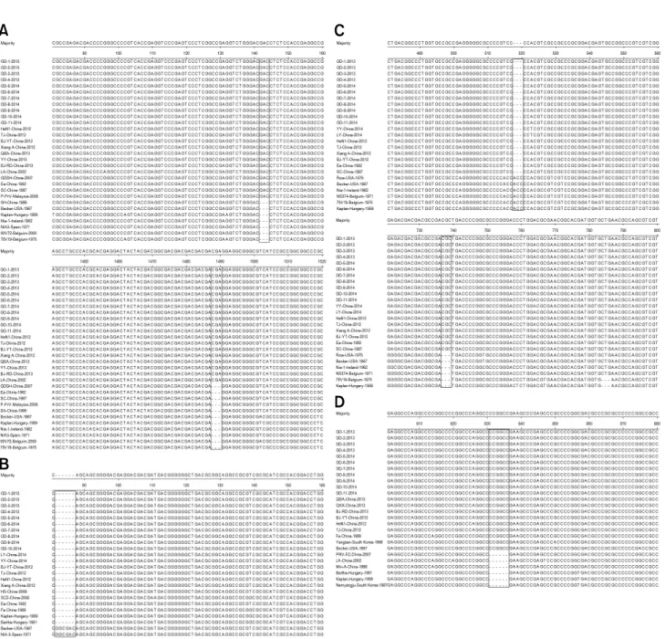
Insertions or deletions were observed in gD, gE, gI and PK genes compared with other PRV isolates from all over the world. Furthermore, sequence alignment showed that insertions in gD and gE were unique molecular characteristics of the new prevalent PRV strains in China.
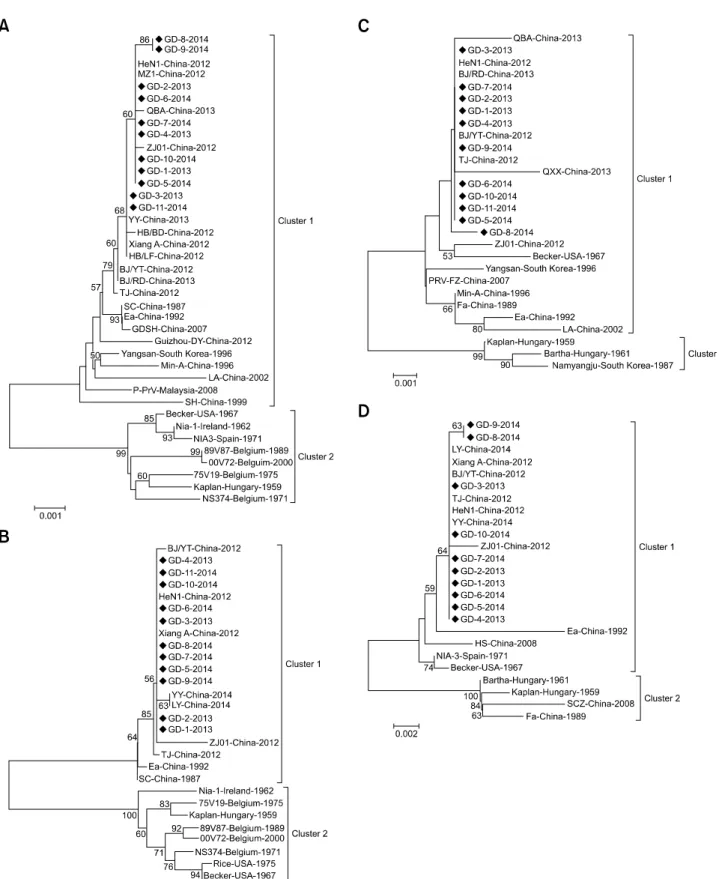
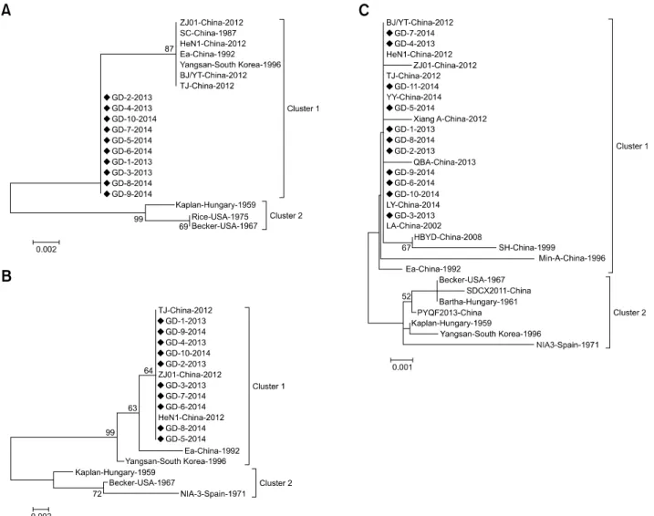
Phylogenetic analysis showed that our isolates were clustered in an independent branch together with other strains isolated from China in recent years, and that they showed a closer genetic relationship with earlier isolates from Asia. Our results suggest that these isolates are novel PRV variants with unique molecular signatures.
Keywords: molecular characterization, phylogenetic analysis, pseudorabies virus
Introduction
Pseudorabies (PR), which is also known as Aujeszky’s disease (AD), has resulted in significant impacts on the swine industry since it was first identified by Aujeszky in 1902. This disease is caused by the pseudorabies virus (PRV), a double- stranded DNA virus that belongs to the Herpesviridae family and the Alphaherpesvirinae subfamily. PRV infection is characterized by respiratory, reproductive, and neurological symptoms, varying according to the age of the pigs and the virulence of the strain. While efforts to eradicate PRV in the United States and Europe have shown great progress, pseudorabies remains an endemic problem in many countries [6,15].
Since late 2011, a few outbreaks of disease in pigs have caused large economic losses to the swine industry in China. It was been shown that the pathogenic PRV was one of the etiologic agents of the epidemic, and PRV variants were isolated from different region of China. New PRV isolates were also reported to have significantly increased virulence and
significant differences in antigenicity compared to previous isolates [1,12,17-19].
Therefore, to elucidate the cause of PR outbreaks among Bartha-K61-vaccinated pigs, clinical samples from pigs with suspected PRV infections were collected in Guangdong province from 2013 to 2014 and 11 PRV strains were isolated in our laboratory. A number of major viral genes were selected for molecular characterization of the new prevalent PRV strains.
Conventional live attenuated vaccine strains, such as Bartha
and Norden, have a natural deletion of a large fragment in the
unique short region of the genome, including the gE, gI, 11K,
and 28K genes [7,13,14]. Moreover, deletion of a large
fragment in the 28K gene was also found in vaccine strain
BUK-TK900 [3]. Based on these findings, these deletions are
the likely cause of attenuated virulence since gE, TK, gI and PK
are known to contribute greatly to the virulence of PRV in pigs
[4,5,8,17]. Furthermore, as a constituent of the viral envelope,
glycoprotein gD is essential for PRV replication and induces
neutralizing antibodies [16]. Thus, the gE, TK, gI, gD, PK, 11K
and 28K genes of the isolated strains were selected for analysis
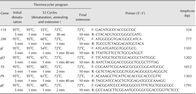
Table 1. Thermocycler programs and primers used in this study for amplification of pseudoravies virus viral genes (11K, 28K, gI, gD,
TK, PK and gE)Gene
Thermocycler program
Primer (5’-3’) Amplicon
Initial (bp) denatu- ration
32 Cycles (denaturation, annealing
and extension )
Final extension
11K