Journal of Korean Radiological Society, November, 1991
공허안 (empty sella) : MRI상 발생 반도와 그 의의
순천 향대 학교 의 과대 학 방사선 과학교실
- Abslract -
안 영·홍현숙·박재성 · 김대호 · 이혜경 · 징무찬 · 최득린 ·김기정
Empty Sella: Incidence and Significance in MR
Young Ahn, M.D., Hyun Sook Hong, M.D., Jae Sung Park‘ M.D., Dae Ho Kim, M.D., Hae Kyung Lee, M.D., Moo Chan Chung, M.D., Deuk Lin Choi, M.D., Ki Jung Kim, M.D.
Departmen o[ Radiology. College o[ Medicine‘ Soonchunhyang University
Anatomic differentiation of the sella turcica has been greatly improved by introducion of MR over the previously used pneumoencephalography and CT. Because we frequenthy encounter the empty sella in MR imaging‘ we retrospectively reviewed and classified 239 incidental empty sellae of 1004 patients who had brain MR. All cases which had initial suspicion of pituitary or hypothalamic lesions were excluded
Although headache is the most common presenting symptom. the associated disease and causes are not speC'ific The incidence of the empty sella(23.8%l is similar to that reported at autopsy cases and increased by the aging process
Therefore‘ we suggest that empty sella can be a normal variant or a degenerative change
lndex Words: Sella turcica. empty‘ 122.373 Sella turcia. MR‘ 122.1214
서 료응 」
과거에는 뇌하수체 질환 특히 뇌하수체 미세선종(pitui tary microadenoma)의 진단 및 감별을 위해 뇌하수체와 터키안(sella turica)에 대한 수많은 연구 노력이 있었고 (1-3) , 한동안 공허안(empty sella)이 증상을 수반하는 질환으로서 empty sella syndrome 으로 명명되고 사용되 어 왔다(4).
Kaufman이 empty sella 의 방사선학적 모양을 기술한 이래(1) 지금까지 뇌기술(pneumoencephalography ), 전 산화 단층촬영(CT)을 포함한 기존의 여러 방사선학적 겁 λ} 방법으로 명확한 구별이 어려웠던 뇌하수체와 터키안 의 해부학적 구조가 최근 자기공명(이하 MR 로 약함) 영 상으로 명확해지면서 두부 MR 상 뇌하수체나 시상하부의 병변과 무관하게 우발적인 empty sella를 자주 접하게 되 었다.
이에 MR상 나타난 empty sella 의 발생 빈도와 유형분 류 및 그 의의에 대하여 후향적으로 분석 하였다.
대상 및 방법
1989 년 5 월부터 1991 년 6 월까지 약 2 년간 순천향대학병 원에서 뇌하수체나 시상하부의 병변을 의심하지 않고 두 부 MR을 시행한 1034 명을 대상으로 하였다
사용된 MR 기기는 자장 0.2Tesla 영구자석형 Hitachl MRP20-2 이 고 routine pulse sequence는 saturatlOn re cvovery( SR)방법으로 반복시간(repetetion time, TR) 500msec, 에코시간(echo time, TE) 38msec의 Tl 강조 영상괴 spine echo( SE) 방법으로 TR2000msec, TE 38 및 l11msec 의 양자농도 강조영상(proton density weighted image)과 T2 강조영상을 얻었다. 시야(field of view, FOV)는 220mm 에서 260mm 로 하고 matrix number는 256X 256 으로 하였으며 절편 두께(slice thickness) 7. 5 또는 10mm로 간격은 두지 않았다.Tl 강조영상의 정중앙 시상 절편상(mid-sagittal imagel을 진단 기준으로 하고 경우 에 따라 Tl 강조영 상, 양자농도, T2 강조영 상의 관상 (coronal image) 및 횡단절편상(axial i mage) 을 분석하 이 논문은 1991년 7 월 2 일 접수하여 1991년 10 월 l일에 채택되었음.
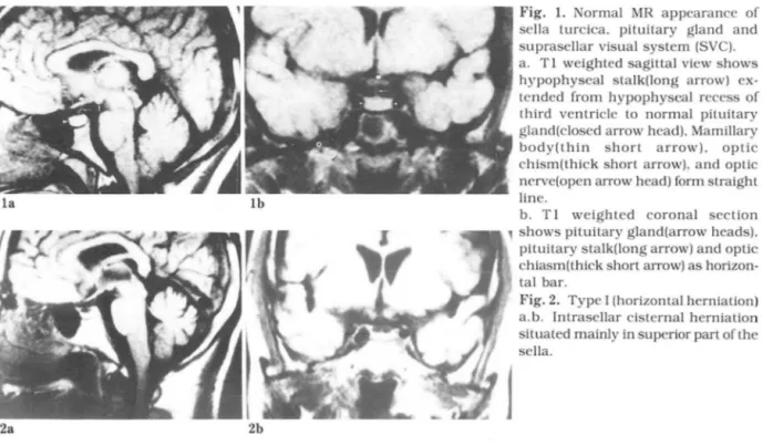
Fig. 1. Normal MR appearance of sella turcica. pituitary gland and suprasellar visual system (SVC) a. T 1 weighted sagittal 、riewshows hypophyseal stalk(long arrow) ex- tended from hypophyseal recess of third ventricle to normal pituitary gland(closed arrow head). Mamillary body(thin short arrow)‘ optic chism(thick short arrow). and optic nerve(open arrow head) form straight line
b. T 1 weighted coronal section
’
shows pituitary gland(arrow heads)‘pituitary stalk(long arrow) and optic chiasm(thick short arrow) as horizon tal bar
Fig.2. Type! (horizontal herniation) a. b. Intrasellar cisternal herniation situated mainly in superior part of the sella.
l
대한방사선의학회지 1991; 27(6): 773~777
la lb
2b 2a
Table 1. Age and Type Distribution of Empty Sellae.
Female Male
-10 11-20 21-30 31-40 41.50 51.60 61.70 71-
(%)
41 ( 0.0) 29 ( 3.5) 41 ( 4.4) 61 (26.2) 60 (38.3) 85 (48.2) 41 (58.5) 22 (50.0)
”
No I “O O O 4 7 낀 퍼
8 II
O
l i
--4
5 2 5 1
-
nu n
]
1i
gu --Qg
n。。ι
-
Type (%)
53 ( 5.7) 56 ( 54) 124 ( 7.3) 113 (22.1) 117 (239) 90 (32.2) 51 (353) 20 (30.0)
m
No 0 0 2 5m
8 6 0 Il
l 3 5 8 7 6 7 l 2
0 2 2 l 5 5 5 --- Type Age
380 (31.1)
No. Number of reviewed patients at the age group
% Incidence of empty seIla at the group
29 53 36
624 (19.4) 31
38 52
Tota!
뇌척수액 함입이 주로 sella 의 전상부를 차지하는 경 우.
3. Type
m
(Extensive herniation)뇌척수액 함입이 대부분의 sella를 차지하고 뇌하수체 가 터키안 바닥에 앓게 눌려 있는 경우로써 저자들은 임 의로 60% 이상의 뇌척수액 함입이 있을때로 정하였다.
Empty sella와 관련된 증상, 질환, 환자의 체형, 이학 적검사 소견을 알아보기 위하여 외래, 입원진료 기록부를 분석하여 후향적 연구를 하였다. 외부에서 의뢰받은 환자
는 여러 정보를 얻기 곤란하여 대상에서 제외하였다.
- 774 -
였다(Fig. 1). Artifact퉁으로 영상의 질이 좋지 않거나 정중앙 시상절편상을 얻지못한 30예는 제외하였다.
터키안 횡격막(diaphragm sella)을 일부 또는 전부 볼 수 있는 경우에 한하여, 뇌하수체에 비정상적인 종괴의 신호(signal)가 없이 sella 내에 항입된 뇌척수액( cerbro.
spinal fluid, CSF)이 보이고, 뇌하수체의 상연이 매끈한 연속상이 며 하방으로 오목 또는 수평 할때 empty sella로 보았고 다음 세가지 유형으로 분류하였다(5)(Fig. 2-4).
1. Type
1
(Horizontal herniation)뇌척수책 함입이 sella 의 전후 상부를 치지하는 경우.
2. Type II (Anterior herniation)
7~ 드프 과
두부 MR 상 분석이 가능했던 총 1004 명중 239 명 (23.
8%)에서 diaphragm sella 하방으로 뇌척수액의 함업(i n trasellar cisternal herniation)을 보인 empty sella로 나타 났다. 여자(31. 1%) 에서 남자(1 9.4%) 보다 발생빈도가
높았고(p
<
0.03), 남여 모두 30세 이전보다 이후에 발생빈도가 급격 히 증가하여 나이 가 들면서 차츰 증가하는 경 향(남여에서 각각 상관계수 0.91, 0.95)을 보였는데, 여 자에서 더 높은 증가율을 보였다(T able 1).
각각 유형별로 보면 총 239 명의 empty sella중 type 1
이 88 예(36.8%), type II 가 67% 예(28.0% ), 그리고 type
m
가 84 예 (35.2% )로 비슷 하였으나 남자에서는안 영 외 : 공 허 안 (emply sella)
partial type~ type 1 파 U 가, 여 자에 서 는 type
m
가 좀더 많았다.
증상으로는 두통이 140 예(58.6% )로 가장 많았고 오심, 구토, 현훈, 호흡 곤란, 안연통 또는 안면강직, 안과적 증상 퉁이 있었으며 이외에도 sella의 wall thinning 으로 뇌척수액 비루 (CSF rhinorrhea)가 type m 에서 l 예 있 었다. 또한 고혈압 74 예, 비만 17예, 당뇨 20 예와 동반되 어 있었다. 두개내 동반질환으로는 다발성 뇌경색 68 예,
외상 71 예, 뇌종양 퉁이 있었고 뇌수술 경력 30예, 두개 내 방사선치료 2 예퉁 이었으나 특이적인 empty sella의 원인을 찾지 못했다.
부수적인 소견으로 터키안상부 시신경계( suprasellar visual system, SVS)의 sella내 함몰이 5 예 있었는데 모 두 중년 여성에서 나타났고 type 1 에서 3 예, type m 에 서 2 예가 있었다(Fig.5). 이들 5 예는 각각 cystlcerCOSls
a b
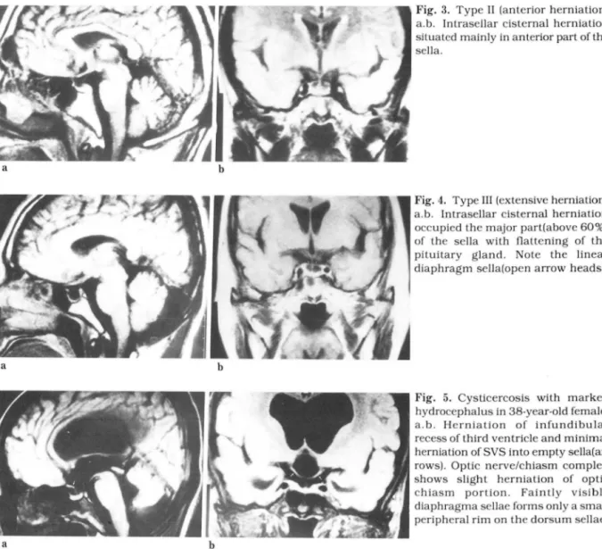
Fig. 3. Type II (anterior herniation) a.b. lntrasellar cisternal herniation situated mainly in anterior part ofthe sella
a b
Fig. 4. Type III (extensive herniation) a.b. lntrasellar cisternal herniation occupied the major part(above 60%) of the seIIa with flattening of the pituitary gland‘ Note the linear diaphragm seIIa(open arrow heads)
a b
Fig. 5. Cysticercosis with marked hydrocephalus in 38-year-old female.
a.b‘ Herniation of infundibular recess of third ventricIe and minimal herniation of SVS into empty seIIa(ar- rows). Optic nerve/chiasm complex shows slight herniation of optic chiasm portion. Faintly visible diaphragma seIIae forms only a smaII peripheral rim on the dorsum seIIae.
대한방사선의학회지 1991; 27(6) : 773~777
2 예, cerebral infarction 1예, meningioma 1 예, 그리고 나머지 1 예에서 양측성 울혈유두 및 안연통을 통반하 였다.
고 *~ E를
1951년 Busch 에 의해 부검상 diaphragm sella가 일부 불완전하거나 작은 테두리만 남게되고 sella 바닥에 뇌하 수체가 눌려있어 일견해서 볼 수 없는 경우로씨, diaphra- gm sella의 결손정도와 뇌하수체의 형태에 따라 empty sella가 해부학적으로 세분되었으나(4) MR 상 diaphragm sella 의 작은 결손여부를 일기엔 곤란하여 지자들은 비교 적 단순한 Bajraktari 분류에 따라 나누었다(5). Empty sella 와 다른 낭종성병변을 감별하기 위해 종괴 효과를 추 정힐 수 있는 infundibular tilt sign을 적용하기도 하였으 나 특이소견이 아닌 것으로 최근 보고되고 있다(6 ).
Intrasellar cisternal herniation 은 지113 뇌 실로부터 CSF 의 만성적인 박동 전달로 sella의 개조(remodelling)에 의 해 형성되띠 위치가 결정펀다고 한다(l ). 일반적으로 뇌 하수체 전엽 (anterior lobe) 이 CSF 압력에 약하여 type
n
가 많으며 type m 보다 type 1 과 typen
가 많은 것으로 알려져왔다(7). 그러나 본 연구에서는 뇌하수체 전상 부가 오목한 모양이 더 라도 MR 에 서 diaphragm sella가 전 혀 보이지 않는 경우는 정상 sella로 간주하여 empty sel la의 대 상에 서 제 외 되 어 type
1
파 typem
가 typeII
보다 더 많았다. 소수의 예에서(7예) 오히려 후상부 뇌척수액 함몰이 있었는데 이는 type 1 으로 분류하였다.과거 뇌수술이나 두개내 방사선치료를 받지않은 사람 에서 임상적으로는 특정이 없은 경우 봉완전한 diaphra- gm sella 를 통해 지주막하 공간(su barachnoid space)이 sella 내 로 획 산되 는 경 우에 primary( idiopathic) empty sella 라 하고(7,8) 뇌수숲이나 두개내 방선치료 시행후 l칼 생한 경우를 secondary empty sella 라 한다(3). 32 예의 empty sella는 두개내 수술이나 빙사선 치료를 시행했던 경우였으나 이틀 모두에서 수승 또는 벙사선치료 시행이 전의 sella를 확인할 수 없었기 때문에 secon dary empty sella 라고 히 기 어 려 웠 다. Empty sella 의 원 인 과 기 전 에 대 한 많은 연구가 있였으나 각각 주장 하는 바가 다양했 고(1, 9-11), 본 연구에서도 특별히 주목할만한 특이원인 을 찾지 못했다. 다만 심한 비만으로 나타나는 Pickwiki- an syndorome 떼 폐환기 감소(hypoventilation)로 혈증 이산화탄소(PCQ,l가 증가하여 뇌혈관 확장을 초래 두개 내압이 싱승되고 empty sella 가 유발 된다는데(1) 관연구 에서는 비만익 예가 17 예 있었고 보다많은 예에서(26 예) 장기 간 호흡곤란을 호소하는 만성 폐 색 성 폐 짐환이 동반 되었는데 고혈압, 당뇨, 울혈성 심부전등과 함께 일부 원
인으로 작용한 것으로 생각펀다.
연령은 8-86 세로 다양했 3 며 평균 띤령은 52 세이고 남 며 공히 60 대에서 가장 많았다 남여비를 보면 여성에서 좀더 많은 분포 (3 : 2) 를 보였으며 지금까지 중년여성 특 히 다산부에서 월동허 많은 것으로(83.7% ) 알려져 왔다 (12)_
전체 적 인 empty sella 의 발생 넨도는 239/1004(23.8% ) 로 Busch 분류상 type
m
B, typem
C 에샤의 27%(4), 기 타 부검예의 5.5-23.5% 경우외 거의 일치하였다 (13-15)_대부분의 대상 환자에서 외상이나 뇌경색, 뇌종양등 다 른 두개내 벙변을 먼저 의심하여 brain MR이 시행되었기 때 문에 두통, 오심 , 구토 등 동반된 증상이 empty sella 만으로 인한 증상으로 보기에는 어려웠으나 두통의 경우 높은 빈도(58.6% )를 보여 empty sella 와 연관성 이 있을 것으로 추측된다. 과거 보고된 63.2-83_ 3%(12, 16, 17) 보다 두통의 빈도가 냥은것은 외래 진료기록부에서 다수 누락된 부분이 있기 때문으로 생각된다. 일반적으로 두통 은 여성에서 더 많고 나이가 틀면서 빈도가 감소하는 경 향이 있으며 전체인구의 약 8% 정도에서 볼수 있다고 한다 08-21 ).
일부 환자에서 호르몬 검사와 뇌하수체 기능검사를 시 행하였지만 extensl ve type으로서 뇌하수체가 가의 보이 지 않는 경우에도 비정상적인 결과는 보이지 않았다.
SVS 의 sella내 함몰은 주로 secondary empty sella에 서 볼 수 있고 SVS 주위의 염증 또는 유착(수술, 방사선치 료, 뇌경색, 감염 동)이나 뇌입상승을 동반한 수두증등으 로 발생힌다고 알려졌다(3 , 22,23). Kaufman 등은 11예의 SVS 함몰중 8 예가 뇌하수체 선종, 수술, 방사선치료 등 의 분명한 원인을 알 수 있는 secondary empty sel!a 였고, 나머지 3 예는 특별한 윈인 없이 hydrocephalus를 동반하 였는데 여기서 뇌압 상승을 하나의 원인기전으로 본다면 이 3예를 secondary empty sella로 포함시키거나 따로 독립된 범주로 분리할 수 있을 것이라 하였다(23). 저자들의 경우 SVS 의 함몰 5 예 중 cysticercosis 2예 와 cerebral infarction 1 예 는 secondary empty sella로 추정 되 며 cysticercosis 2예 중 l 예와 meningioma 1 예에서 심한 hydrocephalus를 동반하 였다. 나머지 1예는 특별한 원인을 찾을 수 없었고 hydroce- phalus도 나타나지 않았다. SVS 함몰에서도 안과적 증상과 별 연관성이 없는 것으로 보고되고 있으며(23) 본 연구에서 도 안과적 증상은 동반하지 않았다.
결콘적으로 보다 발전된 영싱기기인 ìvlR 을 이용하여 empty sella 의 발생빈도(23.8% )가 지금까지 보고된 부김 결과와 거의 일치하였다 많은 예(58.6% )에서 두통을 동 만하였지만 특이적인 원인이나 동반펀 질환과의 연관성
- 776-
은 찾을 수 없었으며, 연령 증가에 따라 발생 빈도가 증 가하는 것으로 보아 empty sella는 정상변이 또는 퇴챙성 변화의 한 형태로 사료된다.
참고문런
1. Kaufman B. The “empty sella“ turcica : A manifesta tion of the intrascllar subarachnoid space. Radiology 1968:90:931-941
2. Gabriele OF. The empty sella syndrome‘ AJR 1968:104: 168-170
3. Lce WM. Adams JE. The cmpty sella syndrome. J Neurosurg 1968:28:351-356
4. Busch W. Die morphologie der sella turcica und ihre beziehungen zur hypophyse. Virchows Arch Path Anat 1951‘320:437 -458
5. Bajraktari X‘ Grepe A. Goulatia RK. Pneu- moencephalographic changes with intrasellar cister- nal herniation(Primary empty sella). Neuroradiology 1977‘ 13:97-105
6. Ahmadi H‘ Larsson EM. Jinkins JR. Normal pituitary gland : Coronal MR imaging of infundibular tilt. Radiology 1990: 177:389-392
7. Weiss SR. Raskind R. Non-neoplastic intrasellar cysts. Int surg 1968:51 :282-286
8. Neelon FA‘ Goree JA. Lebovitz HE. The prirr녕 ry
empty sella Clinical and radiographic characteristics and endocrine function Medicine(BaltimoreJ 1973:52:73-92
9. Raiti S‘ Albrick MJ‘ Maclaren NK‘ Gabricle OF. Chou SM. Empty sella syndrome secondary to intrasellar cyst in adolescence. Am J Dis Child 1976‘130: 1009-1012
10. Obrador S. The empty sella and some related syn- dromes. J Neurosurg 1972:36:162-168
11 김보현, 장기현, 한문희, 한만청, 최길수, 김주완. 터키 안내 낭종의 자발성 파열의 CT 와 MR 소견. 대한방사 선의학회지 1989; 25 : 212-216.
12. Jordan RM. Kendall JW. Kerber CW. The primary
안 영 오1: 공 허 안 (empty 8ella)
empty sella syndrome : Analysis of the clinical characteristics. radiographic features. pituitary function and cerebrospinal nuid adenohypophyseal hormone concentrations. Arn J Med 977:62:569-580 13. Bcrgland RM‘ Ray BS. Torack RM. Anatomical varia-
tions in the pituitary gland and adjacent structures in 225 human autopsy cases. J Neurosurg 1968‘28’93-99
14. Kaufman B. Chamberlin WB Jr. The ubiquitous empty sella turcica. Acta Radiol Diagn 1972: 13:413-425
15 정인혁, 김동익, 서원석, 서정호. 한국 성인 사체 및 전 산화단충황영상에서 뇌하수체와 안격악에 대한 형태계 측학적인 연구 체질인류학회지 1988; 1 : 53-63_
16. Berke JP‘ Buxton LF. Kokmen E. The ‘empty‘ sella Neurology 1975:25: 1137 -1143
17. Foley KM‘ Posner JB. Does pseudotumor cerebri cause the empty sella syndrome? Neurology 1975:25‘565-569
18. Solomon GD. Kunkel RS. Frame J. Demographics of headache in elderly patients. Headache
1990:30:273-276
19. Cook NR. Evans DA. Funkenstein HH. et a1. Cor- relates of headache in a population-based cohort of elderly. Arch Neurol 1989:46:1338-1344
20. Waters WE‘ The Pontypridd headache survey.
Headache 1974:14‘81-90
21. Nikiforow R. Hokkanen E. An epidemiological study of headache in an urban and a rural population in northern Finland. Headache 1978:18:137-145 22. Laws ER. Trautman JC. Hollenhorst RW
Transphenoidal decompression of the optic nerve and chiasm : Visual results in 62 patients. J Neurosurg 1977:46:717-722
23. Kaufman B. Tomsak RL. Kaufman BA. et a1. Her- niation of the suprasellar visual system and third ventricle into empty sellae : Morphologic and clinical considerations. AJR 1989: 152:597 -608