Isolation of the Protease-producing Yeast Pichia anomala CO-1 and Characterization of Its Extracellular Neutral Protease
Ji Yeon Kim*
Liberal Arts College, Inje University, Gimhae-si, Gyeongsangnam-do 50834, Korea Received August 16, 2019 /Revised October 4, 2019 /Accepted October 5, 2019
From a sample of bamboo byproduct, the protease-producing yeast strain CO-1 was newly isolated.
Strain CO-1 is spherical to ovoid in shape and measures 3.1–4.0×3.8–4.4 μm. For the growth of strain CO-1, the optimal temperature and initial pH were 30°C and 4.0, respectively. The strain was able to grow in 0.0–15.0%(w/v) NaCl and 0.0-9.0%(v/v) ethanol. Based on a phylogenetic analysis of its 18S rDNA sequences, strain CO-1 was identified as Pichia anomala. The extracellular protease produced by P. anomala CO-1 was partially purified by ammonium sulfate precipitation, which re- sulted in a 14.6-fold purification and a yield of 7.2%. The molecular mass of the protease was re- corded as approximately 30 kDa via zymogram. The protease activity reached its maximum when 1.0%(w/v) CMC was used as the carbon source, 1.0%(w/v) yeast extract was used as the nitrogen source, and 0.3%(w/v) MnSO
4was used as the mineral source. The protease revealed the highest ac- tivity at pH 7.0 and 30°C. This enzyme maintained more than 75% of its stability at a pH range of 4.0-10.0. After heating at 65℃ for 1 hr, the neutral protease registered at 60% of its original activity.
The protease production coincided with growth and attained a maximal level during the post-ex- ponential phase.
Key words : Characterization, extracellular neutral protease, identification, optimization, Pichia anomala
*Corresponding author
*Tel : +82-55-320-3737, Fax : +82-55-339-3734
*E-mail : [email protected]
This is an Open-Access article distributed under the terms of the Creative Commons Attribution Non-Commercial License (http://creativecommons.org/licenses/by-nc/3.0) which permits unrestricted non-commercial use, distribution, and reproduction in any medium, provided the original work is properly cited.
Journal of Life Science 2019 Vol. 29. No. 10. 1126~1135 DOI : https://doi.org/10.5352/JLS.2019.29.10.1126
Introduction
Protease is one of the important commercial enzymes, ac- counting for approximately 60% of the total market sales in the world. It has numerous applications in detergents, food processing, medical applications, and leather process- ing [26]. In addition, protease is important in biocontrol mechanisms, including mycoparasitism, competition, and antibiosis [9]. On the basis of pH range in which enzyme activity is optimum, protease is categorized as acid, neutral and alkaline protease. Unlike other proteases, neutral pro- tease hydrolyzes hydrophobic amino acid bonds at neutral pH, thereby decreasing the bitter taste of protein hydro- lysates. Thus, neutral protease process has been discovered to be efficient in food industry [29, 33]. Although many dif- ferent proteases have been characterized until now, some of them are not sufficient to meet most of the industrial ap-
plications [17].
Protease is obtained from all organisms. Especially, pro- tease-producing microorganisms are the most appropriate resources for use in industrial production and can be com- mercially exploited. The microorganisms can easily be culti- vated in a large scale, once their biochemical and physical characteristics and physiological functions are established [26]. Extracellular protease from different microorganisms including bacteria, mold, and yeast has been characterized [2, 9, 24]. Despite reports on yeast protease, comparatively little is known about the characteristics of extracellular protease. Nevertheless, some yeast has been studied as pro- tease producers for potential industrial development, be- cause they may be suitable for the biological control of post-harvest diseases of grains and fruits, and as bioremedia- tion agents [5]. Candida humicola [28], Yarrowia lipolytica [13], and Sporidiobolus ruineniae [18] were studied with regard to variability of the characteristics of extracellular protease, ac- cording to the medium composition, pH, and temperature.
The aim of the present work was to identify industrially interesting microorganism for biological control applications.
Accordingly, I isolated and identified a yeast strain from
bamboo-by product that produces extracellular enzymes. In
addition, characteristics of the obtained crude enzyme were
investigated.
Materials and Methods
Growth medium and culture condition
Cultivations were carried out in YM medium composed of 0.3% (w/v) yeast extract (BioShop, USA), 0.3% (w/v) malt extract (BioShop), 0.5% (w/v) Bacto-peptone (Difco), and 1.0% (w/v) dextrose (Merck, Darmstadt, Germany). To avoid bacterial growth, 100 μg/ml penicillin-streptomycin solution (HyClone; Thermo Fisher Scientific, Logan, UT) was added.
To determine the culture conditions, the yeast cells was in- oculated in YM broth and cultivated at 30°C with agitation (200 rpm) for 18-20 hr. Then, the culture suspension (1.0%, v/v) was transferred into an YM broth and incubated stat- ically at 20-40°C, and pH of 2.0-12.0. Salt and ethanol toler- ances were evaluated at 30°C using YM broth added with 0-15.0% (w/v) NaCl, and 0-10.0% (v/v) ethanol, respectively.
The growth of yeast cells was checked by determination of optical density at 600 nm (OD
600) using a spectrophotometer (BioPhotometer 6131, Eppendorf AG, Eppendorf, Germany).
Isolation of yeast strain from bamboo by-products The bamboo by-product sample was obtained from AGRO KOREA, Guri, Korea. Ninety milliliters of sterile 0.85%(w/
v) NaCl was added to 10 g of the bamboo by-product sample and homogenized for 2 min. The homogenate was diluted with 0.85% NaCl (10
1-10
8cells). The suspension was spread onto YM agar and cultivated for 24 hr at 30°C. Colonies were picked randomly, through their different color and shape, and pure cultured by subsequent streaking on YM agar.
Screening of extracellular protease-producing yeast strain
A direct agar plate assay was used for ascertainment of extracellular protease production. Isolated strains were in- oculated in YM agar containing skim milk (1.0%, w/v) [2]
and cultivated for 24 hr at 30℃. Protease production was detected by formation of transparent halos surrounding the colonies on the plates. Positive yeast strains were used to the assay of protease activity.
Characteristics of a newly isolated yeast strain Morphological, cultural, and biochemical characteristics of the bamboo by-product isolate were determined as pre- viously described [3]. Morphology of cells recovered from YM medium was determined using a phase-contrast micro- scope (Model BX51, Olympus, Tokyo, Japan). Biochemical
characteristics of yeast isolates were obtained according to the VITEK 2 system (VITEK 2 Compact 60 apparatus, bio- Mérieux, Hazelwood, MO) and the method explained by Barnett et al. [3].
PCR and DNA sequencing of the 18S rRNA gene Total genomic DNA of the strain was isolated using the genomic DNA extraction kit (GeneAll
TM, GeneAll Biotech- nology, Seoul, Korea). Polymerase chain reaction (PCR) was performed as previously described [10, 34]. The primers NS1 and NS8 were used. The PCR mixture consisted of 10 μl of 2× Prime Taq Premix Solution (GeNet Bio, Cheonan, Korea) containing 1 U/μl Prime Taq DNA polymerase, 0.5 mM dNTP, 4.5 mM MgCl
2, 0.1% gelatin, 1 μl of 10 pM pri- mers, and 1 μl of template, prepared to a final reaction vol- ume of 20 μl. PCR was completed with a total of 30 cycles in the Thermal Cycler (Model PC708 Program Temp Control System, ASTEC, Tokyo, Japan). The amplification program was proceed with an initial denaturation (93°C) for 3 min followed by 30 cycles of denaturation (93℃) for 1 min, pri- mer annealing (57°C) for 1 min, and extension (72℃) for 2 min. PCR was finished with a final extension (72℃) for 5 min and the amplified product was cooled (4℃). Sequencing of amplified DNA fragment was contractually accomplished by Solgent (Daejeon, Korea).
Phylogenetic analysis
The collected DNA sequences were applied for the BLAST gene homology search with the publicly available 18S rDNA sequences. Identification was assigned to the generic level [1]. The 18S rDNA sequences of the isolate were aligned with the sequences of related species obtained from GenBank [31]
using the CLUSTAL X multiple sequence alignment pro- gram. Phylogenetic analysis was achieved using PHYLIP software [11] and a phylogenetic tree was built by the neigh- bor-joining method using TreeView software [25].
GenBank accession number
The partial 18S rDNA sequence of the isolate has been deposited in the DDBJ/EMBL/GenBank nucleotide sequence database with the accession numbers EF427893.
Assay of protease activity
Protease activity with casein as the substrate was estab-
lished by the modified method of Hagihara et al. [15]. The
enzyme solution (1.0 ml) was supplemented to an equal vol-
ume of 0.6% Hammarsten casein (BDH Biochemical, Poole, UK) in 0.1 M phosphate buffer (pH 7.0) and incubated for 10 min at 30℃. The reaction was then stopped by supple- ment of 5 ml of trichloroacetic acid (TCA) mixture (0.11 M TCA, 0.33 M acetic acid, 0.22 M sodium acetate). The mixture was kept at 20-25℃ for 30 min, and the precipitate was re- moved by centrifugation (10,000 rpm) for 5 min. Then, opti- cal density of the recovered supernatant was determined at 280 nm. The blank was run in the same manner, except that the enzyme solution was blended with the TCA before addi- tion of the substrate. One unit of protease activity was de- fined as the amount of enzyme that liberated a digestion product not precipitated by TCA equivalent to 1 μg/ml/
min of tyrosine under the assay conditions. The protein con- tent of the enzyme preparation was estimated by Lowry et al. [23].
Partial purification of protease
The isolate CO-1 was incubated in optimum medium [1.0% (w/v) carboxymethyl cellulose (CMC), 1.0% (w/v) yeast extract, and 0.3% (w/v) MnSO
4, pH 7.0] for 24 hr at 30
oC. The culture broth was centrifuged (12,000 rpm) for 10 min at 4
oC. The enzyme from the cell-free supernatant was precipitated by ammonium sulfate (up to 80% saturation) and kept overnight at 4
oC. The precipitate was collected by centrifugation at 10,000 rpm for 30 min, dissolved in 0.1 M phosphate buffer (pH 7.0), and then dialyzed against the same buffer to eliminate residual ammonium sulfate. This dialyzed soluble fraction was used for protease activity.
Zymogram (activity staining)
Sodium dodecyl sulfate-polyacrylamide gel electrophoresis (SDS-PAGE) in 12% (w/v) polyacrylamide gel containing 1%
gelatin (w/v) was conducted as described by Laemmli [21].
After electrophoresis was completed, zymogram was com- pleted as explained by Bernal et al. [4] with a minor modification. The gel with separated proteins was washed for 30 min at 4℃ in 0.1 M phosphate buffer (pH 7.0) contain- ing 2.5% Triton X-100. Then, the washed gel was soaked at 30°C for 1 hr in pre-warmed 0.1 M phosphate buffer (pH 7.0) and was stained with Coomassie Brilliant Blue R-250 (Sigma, USA). Areas of protease activity occurred as non-stained transparent bands on a dark blue background.
The molecular mass of the proteases was recorded by com- paring the bands of standard molecular mass markers for 220, 97, 66, 45, 30, 20.1 and 14.3 kDa (Amersham Biosciences,
UK).
Effect of various carbon, nitrogen and mineral sources To investigate the effects of various carbon sources on the protease activity, dextrose in YM broth was individually re- placed with 1.0% (w/v) each carbon sources as shown in the Table 3. The effects of various nitrogen sources were ex- amined by individually supplementing a broth containing 1.0% (w/v) CMC with 1.0% (w/v) complex nitrogen sources and 1.0% (w/v) inorganic nitrogen sources (Table 4). To study the effect of various mineral sources, 0.3% (w/v) each mineral sources were individually added to 1.0% (w/v) each CMC and yeast extract broth (Table 5).
Effect of different pH and temperature on protease activity and stability
Conditions for obtaining maximal protease activity were studied by assay of the enzyme at different pH and temper- atures, and the relative enzyme activities were calculated.
Assays were repeated three times, and values presented are averages of the three experiments. For determination of the effect of pH on protease activity, the pH of the reaction mix- ture containing buffer and 0.6% (w/v) Hammarsten casein was varied over the range of 3.0-11.0. Buffers used were 0.05 M citrate buffer (pH 3.0-5.0), 0.1 M phosphate buffer (pH 6.0-8.0), and 0.1 M sodium bicarbonate buffer (pH 9.0-11.0).
pH stability was tested by 24 hr pre-incubation of the en- zyme solutions in the absence of the substrate at different pH values ranging from 3.0 to 11.0 at 4°C. The residual activ- ities (%) of enzyme were determined immediately after this treatment using the standard method described above. The effect of temperature on protease activity was measured by incubation of the reaction mixture at temperatures ranging from 20 to 75℃ in 0.1 M phosphate buffer (pH 7.0) for 10 min. To ascertain the temperature stability, the enzyme re- action was carried out at optimal condition after pre-incubat- ing of the enzyme solution at various temperatures (30-70℃) for 1 hr. And then remaining activity (%) of the enzyme was assayed immediately.
Time course of growth and protease production
The growth and protease production was also studied un-
der the previously determined optimal conditions. The cul-
ture broth was inoculated in optimum medium and in-
cubated at 30℃ with agitation (200 rpm). Culture super-
natants were recovered periodically, and cell growth and
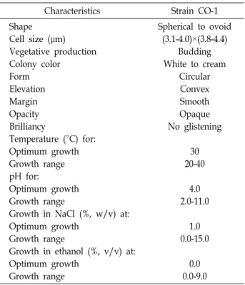
Table 1. Morphological and cultural characteristics of a newly isolated strain CO-1
Characteristics Strain CO-1
Shape Cell size (μm) Vegetative production Colony color
Form Elevation Margin Opacity Brilliancy
Temperature (°C) for:
Optimum growth Growth range pH for:
Optimum growth Growth range
Growth in NaCl (%, w/v) at:
Optimum growth Growth range
Growth in ethanol (%, v/v) at:
Optimum growth Growth range
Spherical to ovoid (3.1-4.0)×(3.8-4.4)
Budding White to cream
Circular Convex Smooth Opaque No glistening
30 20-40
4.0 2.0-11.0
1.0 0.0-15.0
0.0 0.0-9.0
protease activity were monitored as described above.
Results and Discussion
Isolation of protease-producing yeast strain Two yeast strains were isolated from bamboo by-product samples obtained in Guri, Korea, and screened according to their ability to produce extracellular protease based on test- ing for formation of transparent halos. Growth was evident only on YM agar plates supplemented with 100 μg/ml of penicillin-streptomycin after approximately 24 hr of in- cubation. One strain that produced protease (strain CO-1) was selected.
Characteristics of a newly isolated strain CO-1 The isolated CO-1 was characterized morphologically, culturally, and biochemically, as previously described [3].
Morphological and cultural properties of the strain CO-1 are shown in Table 1. After incubation for 24 hr at 30°C on YM agar, all developed colonies displayed a smooth margin and convex elevation, and were opaque, non-glistening, circular and white-to-cream colored. Microscopic examination re- vealed spherical- to ovoid-shaped cells, measuring 3.1-4.0×
3.8-4.4 μm, which occurred singly or with buds. The strain CO-1 grew at temperatures range of 20-40℃, with optimum
growth evident at 30℃. This strain grew in a wide pH range of 2.0-11.0 with optimum growth occurring at 4.0. No growth was detected at pH 12.0. Therefore, in subsequent experiments, the pH of the medium was retained at 4.0.
CO-1 grew in a NaCl concentration ranging up to 15.0%
(w/v), and optimally at 1.0(w/v). In addition, it grew in the presence of 0-9.0%(v/v) ethanol, but showed the greatest exuberance in the absence of ethanol.
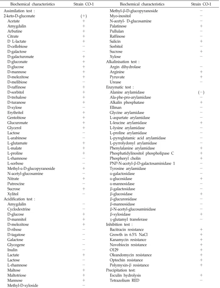
The VITEK system was used for further characterization of the isolated. Biochemical characteristics of CO-1 are dis- played in Table 2. CO-1 was positive in the assimilation test for acetate, amygdalin, arbutine, citrate, lactate, galacturo- nate, gluconate, glucose, mannose, melezitose, raffinose, sor- bitol, trehalose, turanose, erythritol, gentobiose, glucuronate, glycerol, glutamate, malate, proline, methyl-α-D-glucopyr- anoside, nitrate, and sucrose. Positive acidification results were obtained for glucose, mannitol, melezitose, lactate, mal- tose, maltotriose, mannose, palatinose, salicin, and sucrose.
Positive results were also evident in the alkalinization test for arginine and pyruvate, enzymatic test for alkalin pho- phatase, leucine arylamidase, phenylalanine arylamidase, phosphatidylinositol phospholipase C, phosphoryl cholin, α- glucosidase, β-glucosidase, and β-xylosidase, and precip- itation test for esculin hydrolysis. In particular, all inhibition tests showed positive results. The characteristics of P. anom- ala CO-1 differed slightly from those of P. anomala J121 [12], and P. anomala ATCC96603 [14], respectively.
Phylogenetic analysis of a newly isolated strain CO-1
The 18S rDNA sequences analysis was achieved for iden- tification of CO-1. The 1,682 bp sequences obtained were aligned with all of the presently available sequences in the GenBank database. The sequence of CO-1 was highly homol- ogous to P. anomala. The partial 18S rDNA sequence of CO-1 exhibited 99% identity with corresponding sequences of P.
anomala (DQ520880), P. anomala GK1 (AY218895), and P.
anomala (AB054562). A phylogenetic tree was formed based
on the 18S rDNA sequences in order to indicate the com-
parative relationship between CO-1 and other related organ-
isms. Fig. 1 indicates the phylogenetic position of CO-1
based on the almost full-length 18S rDNA sequences. CO-1
belonged to the genus Pichia, and was most closely con-
nected to Pichia anomala. Thus, based on morphological, cul-
tural, biochemical characteristics, and phylogenetic analysis,
the yeast strain CO-1 was identified as P. anomala, and then
Table 2. Biochemical characteristics of a newly isolated strain CO-1
Biochemical characteristics Strain CO-1 Biochemical characteristics Strain CO-1
Assimilation test : 2-keto-D-gluconate Acetate
Amygdalin Arbutine
Citrate D L-lactate
D-cellobiose D-galactose D-galacturonate D-gluconate D-glucose D-mannose D-melezitose D-melibiose D-raffinose D-sorbitol D-trehalose D-turanose D-xylose Erythritol Gentobiose Glucuronate Glycerol Lactose L-arabinose L-glutamate L-malate L-proline
L-rhamnose L-sorbose Methyl-α-D-glucopyranoside N-acetyl-glucosamine Nitrate
Putrescine Sucrose Xylitol
Acidification test : Amygdalin Cyclodextrine D-glucose D-mannitol D-melezitose D-ribose D-tagatose Galactose Glycogene Inulin Lactate Lactose L-rhamnose Maltose Maltotriose Mannose
Methyl-D-xyloside
(+)
+
+
+
+
+
-
-
+
+
+
+
+
-
+
+
+
+
-
+
+
+
+
-
-
+
+
+
-
-
+
-
+
-
+
-
-
-
+
+
+
-
-
-
-
-
+
-
-
+
+
+
-
Methyl-β-D-glucopyranoside Myo-inositol
N-acetyl- D-glucosamine Palatinose
Pullulan Raffinose Salicin Sorbitol Sucrose Xylose
Alkalinisation test : Argin dihydrolase Arginine
Pyruvate Urease Enzymatic test : Alanine arylamidase Ala-phe-pro-arylamidase Alkalin phosphatase Ellman
Glycine arylamidase L-aspartate arylamidase L-leucine arylamidase L-lysine arylamidase L-proline arylamidase
L-pyroglutamic acid arylamidase L-pyrrolydonyl arylamidase Phenylalanine arylamidase
Phosphatidylinositol phospholipase C Phosphoryl cholin
PNP-N-acetyl-β-D-galactosaminidase 1 Tyrosine arylamidase
α-galactosidase α-glucosidase α-mannosidase β-galactosidase β-glucosidase β-glucuronidase β-mannosidase
β-N-acetyl-glucosaminidase β-xylosidase
γ-glutamyl transferase Inhibition test : Bacitracin resistance Growth in 6.5% NaCl Kanamycin resistance Novobiocin resistance O129
Oleandomycin resistance Optochin resistance Polymyxin-β resistance Precipitation test:
Esculin hydrolysis Tetrazolium RED
-
-
-
+
-
-
+
-
+
-
-
+
+
-
(-)
-
+
-
-
-
+
-
-
-
-
+
+
+
-
-
-
+
-
-
+
-
-
-
+
-
+
+
+
+
+
+
+
+
+
-
VITEK 2 system was used. +, positive; -, negative; (+), weakly positive; (-), weakly negative.
Fig. 1. Phylogenetic position of a newly isolated strain CO-1 based on 18S rDNA sequences GenBank accession num- bers are given in parentheses. Scale bar corresponds to 0.001 subscriptions per nucleotide position. Numbers at nodes reveal levels of bootstrap support (%) determined from 100 resampled data.
Fig. 2. SDS-PAGE and zymogram of the protease produced by P. anomala CO-1 Lane 1: molecular mass markers, lane 2, 3, 5 and 7: crude enzyme (culture supernatant), lane 4 and 6: partially purified protease (ammonium sulfate fractionation), lane 8: zymogram of crude enzyme, lane 9: zymogram of partially purified enzyme.
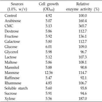
Table 3. Effects of various carbon sources on the growth and activity of the extracellular protease produced by P.
anomala CO-1 Sources (1.0%, w/v)
Cell growth (OD600)
Relative enzyme activity (%) Control
Arabinose CMC Dextrose Fructose Galactose Glucose Glycerol Lactose Maltose Mannitol Mannose Raffinose Rhamnose Soluble starch Sucrose Xylose
4.92 5.07 5.13 5.86 5.92 5.80 6.01 5.98 5.12 5.86 5.88 12.56
5.47 4.85 5.60 5.91 5.56
100.0 160.4 365.7 112.7 136.1 112.7 109.0 96.7 107.3 108.1 90.8 114.7 92.1 106.7 93.8 94.6 187.0
we named P. anomala CO-1.
P. anomala is one of the interesting yeast species, and has shown potential for exploitation in environmental bio- remediation, food fermentation, therapeutic protein pro- duction, and biofuel production [32]. Especially, anti-micro- bial activities of P. anomala make it an appropriate organism for biological control in the agricultural and food sectors [5].
Partial purification and zymogram of the extracellular protease
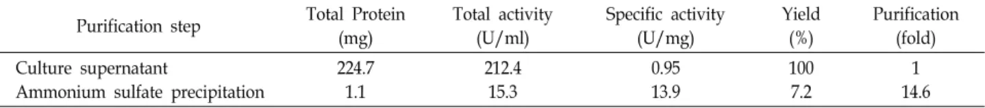
Partial purification of the extracellular protease from P.
anomala CO-1 was carried out by ammonium sulfate precip- itation. Approximately 14.6-fold purification of the crude en- zyme was accomplished with a recovery of 7.2% and specific activity of 13.9 U/mg proteins (Table 6). The protease activ- ity was visualized by zymogram using gelatin as a protease substrate. Zymogram revealed a clear hydrolysis band against dark background for both crude and partial purified enzymes at equivalent positions in SDS-PAGE (Fig. 2). The enzyme indicated a single band equivalent to an obvious molecular mass of 30 kDa. This value is slightly smaller than that studied for some protease from other yeast [24, 27, 28].
Effect of culture conditions on growth and protease activity
The growth and protease activity can also be influenced
by the medium composition (carbon, nitrogen, and mineral
sources). P. anomala CO-1 could utilize different carbon sour-
ces for growth (Table 3). The maximum effect on growth
was observed with mannose. CMC was the significant car-
bon source for protease activity, followed by xylose. On the
other hand, the growth of CO-1 increased in the presence
of glycerol, mannitol, raffinose, soluble starch or sucrose as
individual carbon sources, the activity of the target protease
decreased. The varying results depending on the carbon
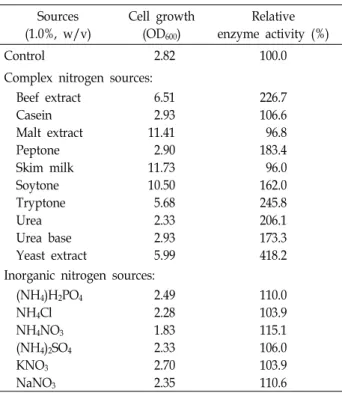
Table 4. Effects of various nitrogen sources on the growth and activity of the extracellular protease produced by P.
anomala CO-1 Sources (1.0%, w/v)
Cell growth (OD600)
Relative enzyme activity (%)
Control 2.82 100.0
Complex nitrogen sources:
Beef extract Casein Malt extract Peptone Skim milk Soytone Tryptone Urea Urea base Yeast extract
6.51 2.93 11.41 2.90 11.73 10.50 5.68 2.33 2.93 5.99
226.7 106.6 96.8 183.4 96.0 162.0 245.8 206.1 173.3 418.2 Inorganic nitrogen sources:
(NH4)H2PO4
NH4Cl NH4NO3
(NH4)2SO4
KNO3
NaNO3
2.49 2.28 1.83 2.33 2.70 2.35
110.0 103.9 115.1 106.0 103.9 110.6
Table 5. Effects of various mineral sources on the growth and activity of the extracellular protease produced by P.
anomala CO-1 Mineral sources
(0.3%, w/v)
Cell growth (OD600)
Relative enzyme activity (%) Control
CaCl2
CaCO3
CoCl2
CuSO4
FeSO4
K2HPO4
KCl KH2PO4
MgSO4
MnSO4
NaCl ZnCl2
2.89 2.82 5.51 1.20 1.39 2.46 2.79 2.74 2.92 2.82 2.10 2.87 1.40
100.0 129.4 148.9 111.7 146.4 77.1 95.5 97.9 98.3 96.0 222.3 98.1 131.3
Fig. 3. Effect of pH on the extracellular neutral protease activity and stability Enzyme activity was recorded at the in- dicated pH (reaction solution) for 10 min at 30℃. The maximum activity of the enzyme was taken as 100%. The error bars represent standard deviations of the means of the three experiments. Symbols: ○, protease activity;
●, protease stability.
source are consistent with a previous description that altered carbon sources have different effects on enzyme activity by various strains [6, 18]. Chi et al. [7] found that soluble starch, and corn starch supported protease activity while sucrose decreased activity. The influence of particular nitrogen sour- ces on protease activity varies according to organism [18, 20]. The influences of various complex and inorganic nitro- gen sources on the growth and protease activity are pre- sented in Table 4. The highest growth was found in the pres- ence of skim milk. Yeast extract had apparent effect on the activity of the extracellular protease. This result is consistent with S. ruineniae CO-3 [18]. However, malt extract and skim milk revealed decrease in the protease activity. The use of inorganic nitrogen sources had no outstanding influence on the protease activity. Chi et al. [7] found that NaNo
3could be stimulatory for alkaline protease activity by Aureobasidium pullulans. The effects of various mineral sources on the growth of the isolate and the protease activity of the crude enzyme are summarized in Table 5. Among the different mineral sources investigated, CaCO
3had the excellent effect on the growth. MnSO
4best enhanced the enzyme activity, followed by CaCO
3, CuSO
4. KH
2PO
4supported the growth but decreased the enzyme activity. Ma et al. [24] reported MnCl
2followed by CuCl
2to be significant mineral sources
while FeCl
3was poor for protease activity by A. pullulans 10. Part of the production cost of industrial enzymes is re- quired to be the cost of the medium [16, 22]. Thus, it is mean- ingful to optimize the medium conditions for cost-effective enzyme production.
Effect of pH and temperature on the protease activity and stability
For determination of the characteristics of the protease
produced by CO-1, influences of pH and temperature on
the protease activity and stability were examined. A strong
reliance on pH for extracellular enzyme activity is an im-
portant characteristic of most microorganisms [20]. Fig. 3
Fig. 5. Time course of the cell growth and extracellular neutral protease production during cultivation Cells were grown aerobically in optimum medium at 30oC. Samples were withdrawn at 3 hr interval for the measurement of cell growth (OD600) and protease activity (U). The error bars represent standard deviations of the means of the three experiments. Symbols: ○, Enzyme activity; ●, cell growth.
Fig. 4. Effect of temperature on the extracellular neutral pro- tease activity and stability Enzyme activity was recorded at different temperatures (20-75℃) for 10 min in 0.1 M phosphate buffer (pH 7.0). The maximum activity of the enzyme was taken as 100%. The error bars represent standard deviations of the means of the three experi- ments. Symbols: ○, protease activity; ●, protease stability.
shows that the maximum activity was observed at pH 7.0, with more than 75% of the maximal activity maintained at pH 3.0-9.0. The protease activity decreased above pH 10.
This indicated that it is an extracellular neutral protease. The results obtained were in corresponded with those reported by other researchers [8, 18]. Contrary to this result, optimum protease activities were observed for A. pullulans 10 (pH 9) [24], Pichia farinosa (pH 3) [19], and Cryptococcus sp. S-2 (pH 5) [27]. The protease showed maximum stability at pH 7.0.
It maintained more than 75% of the maximal activity be- tween pH 4.0-10.0 (Fig. 3). This enzyme was stable within a broad range of pH which is in accordance with the results for P. farinosa CO-2 [19].
Temperature is one of the major factors affecting pro- duction of an enzyme [6]. The influence of temperature was examined by reaction of the enzyme at temperatures range from 20℃to 75℃, which showed that the protease activity was the highest at 30°C. It had more than 80% of the max- imal activity between 20℃ and 50℃, while at 75℃ the en- zyme activity was only 20% (Fig. 4). Similar result was de- tected by Rao et al. [27] for protease produced by Cryptococcus sp. S-2 that showed maximum activity at 30℃.
This temperature is quite different from the optimal values reported for protease from S. ruineniae CO-3 with an optimal temperature at 50℃[18], P. farinosa CO-2 with an optimal temperature at 40℃[19], C. buinensis with an optimal temper- ature at 25℃[8], and A. pullulans with an optimum temper- ature at 45℃[24]. The thermos-stability was analyzed by pre-incubating the enzyme for 1 hr and the residual activity
was evaluated (Fig. 4). The enzyme was stable up to 50°C and showed 60% activity at 65°C, indicating that it was rela- tively stable at high temperature. A similar result was stud- ied by Rao et al. [27] for protease produced by Cryptococcus sp. S-2. The enzyme exhibited more than 80% residual activ- ity at 30-50°C after 1 hr. According to these results, the pro- tease seemed to have thermo-stability.
Cell growth and protease production during cultivation Cultivation time is one of the significant factors to the protease production on an industrial application. The cell growth and extracellular neutral protease production by CO-1 was investigated under optimized conditions for 48 hr (Fig. 5). The highest biomass yield (OD
600= 2.97) was measured after 30 hr of incubation. Under the optimal con- ditions, maximum neutral protease activity was reached at 24 hr of the cultivation when the cell growth attained the post-exponential phase. The relationship between protease production and growth has also been reported in some pub- lished works with yeast strains. In A. pullulans, maximum protease production was found at the mid-exponential phase [7]. Although, S. ruineniae CO-3, optimal enzyme production took place during the early-stationary phase.
Yeast is able to produce enzymes with industrial sig-
nificance; however, several yeast enzymes have been studied
for potential applications [5, 28, 30]. P. anomala is known
for production of several enzymes that have shown potential
for exploitation as biotechnological commodities [32], where-
as the characteristics of extracellular protease under various
Table 6. Purification of the extracellular neutral protease from Pichia anomala CO-1
Purification step Total Protein (mg)
Total activity (U/ml)
Specific activity (U/mg)
Yield (%)
Purification (fold) Culture supernatant
Ammonium sulfate precipitation
224.7 1.1
212.4 15.3
0.95 13.9
100 7.2
1 14.6
conditions have not been intensively studied. More im- portantly, the present study is the first, to the best of our knowledge, to characterize the extracellular neutral protease produced by P. anomala CO-1. In this study, the isolated strain has a tendency to exhibit fast growth with broad growth range of pH, temperature and NaCl concentration, broad range of pH and temperature for optimal protease activities, and pH and thermal stability for extracellular pro- tease activities. Based on these merits, P. anomala CO-1 could be an efficient and economical microorganism with potential applications in industrial production. Accordingly, determi- nation of the influence of culture conditions on growth and protease production is another area of P. anomala CO-1 re- search worthy of future investigation. In the future, statisti- cally designed experiment will be applied for optimal cul- ture conditions in biotechnological processes.
Acknowledgment
This work was supported by a grant from Research year of Inje University in 2016.
References
1. Altschul, S. F., Madden, T. L., Schaffer, A. A., Zhang, J., Zhang, Z., Miller, W. and Lipman, D. J. 1997. Gapped BLAST and PSIBLAST: a new generation of protein data- base search programs. Nucleic Acids Res. 25, 3389-3402.
2. Ali Amoozegar, M., Zahra Fatemi, A., Reza Karbalaei- Heidari, H. and Reza Razavi, M. 2006. Production of an ex- tracellular alkaline metalloprotease from a newly isolated, moderately halophile, Salinivibrio sp. strain AF–2004.
Microbiol. Res. 162, 369-377.
3. Barnett, J. A., Payne, R. W. and Yarrow, D. 2000. In Yeasts:
Characteristics and identification, pp. 1-1139, 3rd ed., Cambridge University Press, Cambridge, UK.
4. Bernal, C., Vidal, L., Valdivieso, E. and Coello, N. 2003.
Keratinolytic activity of Kocuria rosea. World J. Microbiol.
Biotechnol. 19, 255-261.
5. Buzzini, P. and Vaughan-Martini, A. 2006. Yeast bio- diversity and biotechnology, pp. 533–59. In Rosa, C. A. and Peter, G. (eds.), Yeast: Biodiversity and Ecophysiology of Yeasts. Springer-Verlag, Berlin, Germany.
6. Chi, Z. and Zhao, S. 2003. Optimization of medium and cul-
tivation conditions for pullulan production by a new pul- lulan-producing yeast strain. Enzyme Microb. Technol. 33, 206-211.
7. Chi, Z., Ma, C., Wang, P. and Li, H. F. 2007. Optimization of medium and cultivation conditions for alkaline protease production by the marine yeast Aureobasidium pullulans.
Bioresour. Technol. 98, 534-538.
8. de Araújo Viana, D., de Albuquerque Lima, C., Neves, R.
P., Mota, C. S., Moreira, K. A., de Lima-Filho, J. L., Cavalcanti, M. T., Converti, A. and Porto, A. L. 2010. Production and stability of protease from Candida buinensis. Appl. Biochem.
Biotechnol. 162, 830-842.
9. Elad, Y. and Kapat, A. 1999. The role of Trichoderma harzia- num protease in the biocontrol of Botrytis cinerea. Eur. J. Plant Pathol. 105, 177-189.
10. Fell, J. W., Boekhout, T., Fonseca, A., Scorzetti, G. and Statzell-Tallman, A. 2000. Biodiversity and systematics of basidiomycetous yeasts as determined by large-subunit rDNA D1/D2 domain sequence analysis. Int. J. Syst. Evol.
Microbiol. 50, 1351-1371.
11. Felsenstein, J. 1985. Confidence limits on phylogenies: An approach using the bootstrap. Evolution 39, 783-791.
12. Fredlund, E., Druvefors, U., Boysen, M. E., Lingsten, K. J.
and Schnürer, J. 2002. Physiological characteristics of the bi- ocontrol yeast Pichia anomala J121. FEMS Yeast Res. 2, 395- 402.
13. Gonzalez-Lopez, C. I., Szabo, R., Blanchin-Roland,S. and Gaillardin,C. 2002. Genetic control of extracellular protease synthesis in the yeast Yarrowia lipolytica. Genetics 160, 417- 427.
14. Guyard, C., Evrard, P., Corbisier-Colson, A. M., Louvart, H., Dei-Cas, E., Menozzi, F. D., Polonelli, L. and Cailliez, J. 2001. Immuno-crossreactivity of an anti-Pichia anomala kill- er toxin monoclonal antibody with a Williopsis saturnus var.
mrakii killer toxin. Med. Mycol. 39, 395-400.
15. Hagihara, B., Matsubara, H. I., Nakai, M. and Okunuki, K.
1958. Crystalline bacterial protease: I. Preparation of crystal- line protease of Bacillus subtilis. J. Biochem. 45, 185-194.
16. Joo, H. S., Ganesh Kumar, C., Park, G. C., Kim, K. T., Paik, S. R. and Chang, C. S. 2002. Optimization of the production of an extracellular alkaline protease from Bacillus horikoshii.
Process Biochem. 38, 155-159.
17. Karbalaei-Heidari, H. R., Ziaee, A. A., Schaller, J. and Amoo- zegar, M. A. 2007. Purification and characterization of an extracellular haloalkaline protease produced by the moder- ately halophilic bacterium, Salinivibrio sp. strain AF-2004.
Enzyme Microb. Technol. 40, 266-272.
18. Kim, J. Y. 2009. Isolation of Sporidiobolus ruineniae CO-3 and characterization of its extracellular protease. J. Kor. Soc. Appl.
초록:세포 외 중성 단백질분해효소를 생산하는 Pichia anomala CO-1의 분리 동정 및 효소 특성
김지연*
(인제대학교 리버럴아츠칼리지)
세포 외로 단백질분해효소를 생산하는 효모 균주 CO-1을 대나무 부산물에서 분리하였다. CO-1은 원형 또는 타원형(3.1-4.0×3.8-4.4 μm)으로, 생장을 위한 최적 온도는 30℃, 초기 pH는 4.0이었다. 그리고 최대 15.0% (w/v)의 NaCl과 9.0%(v/v)의 ethanol 농도에서 생장하였다. 형태적, 생리·생화학적 특성 및 18S rRNA 유전자 염기서열을 통한 계통분석을 이용하여 동정을 실시한 결과 Pichia anomala로 판명되었다. P. anomala CO-1 단백질분해효소를 부분 정제한 결과 수율은 7.2%였으며, 정제 전에 비해 약 14.6배 정제되었다. Zymogram으로 측정한 효소의 분자 량은 약 30 kDa으로 확인되었다. 본 균주는 배지 중에 탄소원과 질소원, 무기염으로 1.0%(w/v) CMC와 1.0%
(w/v) yeast extract, 0.3%(w/v) MnSO
4를 사용하였을 경우 가장 높은 단백질분해효소 활성을 나타내었다. P.
anomala CO-1이 생산하는 단백질분해효소의 최적 활성 pH와 온도는 각각 7.0과 30℃였다. 또한 본 효소는 pH 4.0-10.0에서 75%의 안정성을 나타내었으며, 65℃에서 1시간 가열하여도 60% 전후의 활성을 유지하였다. 균주의 효소 생산은 생육과 비례하였으며 대수증식기 후반에 최대의 효소 생산을 나타내었다.
Biol. Chem. 52, 1-10.
19. Kim, J. Y. 2010. Isolation of protease-producing yeast, Pichia farinosa CO-2 and characterization of its extracellular enzyme. J. Kor. Soc. Appl. Biol. Chem. 53, 133-141.
20. Kurmar, C. G. and Tagaki, H. 1999. Microbial alkaline pro- tease: from bioindustrial viewpoint. Biotechnol. Adv. 17, 561- 594.
21. Laemmli, U. K. 1970. Cleavage of structural proteins during the assembly of the head of bacteriophage T4. Nature 227, 680-685.
22. Laxman, R. S., Sonawane, A. P., More, S. V., Seetarama Rao, B., Rele, M. V., Jogdand, V. V., Deshpande, V. V. and Rao, M. B. 2005. Optimization and scale up of production of alka- line protease from Conidiobolus coronatus. Process Biochem. 40, 3152-3158.
23. Lowry, O. H., Rosebrough, N., Farr, A. L. and Rondall, R.
L. 1951. Protein measurement with the folin phenol reagent.
J. Biol. Chem. 193, 265-273.
24. Ma, C., Ni, X., Chi, Z., Ma, L. and Gao, L. 2007. Purification and characterization of an alkaline protease from the marine yeast Aureobasidium pullulans for bioactive peptide pro- duction from different sources. Mar. Biotechnol. 9, 343-351.
25. Page, R. D. M. 1996. TREEVIEW: An application to display phylogenetic trees on personal computers. Comput. Appl.
Biosci. 12, 357-358.
26. Rao, M. B., Tanksale, A. M., Ghatge, M. S. and Deshpande, V. V. 1998. Molecular and biotechnological aspects of micro- bial proteases. Microbiol. Mol. Biol. Rev. 62, 597-635.
27. Rao, S., Mizutani, O., Hirano, T., Masaki, K. and Iefuji, H.
2011. Purification and characterization of a novel aspartic
protease from basidiomycetous yeast Cryptococcus sp. S-2.
J. Biosci. Bioeng. 112, 441-446.
28. Ray, M. K., Devi, K. U., Kumar, G. S. and Shivaji, S. 1992.
Extracellular protease from the antarctic yeast Candida humi- cola. Appl. Environ. Microbiol. 58, 1918-1923.
29. Sandhya, C., Sumantha, A., Szakacs, G. and Pandey, A. 2005.
Comparative evaluation of neutral protease production by Aspergillus oryzae in submerged and solid-state fermentation.
Process Biochem. 40, 2689-2694.
30. Strauss, M. L. A., Jolly, N. P., Lambrechts, M. G. and van Resemburg, P. 2001. Screening for the production of ex- tracellular hydrolytic enzymes by non-Saccharomyces wine yeasts. J. Appl. Microbiol. 91, 182-190.
31. Thompson, J. D., Gibson, T. J., Plewniak, F., Jeanmougin, F. and Higgins, D. G. 1997. The ClustalX windows interface:
flexible strategies for multiple sequence alignment aided by quality analysis tools. Nucleic Acids Res. 24, 4876-4882.
32. Walker, G. M. 2011. Pichia anomala: cell physiology and bio- technology relative to other yeasts. Antonie Van Leeuwenhoek 99, 25-34.
33. Wang, L. and Wang, Y. J. 2001. Comparison of protease di- gestion at neutral pH with alkaline steeping method for rice starch isolation. Cereal Chem. 78, 690-692.
34. White, T. J., Bruns, T., Lee, S. and Taylor, J. W. 1990. Ampli- fication and direct sequencing of fungal ribosomal RNA genes for phylogenetics, pp. 315-22. In: Innis, M. A., Gelfand, D. H., Snissky, J. J. and White, T. J. (eds.), PCR Protocols: A Guide to Methods and Applications, Academic Press Inc., NY, USA.