Biomedical Science Letters 2020, 26(3): 149~156 https://doi.org/10.15616/BSL.2020.26.3.149 eISSN : 2288-7415
Simple and Rapid Detection of Vancomycin-Resistance Gene from Enterococci by Loop-Mediated Isothermal Amplification
Yun Hee Baek
1,§,*, Seung Bok Hong
2,§,*and Kyeong Seob Shin
3,4,†
,*1
Department of Microbiology, Chungbuk National University College of Medicine, Cheongju 28644, Korea
2
Department of Clinical Laboratory Science, Chungbuk Health & Science University, Cheongju 28150, Korea
3
Department of Laboratory Medicine, Chungbuk National University Hospital, Cheongju 28644, Korea
4
Department of Laboratory Medicine, Chungbuk National University College of Medicine, Cheongju 28644, Korea We developed a simple and rapid method for detecting vancomycin resistance genes, such as vanA and vanB, using loop-mediated isothermal amplification (LAMP). To identify not only vancomycin resistance genes, but also the genus Enterococcus, primers were designed for vanA, vanB, and 16S rRNA. Screening for vancomycin susceptibility in Enterococcus was performed using Etest (bioMérieux Inc). The results of the LAMP assay were compared to those of real-time RT-PCR. The optimal conditions for the LAMP assay were 65℃ for 60 min. The detection limits of the LAMP assay for vanA, and vanB were 2 × 10
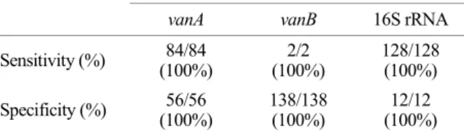
2copies/reaction. Compared to RT-PCR, the sensitivities and specificities of LAMP for 16S rRNA, vanA, and vanB were 100/100%, 100/100%, and 100/100%, respectively. The vanA genotype-vanB phenotype accounted for 57.5% (46/80) of the vancomycin-resistant Enterococci samples collected from 2016 to 2019.
In conclusion, the LAMP assay developed in this study showed high sensitivity and specificity for vancomycin-resistant genes. Moreover, due to the simplicity and rapidity of the LAMP assay, its use can be very useful in clinical microbiology laboratories.
Key Words: Vancomycin-resistant Enterococci (VRE), Loop-mediated isothermal amplification, vanA, vanB, 16S rRNA
INTRODUCTION
Vancomycin-resistant enterococci (VRE) has rapidly spread and emerged as a major nosocomial problem world- wide, since first being isolated in 1986 (Uttley et al., 1988;
Bonten et al., 2001). VRE can cause a variety of invasive infections including intraabdominal infection, bacteremia, and endocarditis. Invasive VRE infections are difficult to treat and are associated with high mortality (Chiang et al.,
2017). Moreover, the transferability of the vanA gene from Enterococci to Staphylococcus aureus can cause serious problems (Niederhäusern et al., 2011).
Rapid and accurate detection of VRE is required for timely antimicrobial treatment and infection control. Culture-based methods to detect VRE are time-consuming, taking several days to complete (2~5 days). Various PCR-based methods have been used for rapid detection of VRE in many hospitals.
PCR-based methods are highly sensitive and specific for vanA-type VRE; however, these methods require special
Original Article
Received: May 26, 2020 / Revised: July 7, 2020 / Accepted: August 10, 2020
*
Professor.
§
These authors contributed equally to this study.
†
Corresponding author: Kyeong Seob Shin. Department of Laboratory Medicine, Chungbuk National University College of Medicine, Cheongju 28644, Korea.
Tel: +82-43-269-6240, Fax: +82-43-271-5243, e-mail: [email protected]
○CThe Korean Society for Biomedical Laboratory Sciences. All rights reserved.
○CCThis is an Open Access article distributed under the terms of the Creative Commons Attribution Non-Commercial License (http://creativecommons.org/licenses/by-nc/3.0/) which permits unrestricted non-commercial use, distribution, and reproduction in any medium, provided the original work is properly cited.
machines and experienced technicians. Moreover, many false-positive results are reported for vanB VRE, mainly due to the non-enterococcal vanB gene, which can be found in anaerobic bacteria in the gut (Ballard et al., 2005; Graham et al., 2008).
Recently, loop-mediated isothermal amplification (LAMP) has been used for the detection of various infections (Hara- Kudo et al., 2005; Misawa et al., 2007; Yamazaki et al., 2008). Compared to PCR, LAMP has the advantage of sim- plicity, rapidity of detection (by the naked eye) and a short amplification time under isothermal conditions. Moreover, LAMP can amplify DNA with high sensitivity and efficacy;
it relies on autocycling strand displacement DNA synthesis performed using the Bst DNA polymerase large fragment.
LAMP shows high specificity when a set of four specifically designed inner and outer primers are used (Notomi et al., 2000; Li et al., 2017).
This study aimed to develop and evaluate a LAMP assay designed for simple and rapid detection of the vanA and vanB genes.
MATERIALS AND METHODS
Bacterial strains
A total of 128 strains of Enterococci, including 88 strains of VRE and 40 strains of vancomycin-susceptible Entero- cocci (VSE), were included in this study. Among 88 VRE, 2 reference strain with vanB genotype and 2 clinical strains with vanC genotype were included. The remaining 84 VRE were clinical strains: 78 vanA E. faecium, 4 vanA E. faecalis, 1 E. avium and 1 E. raffinosus (Table 1). To evaluate the specificity of the LAMP assay for 16S rRNA, 12 reference strains, including 6 gram positive cocci (Staphylococcus aureus ATCC 25923, Staphylococcus epidermidis ATCC 12228, Streptococcus pneumoniae ATCC 49619, Strepto- cocccus agalactiae ATCC 12386, Streptococcus bovis ATCC 49147), 5 gram negative rods (Escherichia coli ATCC 25922, Klebsiella penumoniae ATCC 70063, Enterobacter cloacae ATCC 700323, Pseudomonas aeruginosa ATCC 27853, Aci- netobacter baumannii ATCC 19606) and 2 yeasts (Candia albicans TIMM 3316 and Candida parapsilosis) were used.
DNA extraction from bacterial isolates
A single colony was diluted with 200 μL sterile saline and boiled for 10 min at 100℃. After boiling, the bacteria- containing liquid was centrifuged for 30 sec at 12,000 rpm.
The supernatant was used as the template for real-time RT-PCR and LAMP.
Identification of bacteria and antimicrobial susceptibility test
Identification of bacteria was performed using the VITEK 2 system (bioMérieux Inc., Durham, NC, USA). The min- imum inhibitory concentration (MIC) of vancomycin and teicoplanin was determined by the Etest (bioMérieux Inc.) according to the manufacturer's instructions. The concen- tration of 0.5 MF (1.0×10
8CFU/mL) was inoculated to Muller-Hinton agar and incubated for 24 h at 35℃ in a non-CO
2incubator. The breakpoints were also described in the Clinical and Laboratory Safety Institute guidelines (CLSI, 2018).
Primer design and optimization of reaction conditions for LAMP and RT-PCR assay
Gene sequences of 16S rRNA, vanA, and vanB were searched for in the GenBank database and analyzed with CLC Genomics Workbench (Qiagen, Hilden, Germany) to Table 1. The species of Enterococci used for evaluation of LAMP
assay in this study
Vancomycin
susceptibility (n) Species of Enterococci (n)
Genotype by RT-PCR vanA vanB
VRE (88) E. faecalis (6) 4 2
*E. faecium (78) 78 0
E. avium (1) 1 0
E. raffinosus (1) 1 0 E. casseliflavus (1)
†0 0 E. gallinarum (1)
†0 0
VSE (40) E. faecalis (20) 0 0
E. faecium (20) 0 0
Abbreviations: RT, real-time; VRE, vancomycin-resistant Entero- cocci; VSE, vancomycin-susceptible Enterococci
*
E. faecalis ATCC 700802 and ATCC 51299
†
E. casseliflavus and E. gallinarum has inherent vanC gene
identify highly conserved regions. LAMP primer sets were designed using Explorer V5 software (Eiken Chemical Co.
Ltd., Tokyo, Japan). Primer sets included two external primers (forward outer primer F3 and backward outer primer B3), two internal primers (forward inner primer FIP and back- ward inner primer BIP), and two loop primers (forward loop primer LF and backward loop primer LB). All primers were synthesized by Bionics, Inc. (Seoul, Korea). Detailed infor- mation for the three primer sets used in this study is pre- sented in Table 2 and Fig. 1.
To optimize the reaction conditions of LAMP, various reaction temperatures and times were used. LAMP was carried with a master mix solution containing 5 μL of WarmStart
®colorimetric LAMP master mix (New England Biolabs Inc., Ipswich, MA, USA), 1 μL of F3 and B3 primer, 1 μL of FIP and BIP primer, and 1 μL of LF and LB primer for each reaction. A volume of 2 μL DNA template extracted from the bacterial isolate was added to the master mix and incubated at 65℃ for 60 min. The mixture was then heated
at 80℃ for 10 min for enzyme inactivation. A color change of phenol red pH indicator from pink to yellow, due to a decrease in pH in the presence of extensive amplified DNA, indicated a positive LAMP reaction. LAMP results were also confirmed by 2% agarose gel electrophoresis and the
"ladder-like" amplified DNA products indicated a positive LAMP reaction. To confirm the LAMP results, RT-PCR (CFX96 Touch Real-Time PCR Detection System (Bio-Rad, Hercules, CA, USA) was also performed using SYBR Green (Bio-Rad) with 10 pmol of outer primers (F3 and B3) and 2 μL DNA template. The PCR conditions were as follows:
initial denaturation at 95℃ for 5 min, 35 cycles of dena- turation at 95℃ for 30s, annealing at 60℃ for 30s, elon- gation at 72℃ for 30s, and a final elongation step of 72℃
for 5 min.
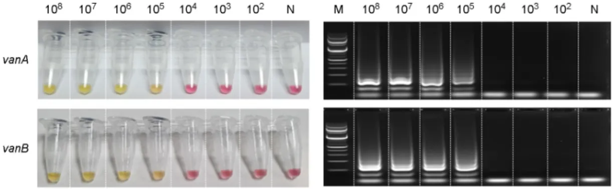
Detection limits of LAMP assay for the 16S rRNA, vanA and vanB genes
To determine the detection limits of the LAMP assay for Table 2. The sequences of primers for LAMP assay used in this study
Target gene Primer Sequence (5' 3')
16S rRNA F3
*GCCGCGGTAATACGTAGG
B3
*TCGCCACTGGTGTTCCTC
FIP CGGGGGCTTTCACATCAGACTT-GTCCGGATTTATTGGGCGTA
BIP CTCAACCGGGGAGGGTCATTG-TTTCACCGCTACACATGGAA
LF AAGAAACCGCCTGCGCTCG
LB GAAACTGGGAGACTTGAGTGC
vanA F3
*GGATTACTTGTTAAAAAGAACCATG
B3
*TCCCAGCATTTTTCGCAA
FIP CCTTGTATGGATCCATCTTCACC-CCATGTTGATGTAGCATTTTCAG
BIP TGTTTGAATTGTCCGGTATCCCTT-CGATGTATGTCAACGATTTGTC
LF TGACTTGCCATGCAAAG
LB TGCGATATTCAAAGCTCAGCAA
vanB F3
*TACGGAATGGGAAGCCGA
B3
*CAAGCTGCGGAGCTTTGA
FIP ACGCCGTGTTTCGTATTCGCTT-GTCTCCCCGCCATACTCTC
BIP CTTTCCCGGTTTTGCATGGCAA-CCCACATAGGGGATACCAGA
LF CCATGCGTTTTCCTATCCGG
LB ATGCGGGGAGGATGGTG
*
Two outer primers F3 and B3 were also used as primers of real-time PCR for 16S rRNA, vanA and vanB
Abbreviations: loop-mediated isothermal amplification; FIP, forward inner primer; BIP, backward inner primer; LF, forward loop primer;
LB, backward loop primer
Fig. 1. Primer designed for 16S rRNA, vanA, vanB loop-mediated isothermal amplification (LAMP) assays. Nucleotide sequences of 16S rRNA (A), vanA (B), vanB (C) and the location of LAMP primers. The forward and backward inner primers are F1c-F2 and B1c-B2 sequences, respectively. The forward and backward outer primers are F3 and B3, respectively.
AACTACGTGCCAGCAGCCGCGGTAATACGTAGGTGGCAAGCGTTGTCCGGATTTATTGGGCGTAAAGCGAGCGCAGGCGGTTTCTTAAGTCTGATGTGAAAGCCCCCGG
CTCAACCGGGGAGGGTCATTGGAAACTGGGAGACTTGAGTGCAGAAGAGGAGAGTGGAATTCCATGTGTAGCGGTGAAATGCGTAGATATATGGAGGAACACCAGTGGCGA A. 16S rRNA
B. vanA
C. vanB
F2
B1
F1c LF
F3
LB B2c B3
711 600 492
601
GGATTACTTGTTAAAAAGAACCATGAATATGAAATCAACCATGTTGATGTAGCATTTTCAGCTTTGCATGGCAAGTCAGGTGAAGATGGATCCATACAAGG
TCTGTTTGAATTGTCCGGTATCCCTTTTGTAGGCTGCGATATTCAAAGCTCAGCAATTTGTATGGACAAATCGTTGACATACATCGTTGCGAAAAATGCTGGGA
TACGGAATGGGAAGCCGACAGTCTCCCCGCCATACTCTCCCCGGATAGGAAAACGCATGGGCTGCTTGTCATGAAAGAAAGCGAATACGAAACACGGCGTATTGATGTG
GCTTTCCCGGTTTTGCATGGCAAATGCGGGGAGGATGGTGCGATACAGGGGCTGTTTGTATTGTCTGGTATCCCCTATGTGGGCTGTGATATTCAAAGCTCCGCAGCTTG F2
B1
F1c LF
F3
LB B2c B3
F2
B1
F1c LF
F3
LB B2c B3
430 329 229
330
386 276 168
277
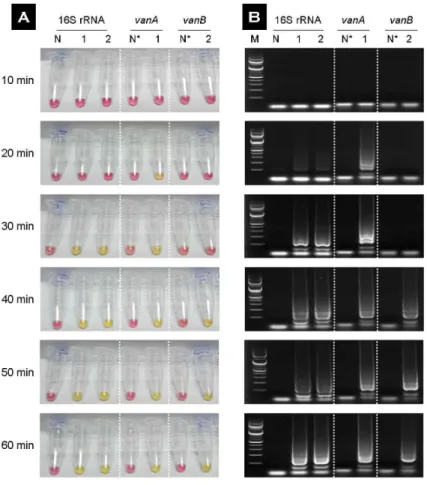
Fig. 2. Visual (A) and agarose gel (B) images of the 16S rRNA, vanA, vanB loop-mediated isother- mal amplification (LAMP) product of vancomycin- resistant Enterococci (VRE) on various reaction times. The yellow color change of pH indicator was interpreted positive for amplification of DNA (A).
The electrophoresis was performed at 2% agarose
gel and amplified products typically showed the
ladder like shape (B). The positive reaction of
LAMP assay for vanA, 16S rRNA and vanB was
observed at 20 min, 30 min, and 40 min, respect-
ively and the best reaction of the three genes was
obtained at 65℃ and 60 min. Abbreviations: N,
negative (non-Enterococcus, Staphylococcus aureus
ATCC 25923); N
*(vancomycin susceptible Entero-
coccus, E. faecalis ATCC 29212).
the 16S rRNA, vanA, and vanB genes, the inoculum with VRE was continuously subjected to 10-fold step dilution from 10
8to 10
2CFU/mL. The experiment was repeated three times.
Specificity of the LAMP assay for 16S rRNA in the genus Enterococcus
The specificity of the LAMP assay for 16S rRNA was evaluated using 12 reference strains (see Bacterial strains section in Material and Methods).
Performance of the LAMP assay using clinical strains To evaluate the performance of the LAMP assay for detection of the vanA and vanB genes, the LAMP assay and RT-PCR were performed simultaneously on 128 strains of
Enterococci with resistance (88) or susceptibility (40) to vancomycin. RT-PCR served as the reference method.
RESULTS
In the reference strains of VRE (vanA and vanB type) and VSE, LAMP to the vanA, 16S rRNA, and vanB genes was observed at 20, 30, and 40 min after amplification, respectively. The optimal conditions for the LAMP assay were 65℃ for 60 min (Fig. 2). Therefore, subsequent tests were performed under those conditions.
Detection limit of LAMP
The detection limits of LAMP were 20 copies/reaction (10
4CFU/mL) for 16S rRNA and 200 copies/reaction (10
5Table 3. The results of loop-mediated isothermal amplification method for the detection of vanA, vanB and 16s rRNA gene from 140 strains
Phenotypic ID
*AST
†Gene
‡LAMP results for vanA, vanB and 16S rRNA
vanA+vanB- vanA-vanB+ vanA-vanB- 16S rRNA
Enterococci (128)
VRE (88) vanA (84) 84 0 0 84
vanB (2) 0 2 0 2
vanC (2) 0 0 2 2
VSE (40) ND 0 0 40 40
Non-Enterococci (12)
§0 0 12 0
Abbreviations: ID, identification; AST, antimicrobial susceptibility testing; VRE, vancomycin-resistant Enterococci; VSE, vancomycin susceptible Enterococci; ND, not detection; LAMP, loop mediated isothermal amplification.
*
Identified by Vitek system and various biochemical reaction test (see text)
†
Antimicrobial susceptibility testing for vancomycin and teicoplanin by E-test
‡
Gene for vancomycin resistance detected by real-time PCR
§