INTRODUCTION
Cyclooxygenases (COXs) are the key enzymes in the con- version of arachidonic acid to prostaglandins. There are two COX isoforms-COX-1 and COX-2. COX-1 is constitutively expressed in most tissues and produces prostaglandins for homeostatic functions (1, 2). COX-2 is highly inducible at inflammation sites and is associated with various human tu- mors such as colonic carcinoma (3), gastric carcinoma (4), prostate carcinoma (5), malignant melanoma (6), or high- grade gliomas (7). COX-2 expression in tumor cell lines is also upregulated during increased proliferation (8), inhibi- tion of apoptosis (9), and neoangiogenesis (10). Recent stud- ies indicate that COX-2 is overexpressed in several thyroid carcinomas, particularly papillary carcinoma (11-14).
Nitric oxide is produced from L-arginine by nitric oxide synthase (NOS). NOS has one inducible isoform, iNOS, and two constitutively expressed isoforms; endothelial NOS and neuronal NOS (15). Among all the three NOS isoforms, iNOS synthesizes the largest amount of nitric oxide released by cells in response to cytokines or other agents (16). The overexpres- sion of iNOS has been demonstrated in chronic inflamma- tory diseases as well as human tumors, including tumors of
the breast, ovary, stomach, head and neck, and central ner- vous system (17-21). It has been reported that iNOS plays an important role in tumorigenesis as a mediator of carcino- genic nitrosamine formation, DNA damage, neovasculariza- tion (22), and apoptosis (23). A few studies have demonstrat- ed that iNOS is overexpressed in several benign and malig- nant thyroid lesions (24, 25).
The aim of the present study was to examine the differences in the immunohistochemical expressions of the COX-2 and iNOS proteins in thyroid neoplasms. Further, we analyzed several clinicopathological findings, patient age, tumor size, thyroid capsular invasion, and lymph node metastasis, in pap- illary carcinoma cases.
MATERIALS AND METHODS Tissue specimens
Samples were selected from 154 patients at the Department of Pathology, Eulji University Hospital (Daejeon, Korea), and Chungnam National University Hospital (Daejeon, Korea) between 1994 and 2003. The diagnoses included papillary
Kyung-Hee Kim, Seong-Ho Kim, Seok Hyung Kim*, Jong-Ho Back, Mee-Ja Park, Jin-Man Kim�
Department of Pathology and Molecular Medicine, Eulji University School of Medicine, Daejeon;
Department of Pathology*, Chungbuk National University College of Medicine, Cheongju; Department of Pathology and Cancer Research Institute�, Chungnam National University College of Medicine, Daejeon, Korea
Address for correspondence Jin-Man Kim, M.D.
Department of Pathology, Chungnam National University College of Medicine, 6 Moonwha-1-dong, Jung-gu, Daejeon 301-131, Korea
Tel : +82.42-580-8237, Fax : +82.42-581-5233 E-mail : [email protected]
*This work was supported by Chungnam National University Hospital Research Fund, 2004.
1064
Thyroid Neoplasms and Their Clinicopathological Correlation
To evaluate the expressions of cyclooxygenase-2 (COX-2) and inducible nitric oxide synthase (iNOS) in thyroid neoplasms in a Korean population, we studied a total of 154 cases: papillary carcinoma of classical type (PTC), 86; follicular adenoma (FA), 21; follicular carcinoma (FC), 35; medullary carcinoma (MC), 3; undifferenti- ated carcinoma (UC), 5; and Hurthle cell neoplasm (HN), 4. Using immunohisto- chemical staining, COX-2 expression was detected in 62 (72.1%) PTC specimens, 5 (23.8%) FA specimens, 10 (28.6%) FC specimens, 0 (0.0%) MC specimens, 1 (20.0%) UC specimen, and 3 (75%) HN specimens. iNOS expression was detect- ed in 66 (76.7%) PTC specimens, 4 (19.0%) FA specimens, 13 (37.1%) FC speci- mens, 0 (0.0%) MC specimens, 3 (60.0%) UC specimens, and 4 (100%) HN speci- mens. The results showed that COX-2 and iNOS were frequently expressed in the PTC and HN specimens, and iNOS was more frequently overexpressed in the FC specimens than in the FA specimens. In PTC, COX-2 and iNOS were significantly overexpressed in patients over 45 yr of age (p=0.029, p=0.041), and iNOS expres- sion was increased in patients with a large primary tumor (p=0.028). These results suggest that the upregulation of COX-2 and iNOS may contribute to the tumor pro- gression of thyroid gland, particularly in PTC and HN, and iNOS may play an adju- vant role during the tumor progression of FC.
Key Words : Thyroid Neoplasms; Cyclooxygenase 2; Nitric Oxide Synthase Type II
Received : 22 September 2005 Accepted : 12 April 2006
carcinoma with classical type (PTC) (n=86), follicular ade- noma (FA) (n=21), follicular carcinoma (FC) (n=35), medu- llary carcinoma (MC) (n=3), undifferentiated carcinoma (UC) (n=5), and Hurthle cell neoplasm (HN) (Hurthle cell ade- noma, n=2; Hurthle cell carcinoma, n=2). Each specimen was subjected to histological evaluation to confirm the diag- nosis, while the relevant clinicopathological information was retrospectively abstracted from patient records. The histopatho- logical slides were reviewed by three pathologists.
Immunohistochemistry
The specimens were formalin-fixed and paraffin-embedded blocks. The tissue blocks were freshly cut into 3 m thick sections onto glass slides. Tissue sections were then deparaf- finized in xylene and rehydrated through graded alcohol and deionized water. Afterwards the sections were heated in a mic- rowave oven in 10 mM citrate buffer (pH 6.0) for 15 min at 100℃for antigen unmasking. Endogenous peroxidase activi- ty of the tissues was inactivated by 10 min incubation in 3%
hydrogen peroxide. To block non-specific binding sites, the slides were then treated with 5% goat serum for 15 min at room temperature. The sections were incubated for one hour
at room temperature with primary antibody to either COX- 2 mouse antihuman monoclonal antibody at a dilution of 1:100 (Mouse anti-COX-2, Clone: COX 229; Zymed labo- ratories Inc., South San Francisco, CA, U.S.A.) or with iNOS epitope specific rabbit antibody at a dilution of 1:100 (Nitric oxide synthase, inducible: RB-9242-P; Lab vision, Fremont, CA, U.S.A.). After incubation with the primary antibody, the sections were processed using standard avidin-biotin im- munohistochemistry, according to the manufacturer’s recom- mendations (Vectastain ABC Elite kit; Vector Laboratories, Burlingame, CA, U.S.A.). Negative controls for the specifici- ty of anti-COX-2 or anti-iNOS antibody were used by omit- ting the primary antibody, and these were prepared using mouse and rabbit immunoglobulins instead of using each primary antibody individually. Thirty paraffin blocks of nor- mal thyroid tissue were used as negative controls.
The intensity of the staining was scored 0-3 (absent, mild, moderate, strong) (Fig. 1, 2) and the area of positivity was estimated as the percentage of the total area of the tumor (<10%, 10-50%, and >50%). The final score was a combi- nation of these two variables and was calculated as follows:
score 0, negative staining or intensity 1+area <10%; score 1, intensity 1+area between 10% and 100% or intensity 2-
Fig. 1. Immunohistochemical staining demonstrates the intensity of COX-2 expression in the cytoplasm of follicular carcinoma cases. (A) score 0; (B) score 1; (C) score 2; (D) score 3 (×400).
A B
C D
3+area <10%; score 2, intensity 2+area >50% or intensity 3+area between 10% and 50%; score 3, intensity 3+area
>50%. A final score of 1-3 was defined as positive immunore- activity. The scoring was performed independently and in a blinded manner by three pathologists who had no knowledge of the clinical data. Discrepant scoring was determined by consensus.
Statistical analysis
The results were analyzed using one-way ANOVA and chi-square tests. Statistical package for the social sciences (SPSS 12.0) was employed. A p value <0.05 was regarded as statistically significant.
RESULT
Cytoplasmic COX-2 expression (final scores 1-3) was de- tected in 62 (72.1%) PTC specimens, 5 (23.8%) FA speci- mens, 10 (28.6%) FC specimens, 0 (0.0%) MC specimen, 1 (20.0%) UC specimen, and 3 (75%) HN specimens. Cyto- plasmic iNOS expression (final scores 1-3) was detected in
Fig. 2. Immunohistochemical staining demonstrates the intensity of iNOS expression in the cytoplasm of papillary carcinoma cases. (A) score 0; (B) score 1; (C) score 2; (D) score 3 (×400).
A B
C D
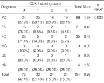
COX-2 staining score
0 1 2 3
Diagnosis Total Mean p-
value*
PC 24 25 18 19 86 1.37 0.000
(27.9%) (29.1%) (20.9%) (22.1%)
FA 16 2 2 1 21 0.43
(76.2%) (9.5%) (9.5%) (4.8%)
FC 25 5 3 2 35 0.49
(71.4%) (14.3%) (8.6%) (5.7%)
MC 3 0 0 0 3 0.00
(100%) (0.0%) (0.0%) (0.0%)
UC 4 0 0 1 5 0.60
(80.0%) (0.0%) (0.0%) (20.0%)
HN 1 1 1 1 4 1.50
(25.0%) (25.0%) (25.0%) (25.0%)
Total 73 33 24 24 154 0.99
(47.4%) (21.4%) (15.6%) (15.6%)
Table 1.COX-2 protein staining in thyroid tumors by immuno- histochemistry
*Statistical analysis was performed by one-way ANOVA.
PC, Papillary carcinoma of classical type; FA, Follicular adenoma; FC, Follicular carcinoma; MC, Medullary carcinoma; UC, Undifferentiated carcinoma; HN, Hurthle cell neoplasm.
66 (76.7%) PTC specimens, 4 (19.0%) FA specimens, 13 (37.1%) FC specimens, 0 (0.0%) MC specimen, 3 (60.0%) UC specimens, and 4 (100%) HN specimens. The COX-2 and iNOS proteins were frequently expressed in the PTC and HN specimens. The mean of the COX-2 final scores was 1.37 in the PTC, 0.43 in the FA, 0.49 in the FC, 0.00 in the MC, 0.60 in the UC, and 1.50 in the HN specimens (Table 1). The mean of the iNOS final scores was 1.73 in the PTC, 0.19 in the FA, 0.74 in the FC, 0.00 in the MC, 0.80 in the UC, and 1.75 in the HN specimens. The mean value of the iNOS final
scores in the FC specimens was greater than that in the FA specimens (Table 2). In the normal thyroid tissue, each final score with regard to COX-2 and iNOS expressions was zero.
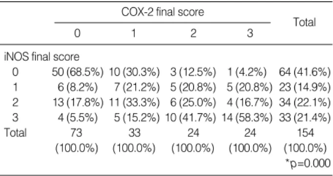
Significant positive correlations were detected between COX- 2 and iNOS expressions (Table 3). In PTCs, COX-2 and iNOS expressions increased in the patients over 45 yr of age, and high iNOS expression correlated with the primary tumor size. However, COX-2 and iNOS expressions have not been shown to be associated lymph node metastasis at operation (Table 4, 5).
DISCUSSION
In this study, the final immunoreactive scores of COX-2 and iNOS expressions were significantly high in the PTC and HN specimens. In contrast, the FA, FC, MC, and UC speci- mens showed lower COX-2 and iNOS expressions. COX-2
iNOS staining score
0 1 2 3
Diagnosis Total Mean p-
value*
PC 20 13 23 30 86 1.73 0.000
(23.3%) (15.1%) (26.7%) (34.9%)
FA 17 4 0 0 21 0.19
(81.0%) (19.0%) (0.0%) (0.0%)
FC 22 3 7 3 35 0.74
(62.9%) (8.6%) (20.0%) (8.6%)
MC 3 0 0 0 3 0.00
(100.0%) (0.0%) (0.0%) (0.0%)
UC 2 2 1 0 5 0.80
(40.0%) (40.0%) (20.0%) (0.0%)
HN 0 1 3 0 4 1.75
(0.0%) (25.0%) (75.0%) (0.0%)
Total 64 23 34 33 154 1.23
(41.6%) (14.9%) (22.1%) (21.4%)
Table 2.iNOS protein staining in thyroid tumors by immunohis- tochemistry
*Statistical analysis was performed by one-way ANOVA.
PC, Papillary carcinoma of classical type; FA, Follicular adenoma; FC, Follicular carcinoma; MC, Medullary carcinoma; UC, Undifferentiated carcinoma; HN, Hurthle cell neoplasm.
COX-2 final score
0 1 2 3 Total
iNOS final score
0 50 (68.5%) 10 (30.3%) 3 (12.5%) 1 (4.2%) 64 (41.6%) 1 6 (8.2%) 7 (21.2%) 5 (20.8%) 5 (20.8%) 23 (14.9%) 2 13 (17.8%) 11 (33.3%) 6 (25.0%) 4 (16.7%) 34 (22.1%) 3 4 (5.5%) 5 (15.2%) 10 (41.7%) 14 (58.3%) 33 (21.4%)
Total 73 33 24 24 154
(100.0%) (100.0%) (100.0%) (100.0%) (100.0%)
*p=0.000 Table 3.Correlation between COX-2 and iNOS expression in the thyroid neoplasms
*, iNOS final score 0+1 vs. score 2+3.
COX-2 final score
0 1 2 3
Total
Age (yr)
<45 19 (39.6%) 14 (29.2%) 8 (16.7%) 7 (14.6%) 48 (100%)
≥45 5 (13.2%) 11 (28.9 %) 10 (26.3%) 12 (31.6%) 38 (100%) p=0.029 Tumor size
T1 7 (25.0%) 8 (28.6%) 7 (25.0%) 6 (21.4%) 28 (100%) T2 3 (17.6%) 7 (41.2%) 3 (17.6%) 4 (23.5%) 17 (100%) T3 0 (0%) 1 (100%) 0 (0%) 0 (0%) 1 (100%) T4 14 (35.0%) 9 (22.5%) 8 (20.0%) 9 (22.5%) 40 (100%) NS (p=0.770) LN metastasis
Absent 12 (24.0%) 14 (28.0 %) 12 (24.0%) 12 (24.0%) 50 (100%) Present 12 (33.3%) 11 (30.6%) 6 (16.7%) 7 (19.4%) 36 (100%) NS (p=0.697) Table 4.Relationship between COX-2 final scores by immuno- histochemical staining and clinicopathological features of papil- lary carcinoma of classical type
LN, lymph node; NS, not significant; T1, ≤2 cm; T2, >2 cm, ≤4 cm; T3,
>4 cm; T4, tumor of any size extending beyond the thyroid capsule inva- sion.
iNOS final score
0 1 2 3 Total
Age (yr)
<45 15 (31.3%) 8 (16.7%) 13 (27.1%) 12(25.0%) 48 (100%)
≥45 5 (13.2%) 5 (13.2%) 10 (26.3%) 18 (47.4%) 38 (100%)
*p=0.041 Tumor size
T1 7 (25.0%) 2 (7.1%) 13 (46.4%) 6 (21.4%) 28 (100%) T2 6 (35.3%) 5 (29.4%) 2 (11.8%) 4 (23.5%) 17 (100%) T3 0 (0%) 0 (0%) 1 (100%) 0 (0%) 1 (100%) T4 7 (23.3%) 6 (15.1%) 7 (26.7%) 20 (34.9%) 40 (100%)
�p=0.028 LN metastasis
Absent 10 (20.0%) 7 (14.0 %) 16 (32.0%) 17 (34.0%) 50 (100%) Present 10 (27.8%) 6 (16.7%) 7 (19.4%) 13 (36.1%) 36 (100%) NS (p=0.593) Table 5.Relationship between iNOS final scores by immunohis- tochemical staining and clinicopathological features of papillary carcinoma of classical type
LN, lymph node; T1, ≤2 cm; T2, >2 cm, ≤4 cm; T3, >4 cm; T4, tumor of any size extending beyond the thyroid capsule invasion; *, iNOS final score 0+1 vs. score 2+3; �, T1 vs. T2 vs. T3+T4; NS, not significant.
prostaglandins (products of COX-2) may affect iNOS expres- sion (26, 27). It has been observed that COX-2 plays impor- tant roles in tumor progression as mediators for the develop- ment of tumor vasculature (10), inhibition of apoptosis (9) and increasing tumor cell invasiveness (28, 29). Although the exact signaling pathways of COX-2-promoted tumori- genesis are yet unknown, several animal studies support a direct role of COX-2 in tumorigenesis since the genetic dele- tion of COX-2 suppresses tumor growth and multiplicity in mice (30).
Both COX-2 and iNOS overexpressions showed an age- associated increase in PTCs. Many authors have demonstrat- ed an age-associated increase in COX-2 activity and prosta- glandin E2 production in animal models (31), and Siironen et al. reported that COX-2 expression increased in older pati- ents with PTC (11). They divided the patients into two groups;
patients less than 35 yr of age and those older than 55 yr of age. Initially, Ito et al. had reported that COX-2 expression decreased with age in PTCs (32). They divided patients into those under and over 54 yr of age. But the Union Internati- onale Contre le Cancer TNM system classifies all PTC patients under 45 yr in a low-risk category, independent of tumor size, extent of tumor, lymph node metastasis, and distant meta- stasis (33). In our study, we divided PTC patients into two groups, namely, those under and over 45 yr of age and detect- ed an age-associated increase in COX-2 and iNOS expressions.
The results were in agreement with the report of Siironen et al. and demonstrated that the overexpression of COX-2 and iNOS could correlate with the more aggressive behavior of PTC.
Similar to the COX-2 expression, we detected iNOS over- expression in PTC and HN specimens and iNOS expression was increased in patients with a large primary tumor. The iNOS overexpression in the PTC specimens is in line with the previous reports (13, 25). Previous reports suggested that the nitric oxide released by iNOS might be involved in tumo- rigenesis as an inhibitor of epithelial cell apoptosis (23) and neoangiogenesis (34). With regard to the FA and FC speci- mens, iNOS overexpression was more frequently observed in the FC specimens than in the FA specimens. Among the FC specimens, there were 37.1% positive cases, whereas among the FA specimens, there were only 19.0% positive cases. Al- though this result is insufficient to arrive at a conclusion, we suggest that iNOS expression analysis could be helpful in understanding the tumor progression of FC.
Overexpression of the COX-2 protein in HN specimen has been reported previously (35), but the role played by COX- 2 and iNOS in HN is unknown. A previous study has shown that COX-2 and iNOS are overexpressed in the swollen cyto-
We have not demonstrated COX-2 and iNOS expression in the three cases of MCs. However, Bell et al. reported COX- 2 and iNOS expression in nine cases of MC along with im- munohistochemical reactivity for COX-1 and COX-2 enzymes (36). Kim et al. reported that one of two MC cases showed immunohistochemical overexpression for COX-2 (35), and Choe et al. reported that all the five MC cases showed immu- nohistochemical overexpression for iNOS (24). The number of MC cases that we have reported here was too small for sta- tistical analysis. The association of COX-2 and iNOS expres- sions with MC should be further investigated for better under- standing of MC tumorigenesis.
In conclusion, the present immunohistochemical study has shown that overexpressions of the COX-2 and iNOS proteins were more frequently observed in PTC and HN specimens than other thyroid neoplasms. Further, FCs tended to show iNOS overexpression more frequently than FAs. The expres- sions of both COX-2 and iNOS increased in older PTC pati- ents, and iNOS overexpression was correlated with the pri- mary tumor size of PTC. These preliminary data suggest that COX-2 and iNOS expressions could be important molecu- lar events for the tumor progression of PTC and HN, and iNOS expression could play a role in the tumor progression of FC.
REFERENCES
1. Dubois RN, Abramson SB, Crofford L, Gupta RA, Simon LS, van de Putte LB, Lipsky PE. Cyclooxygenase in biology and disease.
FASEB J 1998; 12: 1063-73.
2. Vane JR, Bakhle YS, Botting RM. Cyclooxygenases 1 and 2. Annu Rev Pharmacol Toxicol 1998; 38: 97-120.
3. Eberhart CE, Coffey RJ, Radhika A, Giardiello FM, Ferrenbach S, DuBois RN. Up-regulation of cyclooxygenase 2 gene expression in human colorectal adenomas and adenocarcinomas. Gastroenterol- ogy 1994; 107: 1183-8.
4. van Rees BP, Saukkonen K, Ristimaki A, Polkowski W, Tytgat GN, Drillenburg P, Offerhaus GJ. Cyclooxygenase-2 expression during carcinogenesis in the human stomach. J Pathol 2002; 196: 171-9.
5. Madaan S, Abel PD, Chaudhary KS, Hewitt R, Stott MA, Stamp GW, Lalani EN. Cytoplasmic induction and over-expression of cyclooxy- genase-2 in human prostate cancer: implications for prevention and treatment. BJU Int 2000; 86: 736-41.
6. Denkert C, Kobel M, Berger S, Siegert A, Leclere A, Trefzer U, Ha- uptmann S. Expression of cyclooxygenase 2 in human malignant melanoma. Cancer Res 2001; 61: 303-8.
7. Joki T, Heese O, Nikas DC, Bello L, Zhang J, Kraeft SK, Seyfried NT, Abe T, Chen LB, Carroll RS, Black PM. Expression of cyclooxy-
genase 2 (COX-2) in human glioma and in vitro inhibition by a spe- cific COX-2 inhibitor, NS-398. Cancer Res 2000; 60: 4926-31.
8. Fujita H, Koshida K, Keller ET, Takahashi Y, Yoshimito T, Namiki M, Mizokami A. Cyclooxygenase-2 promotes prostate cancer pro- gression. Prostate 2002; 53: 232-40.
9. Kamijo T, Sato T, Nagatomi Y, Kitamura T. Induction of apoptosis by cyclooxygenase-2 inhibitors in prostate cancer cell lines. Int J Urol 2001; 8: S35-9.
10. Fosslien E. Review: molecular pathology of cyclooxygenase-2 in can- cer-induced angiogenesis. Ann Clin Lab Sci 2001; 31: 325-48.
11. Siironen P, Ristimaki A, Nordling S, Louhimo J, Haapiainen R, Haglund C. Expression of COX-2 is increased with age in papillary thyroid cancer. Histopathology 2004; 44: 490-7.
12. Cornetta AJ, Russell JP, Cunnane M, Keane WM, Rothstein JL. Cy- clooxygenase-2 expression in human thyroid carcinoma and Hashi- moto’s thyroiditis. Laryngoscope 2002; 112: 238-42.
13. Nose F, Ichikawa T, Fujiwara M, Okayasu I. Up-regulation of cy- clooxygenase-2 expression in lymphocytic thyroiditis and thyroid tumors: significant correlation with inducible nitric oxide synthase.
Am J Clin Pathol 2002; 117: 546-51.
14. Specht MC, Tucker ON, Hocever M, Gonzalez D, Teng L, Fahey TJ 3rd. Cyclooxygenase-2 expression in thyroid nodules. J Clin Endo- crinol Metab 2002; 87: 358-63.
15. Nathan C, Xie QW. Nitric oxide synthases: roles, tolls, and controls.
Cell 1994; 78: 915-8.
16 Assurey J, Canha FQ, Lew FY, Moncada S. Feedback inhibition of nitric oxide synthase activity by nitric oxide. Br J Pharmacol 1993;
108: 833-7.
17. Duenas-Gonzalez A, Isales CM, del Mar Abad-Hernandez M, Gon- zalez-Sarmiento R, Sangueza O, Rodriguez-Commes J. Expression of inducible nitric oxide synthase in breast cancer correlates with metastatic disease. Mod Pathol 1997; 10: 645-9.
18. Thomsen LL, Lawton FG, Knowles RG, Beesley JE, Riveros-Moreno V, Moncada S. Nitric oxide synthase activity in human gynecologi- cal cancer. Cancer Res 1994; 54: 1352-4.
19. Son HJ, Kim YH, Park DI, Kim JJ, Rhee PL, Paik SW, Choi KW, Song SY, Rhee JC. Interaction between cyclooxygenase-2 and in- ducible nitric oxide synthase in gastric cancer. J Clin Gastroenterol 2001; 33: 383-8.
20. Gavilanes J, Moro MA, Lizasoain I, Lorenzo P, Perez A, Leza JC, Alvarez-Vicent JJ. Nitric oxide synthase activity in human squamous cell carcinoma of the head and neck. Laryngoscope 1999; 109: 148- 52.
21. Cobbs CS, Brenman JE, Aldape KD, Bredt DS, Israel MA. Expres- sion of nitric oxide synthase in human central nervous system tumors.
Cancer Res 1995; 55: 727-30.
22. Thomsen LL, Miles DW. Role of nitric oxide in tumor progression:
lessons from human tumors. Cancer Metastasis Rev 1998; 17: 107-18.
23. Kim YM, Talanian RV, Billiar TR. Nitric oxide inhibits apoptosis by preventing increases in caspase-3-like activity via two distinct mechenisms. J Biol Chem 1997; 272: 31138-48.
24. Choe W, Kim S, Hwang TS, Lee SS. Expression of inducible nitric oxide synthase in thyroid neoplasms: immunohistochemical and mo- lecular analysis. Pathol Int 2003; 53: 434-9.
25. Kitano H, Kitanishi T, Nakanishi Y, Suzuki M, Takeuchi E, Yaza- wa Y, Kitajima K, Kimura H, Tooyama I. Expression of inducible nitric oxide synthase in human thyroid papillary carcinomas. Thy- roid 1999; 9: 113-7.
26. Perez-Sala D, Lamas S. Regulation of cyclooxygenase-2 expression by nitric oxide in cells. Antioxid Redox Signal 2001; 3: 231-48.
27. Kobayashi O, Miwa H, Watanabe S, Tsujii M, Dubois RN, Sato N.
Cyclooxygenase-2 downregulates inducible nitric oxide synthase in rat intestinal epithelial cells. Am J Physiol Gastrointest Liver Physi- ol 2001; 281: 688-96.
28. Ermert L, Dierkes C, Ermert M. Immunohistochemical expression of cyclooxygenase isoenzymes and downstream enzymes in human lung tumors. Clin Cancer Res 2003; 9: 1604-10.
29. Sharma S, Stolina M, Yang SC, Baratelli F, Lin JF, Atianzar K, Luo J, Zhu L, Lin Y, Huang M, Dohadwala M, Batra RK, Dubinett SM.
Tumor cyclooxygenase 2-dependent suppression of dendritic cell function. Clin Cancer Res 2003; 9: 961-8.
30. Oshima M, Dinchuk JE, Kargman SL, Oshima H, Hancock B, Kwong E, Trzaskos JM, Evans JF, Taketo MM. Suppression of intestinal polyposis in Apc delta716 knockout mice by inhibition of cyclooxy- genase 2 (COX-2). Cell 1996; 87: 803-9.
31. Hayek MG, Mura C, Wu D, Beharka AA, Han SN, Paulson KE, Hwang D, Meydani SN. Enhanced expression of inducible cyclooxy- genase with age in murine macrophages. J Immunol 1997; 159: 2445- 51.
32. Ito Y, Yoshida H, Nakano K, Takamura Y, Miya A, Kobayashi K, Yokozawa T, Matsuzuka F, Matsuura N, Kuma K, Miyauchi A. Cy- clooxygenase-2 expression in thyroid neoplasms. Histopathology 2003; 42: 492-7.
33. Sobin L, Wittekind C, eds. TNM classification of malignant tumors, 5th edn. New York: Wiley-Liss, Inc. 1997.
34. Ziche M, Morbidelli L, Choudhuri R, Zhang HT, Donnini S, Granger HJ, Bicknell R. Nitric oxide synthase lies downstream from vascular endothelial growth factor induced but not basic fibroblast growth factor-induced angiogenesis. J Clin Invest 1997; 99: 2625-34.
35. Kim SJ, Lee JH, Yoon JS, Mok JO, Kim YJ, Park HK, Kim CH, Byun DW, Suh KI, Yoo MH. Immunohistochemical expression of COX-2 in thyroid nodules. Korean J Intern Med 2003; 18: 225-9.
36. Bell CD, Vidal S, Kovacs K, Horvath E, Rotondo F. An immunohisto- chemical survey of nine cases of medullary carcinoma of thyroid including reactivity for Cox-1 and Cox-2 enzymes. Endocr Pathol 2002; 13: 331-40.