Available at http://www.formulastudy.com
HFS
Original Article / 원저
죽엽 열수추출물의 염증억제 효과
손진원
#, 박상미
#, 정지윤, 황보민, 조일제, 정태영, 박정아, 김상찬, 지선영
*대구한의대학교 한의과대학
Anti-inflammatory Effects of the Water Extract of Phyllostachys Folium via NF-κB Inhibition
Jin Won Son
#, Sang Mi Park
#, Ji Yun Jung, Min Hwangbo, Il Je Cho, Tae Young Jung, Chung A Park, Sang Chan Kim, Seon Young Jee
*College of Korean Medicine, Daegu Haany University, Gyeongsan 38610, Republic of Korea.
ABSTRACT
Objectives : Phyllostachys Folium is leaves of Phyllostachys nigra var. henesis STAPF. In the East Asian traditional medicine, the herb has been used to treat nasal bleeding, dysuria, epilepsy and etc. The present study was conducted to evaluate the anti-inflammatory effects of the Phyllostachys Folium water extracts (PFE) in vitro and in vivo model.
Methods : Cell viability was measured by MTT assay after the treatment of PFE and NO production was monitored by measuring the nitrite content in culture medium. iNOS, COX-2, IκB, p-IκBα amd NFκB were detected by immunoblot analysis, and levels of cytokine were analyzed by sandwich ELISA kit. Anti-edema effect of PFE was determined in the carrageenan-induced paw edema model in rats.
Results : LPS increased NO and cytokines levels compared with control, these increases were attenuated by PFE. In addition, LPS-induced pro-inflammatory proteins such as iNOS, COX-2 were down regulated by PFE. These anti-inflammatory effect of PFE results from inhibition of phosphorylation of IκB and translocation of NF-κB.
Conclusion : These results show that PFE has some anti-inflammatory effects which might play a role in gram-negative
ⓒ 2016 The Korean Medicine Society For The Herbal Formula Study
This paper is available at http://www.formulastudy.com which permits unrestricted non-commercial use, distribution, and reproduction in any medium, provided the original work is properly cited.
bacterial infection inflammation and NFκB activated diseases.
Key Words : Phyllostachys Folium, Anti-inflammatory effects, NF-κB.
Ⅰ. 서론
*죽엽 (竹葉; Phyllostachys Folium)은 禾本科 (벼 과; Gramineae)에 속한 여러 해 살이 常綠喬木인 솜 대 Phyllostachys nigra var. henonis STAPF의 잎을 건조한 것으로1-3), 性味는 甘淡寒하며, 心肺膽胃에 작 용한다. 淸熱除煩, 生津利尿의 효능으로 熱病煩渴, 小 兒驚癎, 咳逆吐衄, 面赤, 小便短赤, 口糜舌瘡 등을 치 료한다3).
죽엽에는 friedelin, glutinol, luteolin 6-C-α -L-arabinopyranoside, tricin, isoorientin 등의 성 분이 있는 것으로 알려져 있으며4), 생리활성에 대한 연구로는 죽엽추출물이 고지방식이 및 알콜로 유도된 동물모델에서의 고지질혈증 개선효과2,5), 죽엽의 항산화 화합물인 methyl chlorogenic acid 유도체 (MCGA)-3의 항산화효과6), 죽엽물추출물의 GABA신경계를 통한 항 불안효과7), 죽엽 물추출물 및 에탄올추출물의 항균효 과8), 죽엽물추출물의 항암효과9), CCl4로 유도된 간손 상 및 알콜의 반복투여로 유도된 간손상에 대한 간보
호효과2,10)가 보고되었다. 그러나 아직까지 죽엽의 항
염증효과에 대한 연구는 매우 미흡한 실정이다.
대식세포는 염증 반응시 interleukin (IL)-1β, -6, tumor necrosis factor-α (TNF-α) 등의 다양한 사 이토카인을 생산하여 생체 방어에 중요한 역할을 한다
11,12). Lipopolysaccharide (LPS)는 그람-음성 박테리 아의 세포벽 구성성분으로, 국소 및 전신의 염증반응 을 유발하며, 대식세포의 toll-like receptor 4 (TLR4)와 결합하여 IκB kinase cascade를 통하여 다양한 전염 증성물질들을 생성시키므로 염증반응 및 항염증약물의 연구에서 빈용되는 연구모델이다13). 홍조류(red seaweed) 에서 추출되는 carrageenan (CA)은 급성염증반응을 유 도하므로, 약물의 항염증효과 평가와 염증매개물질의 조절에 관한 연구의 동물모델로 사용되고 있다14,15).
본 연구에서는 죽엽이 淸熱瀉火藥임에 근거하여 죽 엽추출물의 항염증활성을 평가하고자 하였다. In vitro 연구로 LPS로 활성화된 설치류 대식세포주인 Raw 264.7 cells 모델에서 염증의 기전 및 염증관련 cytokine 의 발현에 대한 평가를 하였으며, in vivo 연구로 CA 로 유도된 rat의 족부종에 대한 부종억제효과를 평가 하였다.
Ⅱ. 재료 및 방법
1. 추출물(PFE)의 제조
죽엽 (Phyllostachys Folium)은 대원약업사(대구)에 서 구입하였으며, 죽엽 300 g에 물 1.5 ℓ를 넣고 약탕 기 (대웅, 한국)로 3시간 추출한 후, 추출물을 거즈로 1 차 여과하고 3000×g에서 3분간 원심분리하였다. 원심 분리 후의 상층액만을 취하여 0.2 ㎛ filter (Nalgene, New York, USA)로 여과하였다. 이 여과액을 rotary evaporator (EYELA, Tokyo, Japan)로 동결건조하여 9.08 g을 얻었으며, 사용 때까지 -20℃에서 보관하였 다. 죽엽추출물 (Phyllostachys Folium Extract; PFE)의 수율은 3.0%였으며 in vitro 처치시에는 DMEM에 녹여 사용하였으며, in vivo 실험에서는 생리식염수에 녹여 사용하였다.
2. 시약
3-(4,5-dimethylthiazol-2-yl)-2,5-diphenyltetraz oleum (MTT)와 LPS (Escherichia coli 026:B6;
Difco, Detroit, MI, USA)는 Sigma (St. Louis, MO, USA)에서 구입하였고, fetal bovine serum (FBS), Penicillin 및 streptomycin은 Gibco/BRL (Eggenstein, Germany)로부터 구입하였다. Western blot에 사용된 antibody들은 BD Bioscience (San Jose, CA, USA), Cayman (Ann Arbor, Mi, USA), Zymed (San Francisco, CA, USA)에서 각각 구입하였고, NC paper
# Authors contributed equally
* Corresponding author: Seon Young Jee, College of Korean Medicine, Daegu Haany University, 1, Hanuidae-ro, Gyeongsan-si, Gyeongsangbuk-do, 38610, Republic of Korea.
Tel:+82-53-770-2272 Fax:+82-53-819-1860, E-mail : [email protected]
∙Received : September 29, 2016 / Revised : November 5, 2016 / Accepted : November 8, 2016
는 Schleicher & Schuell (Dassel, Germany)에서 구 입하였다. TNF-α, IL-1β, IL-6의 ELISA kit는 Pierce endogen (Rockford, IL, USA)에서 구입하였다.
3. 세포배양
Murine macrophage cell line인 Raw 264.7 cells는 한국세포주은행 (Seoul, Korea)에서 구입하였으며, Dulbecco’s modified Eagle’s medium (DMEM)에 10% FBS, 100 U/㎖ penicillin 및100 ㎍/㎖
streptomycin을 혼합한 배지를 사용하여 37℃, 5%
CO2환경이 유지되는 배양기에서 배양하였다. 실험과 정의 모든 cells은 80-90%의 confluence에서 실험하였 고, 20 passages를 넘기지 않고 사용하였다.
4. 세포생존율의 측정
Raw 264.7 cells를 96 well plate에 5×10⁴ cells/well 로 분주한 다음 PFE를 농도별로 처리하여 MTT assay 를 시행하였다. 세포에 10-100 ㎍/㎖의 농도로 PFE를 처치한 후 37℃, 5% CO2의 배양기에서 24시간 배양하 고, 각 well당 MTT (0.1 ㎎/㎖)용액을 50 ㎕씩 넣고 4 시간 배양한 후 배지를 제거하고 생성된 formazan crystals을 dimethylsulfoxide (DMSO)에 녹여 Titertek Multiskan automatic microplate reader (Model MCC/340, Huntsville, AL)를 사용하여 570 ㎚에서 흡 광도를 측정하였다. 또한, LPS와 PFE의 동시처리에 의 한 세포생존율 역시 동일한 방법으로 세포생존율을 구하였 다. 세포생존율은 control cells에 대한 백분율로 나타내었 다. [i.e. viability (% of control) = 100 ×/(absorbance of treated sample)/(absorbance of control)].
5. NO 생성량 측정
Raw 264.7 cells로부터 생성된 nitric oxide (NO) 의 양은 세포 배양액에 Griess 시약을 처치하여 측정 하였다. PFE를 1시간 전처치한 후, LPS를 24시간 처 치하고, 각 군별 세포배양 상등액 50 ㎕을 96 well plate에 넣고, Griess시약 (1% sulfanilamide in 5%
phosphoric acid + 1% α-naphthylamide in H2O) 50 ㎕를 혼합하고 10분 동안 반응시킨 후 540 ㎚에서 Titertek Multiskan automatic microplate reader로 흡광도를 측정하였다. NO의 생성량은 control cell의 NO에 대한 비율로 나타내었다.
[i.e. NO production (fold) = (absorbance of treated
sample)/(absorbance of control)].
6. Immunoblot analysis
염증관련 단백질의 발현을 관찰하기 위하여, 처치된 세포들을 PBS로 수거한 후, 20 mM Tris Cl (㏗ 7.5), 1% Triton X-100, 137 mM sodium chloride, 10%
glycerol, 2 mM EDTA, 1 mM sodium orthovanadate, 25 mM β-glycerophosphate, 2 mM sodium pyrophosphate, 1 mM phenylmethylsulfonyl fluoride (PMSF)와 1 ㎎/㎖ leupeptin을 함유하는 buffer를 사 용하여 세포를 용해시켰다. 전세포추출액을 10,000×g 로 10분간 원심분리하여 찌꺼기를 제거하였다. Sodium dodecyl sulfate-polyacrylamide gel electrophoresis (SDS-PAGE)로 전기영동 후, 단백질을 nitrocellulose membrane으로 전이하였다. 이 membrane에 iNOS, COX-2, p-IκB, NFκB, actin, lamin 등의 항체에 반응 및 blocking과 세척 후 horseradish peroxidase conjugated secondary antibody를 결합시키고, 이를 ECL western blotting detection reagents (Amersham) 를 사용하여 발색하였다. β-actin과 lamin A/C는 각 단백질의 loading control로 사용되었다.
7. 핵분획의 준비
LPS 및 PFE가 처치된 Raw 264.7 cells를 PBS로 세척한 후, PBS 1 ㎖을 가하여 수거하고, 원심분리하 여 세포들을 microtubes에 수거하였다. 10 mM HEPES-KOH (㏗ 7.6), 10 mM KCl, 1.5 mM MgCl2, 0.1% Nonidet P-40, 1 mM dithiothreitiol, 4 ㎍/㎖
leupeptin, 0.5 mM PMSF를 함유하는 hypotonic buffer 100 ㎕를 가하여 vortex하고, 얼음 위에서 10 분간 배양하였다. 이를 4°C에서 7,800×g로 10분간 원심분리한 후, pellet에 10 mM HEPES (㏗ 7.6), 400 mM KCl, 0.1 mM EDTA, 25% glycerol, 4 ㎍/
㎖ leupeptin, 1 mM PMSF을 함유하는 extraction buffer를 50 ㎕을 가하여, vortex한 후 얼음 위에서 1 시간 동안 방치하였다. 이를 다시 14,000×g로 15분간 원심분리하여, 핵분획을 포함하고 있는 상등액을 취 하였다.
8. Cytokine 및 PGE2의 측정
Cytokine 및 prostaglandin E2 (PGE2)를 측정하기 위하여 6 well plate에 cells (5×10⁵/㎖)을 분주하고
PFE를 농도별로 처치한 다음, 1시간 후에 LPS를 처치 하였다. LPS 처치 후 특정 시간에 배지를 수거하여 cytokine과 PGE2를 측정하였다. Tumor necrosis factor-α (TNF-α), IL-6와 PGE2는 ELISA kit를 사 용하여 측정하였다.
9. 실험동물 및 처치
실험동물은 4주령 된 Sprague-Dawley계 수컷 흰쥐 (130-160 g)를 1주일 동안 환경에 적응시킨 후 실험 에 사용하였으며, 사육실 환경은 온도 20-23℃, 습도 60%, 12시간 light/dark cycle을 유지하고, 사료 (Nestle Purina Petcare Korea, Seoul, Korea)와 음 료는 자유롭게 섭취하도록 하였다. 동물실험은 대구한 의대학교 동물실험윤리위원회 (IACUC)의 규정에 따라 윤리위원회의 승인 (DHU2013-091)을 획득한 후 시행 하였다.
실험은 아무런 처치를 하지 않은 군을 Control군, carrageenan (100 ㎕/rat)만을 피하주사한 CA군, carrageenan과 dexamethasone (1 ㎎/㎏, p.o.)을 투 여한 DEXA군, carrageenan과 0.3 g/㎏의 PFE를 투 여한 0.3 g/㎏ PFE군, carrageenan과 1.0 g/㎏의 PFE를 투여한 1.0 g/㎏ PFE군으로 나누었으며, 각 군당 수는 5마리로 하였다. Dexamethasone과 PFE는 4일 동안 매일 1회 투여하였으며, 마지막 약물투여 1 시간 후 carrageenan을 100 ㎕/rat로 rat의 오른쪽 발바닥에 주입하여 족부종을 유발하였다. 족부종의 측 정은 carrageenan 주입 후 0, 1, 2, 3, 4시간에 부종 측정기 (Plethysmometer, LE 7500; LETICA Scientific Instruments, Spain)를 이용하여 부종 정도를 측정하 였다.
10. 통계적 검증
실험 결과는 mean ± SD로 나타내었으며, 처치군 간의 유의성은 one way analysis of varience (ANOVA) 로 검정한 후 Newman-Kleuls test로 검정하였다. 통 계적 유의성 검정은 p<0.05 또는 p<0.01로 하였다.
Ⅲ. 실험결과
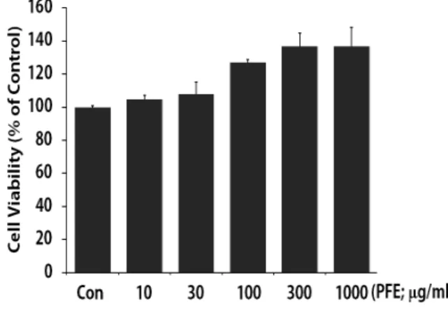
1. PFE가 Raw 264.7 cells의 세포생존율에 미치는 영향 Raw 264.7 cells에 PFE를 10-1000 ㎍/㎖의 농도로 처치하고 24시간 배양후 세포생존율을 측정하였다.
Control을 100.00 ± 0.98%로 하였을 때, PFE 10, 30, 100, 300, 1000 (㎍/㎖)은 각각 104.66 ± 2.51, 107.90 ± 7.17, 126.81 ± 1.96, 136.89 ± 7.83, 136.64 ± 11.66 (%)의 세포생존율을 보여 PFE는 세포 독성을 나타내지 않았다 (Fig. 1).
Fig. 1. The effects of PFE on the cell viability on Raw 264.7 cells.
Raw 264.7 cells were treated with 10-1000
㎍/㎖ of PFE dissolved in media for 24 h, and the cells were treated with MTT solution for 4 h. The method of MTT assay was described in the materials and methods section. Data represent the mean
± SD with eight separate experiments.
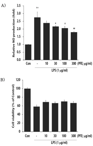
2. PFE가 LPS로 유도된 Raw 264.7 cells의 NO 생 성에 미치는 영향
PFE를 10-300 ㎍/㎖의 농도로 24시간 세포에 처리 하여 생성되는 NO양을 측정하였다. LPS군에서는 Control군에 비교하여 NO의 생성량이 2.76 ± 0.35 배로 유의하게 증가하였으며, 이러한 NO의 증가는 PFE 30, 100, 300 (㎍/㎖)의 농도에서 각각 2.17 ± 0.06, 2.06 ± 0.11, 1.80 ± 0.04배로 유의성 있게 감 소하였다 (Fig. 2A).
또한 PFE와 LPS의 병용처치에 의한 세포생존율은 control군을 100.00 ± 2.21%로 하였을 때, LPS 단독 처치군은 58.39 ± 3.56%로 유의하게 감소하였다. 그 러나 PFE와 LPS의 병용처치는 PFE 10, 30, 100, 300 (㎍/㎖)의 농도에서 각각 69.06 ± 3.70, 66.35
± 3.16, 70.36 ± 3.15, 66.53 ± 3.76 (%)으로서 LPS의 단독처치에 비교하여 유의한 세포독성을 나타 내지 않았다 (Fig. 2B).
A)
B)
Fig. 2. The effects of PFE on production of nitric oxide (A) and cell viability (B) on Raw 264.7 cells
Raw 264.7 cells were treated with 10-300
㎍/㎖ of PFE dissolved in media for 1 h prior to the addition of LPS (1 ㎍/㎖), and the cells were further incubated for 24 h.
The concentrations of NO in culture medium were monitored as described in the materials and methods section (A).
Effect of cell viability on LPS+PFE-treated cells were determined by MTT assay (B).
Data represent the mean ± SD with eight separate experiments. (*, significant as compared to control. **P<0.01; #, significant as compared to LPS alone, #P<0.05, ##P
<0.01)
3. PFE가 iNOS의 발현에 미치는 영향
PFE가 유의한 세포독성 없이 NO를 억제하였으므 로, NO의 생성과 관련되는 inducible nitric oxide synthase (iNOS) 단백질의 발현을 평가하였다. LPS 처치 시에는 iNOS 단백질의 발현이 증가되었으나, LPS에 PFE를 처치한 실험군에서는 iNOS의 발현량이 뚜렷하게 감소되었다 (Fig. 3).
Fig. 3. The effects of PFE on the induction of iNOS expression on LPS-activated Raw 264.7 cells.
The levels of iNOS expression were monitored 24 h after treatment of LPS (1
㎍/㎖) with or without PFE pretreatment (i.e. 1 h before LPS). The β-actin was used as a loading control in total cell lysate.
4. PFE가 COX-2의 발현과 PGE2에 미치는 영향 염증반응에서 중요한 역할을 하는 cyclooxygenase-2 (COX-2)는 LPS 처치 시에는 COX-2 단백질이 현저 하게 발현 증가되었으나, PFE를 전처리한 실험군에서 는 COX-2의 발현이 뚜렷하게 감소하였다 (Fig. 4A).
Cyclooxygenase pathway를 통하여 생성되는 PGE2는 Control군에서 24.55 ± 12.61 pg/㎖를 나타내었고, LPS는 3188.66 ± 237.74 pg/㎖로 유의성 있게 증가하 였으며, PFE는 100과 300 ㎍/㎖에서 PGE2를 각각 2317.59 ± 151.86, 1590.59 ± 120.37 (pg/㎖)로 유의 성 있게 감소시켰다 (Fig. 4B).
A)
B)
Fig. 4. The effects of PFE on the induction of COX-2 expression (A) and PGE2 production (B) on LPS-activated Raw 264.7 cells.
The levels of COX-2 were monitored 24 h after treatment of LPS (1㎍/㎖) with or without PFE pretreatment (i.e. 1 h before LPS). The β-actin was used as a loading control (A). Cells were cultured with LPS (1 ㎍/㎖) in the presence or absence of PFE for 24 h to determine the level of PGE2. The cultured medium was collected and directly assayed for PGE2 (B). The data represent the mean ± SD of three separate experiments.
(*: significant compared with the control,
**P<0.01, #: significant compared with the LPS alone, ##P<0.01).
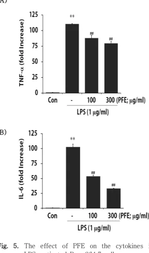
5. PFE가 LPS로 유도된 Raw 264.7 cells의 cytokine 생성에 미치는 영향
활성화된 대식세포는 TNF-α, IL-1β, IL-6, chemokine 등을 생산하며, TNF와 IL은 패혈성 쇼크의 중요한 매개체로서 염증반응의 핵심적 역할을 한다. 본 연구에서 LPS는 control 대비 TNF-α의 분비를 108.65 ± 1.46배 유의성 있게 증가시켰으며, PFE는
100, 300 (㎍/㎖)의 농도에서 TNF-α의 생성량을 각각 85.84 ± 4.10배, 77.72 ± 2.55배로 유의성 있게 감소 시켰다 (Fig. 5A). 또한 LPS는 control 대비 IL-6의 생성을 102.83 ± 4.90배로 유의성 있게 증가시켰으 며, PFE는 100, 300 (㎍/㎖)에서 각각 53.67 ± 1.95, 33.16 ± 0.36배로 LPS로 증가된 IL-6를 유의성 있 게 감소시켰다 (Fig. 5B).
A)
B)
Fig. 5. The effect of PFE on the cytokines in LPS-activated Raw 264.7 cells.
Production of cytokines was measured in the media of Raw 264.7 cells cultured with LPS (1 ㎍/㎖) in the presence or absence of PFE for 12 h (TNF-α) and 6 h (IL-6), respectively. The amount of cytokines was measured by immunoassay as described in materials and methods. Data represent the mean ± SD with three separate experiments. (*, significant as compared to control. **P<0.01; #, significant as compared to LPS alone, ##P<0.01)
6. PFE가 LPS로 활성화된 Raw 264.7 cells에서 NF-κB pathway에 미치는 영향
비활성 대식세포에서는 NF-κB가 세포질에서 inhibitory-κB와 결합형으로 존재하나, 활성화되면 I-κB가 인산화되고, NF-κB가 핵으로 들어가 iNOS, COX-2 등의 전사를 유도하게 된다. 본 연구에서는 세 포질에서 IκBα의 발현과 인산화를, 핵분획에서 NF- κB의 발현을 평가하였다.
세포질에서 p-IκBα는 LPS처치에 의하여 뚜렷하게 증가하였으며, PFE 300 ㎍/㎖의 전 처치에 의해 IκB α의 인산화가 현저하게 감소되었다. 또한 IκBα는 LPS에 의하여 감소되었고, 이러한 감소는 PFE의 전 처 치에 의하여 증가되었다 (Fig. 6A). 핵내에서의 NF-κ B는 LPS 처치에 의해 증가하였으며, PFE는 핵분획에 서의 NF-κB를 감소시켰다 (Fig. 6B).
A)
B)
Fig. 6. The effect of PFE on the NF-κB pathway in LPS-activated Raw 264.7 cells.
The levels of p-I-κBα, I-κBα (A) and nuclear NF-κB proteins (B) were monitored after treatment of cells with LPS (1㎍/㎖) with or without PFE pretreatment (i.e. 1 h before LPS). β -actin and lamin A/C were used as a loading control for lysate and nuclear fraction, respectively.
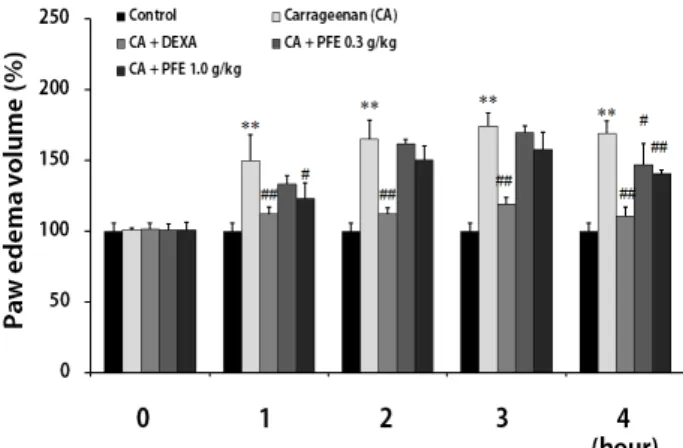
7. PFE가 carrageenan으로 유도된 rat의 족부종에 미치는 영향
PFE가 LPS에 의해 활성화된 NF-κB를 억제하여
in vitro에서 염증매개물질을 억제함을 확인하고,
carrageenan (CA)으로 유도된 염증동물모델에서 족부 종을 측정하였다. CA군의 족부종은 control군에 비교 하여, 1, 2, 3, 4 시간에 각각 149.61 ± 18.22, 164.85 ± 13.42, 174.25 ± 9.02, 169.01 ± 8.66 (%)로 유의한 발 부종을 유발하였다. Dexamethasone (DEXA)군에서는 각각 112.32 ± 4.77, 112.07 ± 4.17, 118.69 ± 5.22, 110.26 ± 6.84 (%)로 유의한 족부종억제를 나타내었다. PFE 0.3 g/㎏를 투여한 군 에서는 1-4 시간에 각각 133.31 ± 6.00, 161.51 ± 3.07, 169.68 ± 4.79, 146.90 ± 14.71 (%)를 나타내 어 4 시간에 유의한 족부종의 억제를 나타내었으며, PFE 1.0 g/㎏를 투여한 군에서는 각각 122.97 ± 11.00, 150.11 ± 9.79, 157.74 ± 12.24, 140.59 ± 2.65 (%)로 1, 4시간에 유의한 족부종의 억제를 나타 내었다 (Fig. 7).
Fig. 7. The effect of PFE on the carrageenan- induced paw edema.
PFE was administered to rats at an oral dose of 0.3 and 1.0 g/㎏/day for 4 days before the induction of carrageenan (CA) injection. Paw edema was induced by subcutaneously injecting a 1% solution of CA dissolved in saline (0.1 ㎖ per animal) into the right hind paw. The swelling of the paw was measured 0-4 h after CA injection. Dexamethasone (DEXA; 1 ㎎/㎏
p.o.) was used as a positive control.
Data represent the mean ± SD of five animals. (*: significant compared with the Control, **P<0.01, #: significant compared with the carrageenan alone,
#P<0.05, ##P<0.01)
Ⅳ. 고찰
본 연구는 죽엽의 항염증효과를 평가하기 위하여, LPS로 활성화된 Raw 264.7 cells에서 죽엽의 처치가 염증매개물질에 미치는 영향과 carrageenan으로 유도 된 염증성 족부종에 대한 죽엽의 효능을 평가하고자 수행하였다.
죽엽은 甘淡寒하며, 心肺膽胃에 작용하고, 淸熱除 煩, 生津利尿의 효능이 있어 熱病煩渴, 小兒驚癎, 咳 逆吐衄, 面赤, 小便短赤, 口糜舌瘡 등의 증상을 치료 한다3).
근래까지 죽엽의 생리활성에 대한 연구로는 죽엽추 출물 투여가 고콜레스테롤 투여에 따른 고지방식이 및 지질식이에 의한 혈청중 중성지질 감소 효과가 보고되 었으며5), 또한 이 등2)은 죽엽물추출물이 장기간의 알 콜 투여로 증가된 흰쥐의 triglyceride를 유의하게 감 소시킴을 보고하였다. 또한 죽엽의 세포보호효과로서, 권6)은 죽엽의 항산화 화합물인 methyl chlorogenic acid 유도체 (MCGA)-3가 bovine aortic endothelial cell에서 ROS로 생성되는 세포괴사에 대하여 heme oxygenase 1, ferritin 및 glutathione 등을 활성화시 켜 세포보호효과를 나타냄을 보고하였으며, 김 등10)은 왕대잎 (Phyllostachys bambusoides) 물추출물이 흰 쥐 肝均質液에 대하여 지질과산화물 형성을 저해함을 제시하고, in vivo로써 CCl4로 유도된 간독성을 억제 함을 보고하였다. 또한 이 등2)도 죽엽 물추출물이 장 기간의 알콜 투여로 증가된 흰쥐의 간손상을 유의하게 억제함을 보고하였다. 한편 김 등7)은 죽엽 물추출물이 elevated plus-maze에서 항불안효과가 우수하였으며, 이러한 효능은 선택적으로 GABA 신경계에 적용함으 로서 효능을 나타냄을 확인하였다. 도 등8)은 죽엽이 Staphylococcus aereus, S. aereus, Salmonella typhimurium에 항균효과가 있음을 보고하였고, 송 등
9)은 죽엽 물추출물이 Sarcoma-180을 처치한 생쥐의 생존율을 연장시키며, 암종/체중의 비율을 유의성 있 게 감소시킴을 밝혀 죽엽의 항암활성을 제시하였다.
그러나 죽엽의 항염증효과에 관한 연구는 매우 제한적 인 실정으로, 본 연구에서는 죽엽 열수추출물 (PFE)의 염증억제 기전 및 염증관련 cytokine의 발현에 대한 평가를 하였으며, in vivo 연구로 carrageenan으로 유도된 rat의 족부종에 대한 부종억제효과를 평가하였 다.
산화질소 (nitric oxide; NO)는 L-arginine과 O2로 부터 nitric oxide synthase (NOS)를 경유하여 생성 되는 radical로, 세포내에서 신호전달자로서 중요한 역할을 한다. NOS는 constituent NOS (cNOS)와 inducible NOS (iNOS)가 있으며, cNOS에는 신경세 포의 neuronal constituent NOS (ncNOS)와 내피세 포의 endotherial constitute NOS (ecNOS)가 있다.
cNOS는 일반적으로 생리적으로 작용하는데 반해, iNOS는 LPS, interferon-γ, IL-1, TNF-α 등의 자 극에 의해 대식세포, 혈관평활근세포, 내피세포 등에 서 장시간 다량의 NO를 생성한다. 고농도의 NO 생성 은 혈관확장, 염증반응 유발에 의한 조직의 상해를 초 래할 수 있다16-21).
본 연구에서는 PFE의 30, 100 그리고 300 ㎍/㎖의 농도는 LPS에 의해 증가된 NO의 생성량을 LPS 단독 처치군 대비 78.62, 74.64, 54.35(%)로 유의성 있게 감소시켰다. 또한 PFE 10-1000 ㎍/㎖ 및 PFE (10-300 ㎍/㎖)+LPS의 처치에서 어떠한 세포독성도 나타내지 않았다. 이러한 결과는 PFE가 LPS로 활성 화된 Raw 264.7 cells에서 NO의 생성을 억제할 수 있음을 의미한다. PFE가 NO를 억제함을 확인하고, NO를 생성함에 핵심적인 역할을 하는 iNOS 단백질의 발현을 살펴보았다. iNOS는 LPS에 의해 발현이 현저 히 증가되었고, PFE에 의해 LPS로 유도된 iNOS의 발 현량이 뚜렷하게 감소되었다. 이러한 결과는 PFE가 NO를 억제함에 있어 iNOS단백질의 발현억제가 매개 됨을 시사한다.
한편, COX-2는 유도형 효소로서 염증반응 및 발열 통증 등을 유발하는 prostaglandins을 생성하여 염증 성 질환에서 중요한 역할을 하는 것으로 알려져 있다.
COX-1은 위장에서 위점막보호, 신장혈류량조절 등을 통하여 신체의 항상성유지에 중요한 역할을 한다22). Cyclooxygenase pathway를 통하여 이루어지는 PGE2
는 arachidonic acid로부터 PGG2, PGH2를 거쳐 생성 되는 염증매개물질로서 염증반응과 종양의 발생에 관 여한다11).
본 연구에서 PFE는 LPS에 의해 발현이 증가된 COX-2 단백질을 현저하게 감소시켰으며, PGE2는 control에서 24.55 ± 12.61 pg/㎖을 나타내었고, LPS 는 3188.66 ± 237.74 pg/㎖로 PGE2를 유의성 있게 증 가시켰으며, PFE는 100과 300 ㎍/㎖에서 PGE2를 각각 2317.59 ± 151.86, 1590.59 ± 120.37 (pg/㎖)로 유의
성 있게 감소시켜 COX-2의 억제와 동일한 결과를 나 타내었다.
Cytokine은 면역세포에서 분비되어 면역체계를 조절 하는 단백질로서, 활성화된 대식세포에서는 TNF, IL 등의 cytokine이 분비된다. TNF와 IL은 패혈성 쇼크의 중요한 매개체로서 염증반응의 핵심적 역할을 한다.
TNF-α는 pro-inflammatory cytokine으로서 blood monocytes, macrophages, mast cells, endothelial cells 등으로부터 분비된다. 근래에는 TNF-α가 많은 자가면역질환에서 염증의 개시 및 유지에 중요한 역할 을 하는 것으로 알려져 있다23,24).
본 연구에서 LPS는 control 대비 TNF-α의 분비를 108.65 ± 1.46배 유의성 있게 증가시켰으며, PFE는 100, 300 (㎍/㎖)의 농도에서 TNF-α의 생성량을 각각 85.84 ± 4.10배, 77.72 ± 2.55배로 유의적으로 감소 시켰다. 또한 LPS는 control군 대비 IL-6의 생성을 102.83 ± 4.90배로 유의성 있게 증가시켰으며, PFE 는 100, 300 (㎍/㎖)에서 각각 53.67 ± 1.95, 33.16
± 0.36배로 LPS로 증가된 IL-6를 유의성 있게 감소 시켰다.
iNOS와 COX-2의 발현에 관여하는 전사인자로는 NF-κB, AP-1, C/EBP 등이 있고, 이중 핵심적 전사 조절인자로는 NF-κB가 대표적인 전사조절인자로 알 려져 있으며17,23,25,26), LPS 등의 염증유발 자극에 의해 NF-κB pathway가 활성화되어 iNOS나 TNF-α 등 의 유전자 발현에 관련한다23).
본 연구에서는 PFE의 염증매개물질억제 작용이 NF- κB pathway와 관련됨을 평가하기 위하여 세포질에서 I-κBα의 발현과 인산화를 평가하고, 핵분획으로의 NF-κB의 이동을 평가하였다. 세포질에서 p-I-κBα 는 LPS처치에 의하여 뚜렷하게 증가하였으며, PFE 300 ㎍/㎖의 전 처치는 p-I-κBα가 현저하게 감소되 었다, 또한 I-κBα는 LPS에 의하여 감소되었고, 이러 한 감소는 PFE의 전 처치에 의하여 현저하게 증가되었 다. 이러한 결과는 LPS에 의해 p-I-κBα가 증가한 만 큼 I-κBα는 감소하고, PFE가 p-I-κBα를 억제하는 만큼 I-κBα가 증가함을 의미한다. 핵내에서는 PFE가 LPS에 의해 증가된 NF-κB의 발현을 현저하게 감소시 켰다.
이러한 연구결과는 PFE가 NF-κB의 억제단백질중 의 하나인 I-κBα의 인산화를 억제하고, 이로 말미 암아 NF-κB의 핵으로의 전위를 억제하며, 그러한
결과로 iNOS, COX-2, TNF-α, IL-6, NO, PGE2의 감소가 유도됨을 의미한다.
PFE가 in vitro에서 LPS에 의해 활성화된 NF-κB pathway를 억제하여 염증매개물질을 억제함을 확인하 고, in vivo로써 carrageenan으로 유도된 염증동물모 델에서 PFE가 염증성 족부종을 억제할 수 있는지 평 가하였다.
CA군의 족부종은 1-4 시간에 각각 149.61 ± 18.22, 164.85 ± 13.42, 174.25 ± 9.02, 169.01 ± 8.66 (%)로서 유의한 발 부종을 유발하였으며, PFE 0.3 g/㎏를 투여한 군에서는 1-4 시간에 각각 133.31
± 6.00, 161.51 ± 3.07, 169.68 ± 4.79, 146.90 ± 14.71 (%)을 나타내어 4 시간에 유의한 족부종의 억제 를 나타내었으며, PFE 1.0 g/㎏를 투여한 군에서는 각각 122.97 ± 11.00, 150.11 ± 9.79, 157.74 ± 12.24, 140.59 ± 2.65 (%)로 1, 4시간에 유의한 족부 종의 억제를 나타내었다.
이상의 in vitro 및 in vivo에서의 연구결과는 죽엽 열수추출물 (PFE)에는 항염증작용이 있음을 의미하며, 임상적으로 염증성 질환에 죽엽이 광범위하게 활용될 수 있는 것으로 사료된다.
Ⅴ. 결론
죽엽 열수추출물(PFE)의 항염증효능을 평가하기 위 하여, LPS로 활성화된 Raw 264.7 cells에서 NO의 생 성량, iNOS, COX-2의 발현 및 TNF-α, IL-6, PGE2, NF-κB에 미치는 PFE의 영향 및 carrageenan으 로 유도된 rat의 족부종에 미치는 PFE의 영향을 살펴 본바 다음과 같은 결론을 얻었다.
1. PFE는 LPS로 증가된 NO를 유의적으로 억제하였으 며, iNOS의 발현 역시 억제하였다. 또한, PFE는 LPS에 의해 활성화된 Raw 264.7 cells에서 분비되 는 cytokine을 유의하게 억제하였다.
2. PFE는 LPS에 의해 발현이 증가된 COX-2 단백질 의 발현을 감소시켰으며, PGE2 역시 PFE의 처치에 의하여 유의하게 감소하였다.
3. PFE는 LPS로 활성화된 p-I-κBα의 발현을 억제 하고, 핵분획에서의 NF-κB의 발현을 유의하게 억 제하였다.
4. Carrageenan으로 유도된 rat의 족부종에 대하여
PFE (0.3g/kg)은 4시간에, PFE (1.0g/kg)은 1, 4 시간에 염증성 부종을 유의성 있게 억제하였다.
이러한 결과로 보아, 淸熱瀉火藥으로 사용되는 죽엽 이 그람 음성균의 감염이나 NF-κB가 활성화되어 나 타나는 염증성 질환의 치료에 활용될 수 있을 것으로 사료된다.
* 이 논문은 2016년도 정부(미래창조과학부)의 재 원으로 한국연구재단의 지원을 받아 수행된 연구임 (No.2012R1A5A2A42671316)
References
1. Kim JH, Joo YS. A Literature Study on the Origin of Herba Lopharheri and Folium Phyllostachys.
K.O.M.S. 1996:17(2):5-16.
2. Lee J, Seo B, Park J, Roh S. Effects of water ex- tracts from Phyllostachys Folium on hyper- lipidemia and liver damage induced by alcohol.
Kor. J. Herbology. 2011:26(3):31-6.
3. Professors of Herbology. Herbology. Seoul.
Younglimsa. 1992:166.
4. Yoon KD, Kim Y, Huh H. The flavone glycosides of Sasa borealis. Kor. J. Pharmacogn. 2000:31(2):
224-7.
5. Shin MK, Han SH, Kim KS, Kim YH, Jung WH, Yoon HS, Jung YH. Antihyperlipidemic Effects of Extract from Phyllostachys Folium in Cholesterol- Induced Hyperlipidemia Rats. The East Asian
Society of Dietary Life 2000 Spring Conference.
2000:132.
6. Kwon MH. Effects of Antioxidant Phytochemicals on Stress Protein Expression in Cultured Animal Cell Line. Korea Research Foundation. 2004.
7. Kim SY, Ryu JH, Jang CG. Development of food and medicinal substances possessing tranquiliz- ing effects from natural resources. Korea Science and Engineering Foundation. 2006.
8. Do J, Kim K, Jo J, Kim Y, Kim B, Kim H, Lim S, Lee S. Antimicrobial, Antihypertensive and Anticancer Activities of Medicinal Herbs. Korean
J. Food Sci. Technol. 2005:37(2):206-13.
9. Song J, Park S, Choi J, Kim J. Effects of Phyllostachyos Folium (PF) on solid tumor in mice. The Journal of Korean Oriental Medical Ophthalmology & Otolaryngology & Dermatology.
2009:22(2):39-49.
10. Kim N, Lee S, Kwon C, Hong N. Antilipoperoxidant Effects of Leaves of Phyllostachys bambusoides S. et Z. Kor. J. Pharmacogn. 1995:26(4):368-76.
11. Lee D, Park S, Hwangbo M, Jung T, Kim S, Jee S. Roots of Daucus carota sativa abrogates acute phase of Inflammation by the Inhibition of NO and Pro-Inflammatory Cytokine Production.
The Journal of Korean Oriental Medical Ophthalmology & Otolaryngology & Dermatology.
2013:26:45-57.
12. Higuchi M, Higashi N, Taki H, Osawa T. Cytolytic mechanism of activated macrophages. Tumor necrosis factor and L-arginine-dependent mechanism acts as synergistically as the major cytolytic mechanism of activated macrophages.
J Immunol. 1990:144:1425-31.
13. Kook Y. Effect of Hwangryunhaedok-tang on Blood Pressure and Renal Functions in Spontaneously Hypertensive Rats. The Korean Journal of Oriental Medical Prescription. 2002:10(1):
113-29.
14. Di Rosa M. Review: Biological properties of carrageenan. J Pharm Pharmacol. 1972:24:89-102.
15. Nicklin S, Miller K. Effect of orally administered foodgrade carrageenans on antibody-mediated and cellmediated immunity in the inbred rat.
Food Chem Toxicol. 1984:22:615-21.
16. Koskenkorva-Frank TS, Weiss G, Koppenol WH, Burckhardt S. The complex interplay of iron me- tabolism, reactive oxygen species, and reactive nitrogen species: Insights into the potential of various iron therapies to induce oxidative and nitrosative stress. Free Radic Biol Med.
2013:65:1174-94.
17. Bove PF, van der Vliet A. Nitric oxide and re- active nitrogen species in airway epithelial sig-
naling and inflammation. Free Radic Biol Med.
2006:41(4):515-27.
18. Feng C, Chen L, Li W, Elmore BO, Fan W, Sun X. Dissecting regulation mechanism of the FMN to heme interdomain electron transfer in nitric oxide synthases. J Inorg Biochem. 2014:130:130-40.
19. Chiou WF, Chou CJ, Chen CF. Camptothecin sup- presses nitric oxide biosynthesis in RAW 264.7 macrophages. Life Sci. 2001:69:625-35.
20. Kawamata H, Ochiai H, Mantani N, Terasawa K.
Enhanced expression of inducible nitric oxide synthase by Juzen-taiho-to in LPS-activated RAW 264.7 cells, a murine macrophage cell line.
Am J Chin Med. 2000:28:217-26.
21. Lee BG, Kim SH, Zee OP, Lee KR, Lee HY, Han JW, Lee HW. Suppression of inducible nitric ox- ide synthase expression in RAW 264.7 macro- phages by two-carboline alkaloids extracted from Melia azedarach. Eur J Pharmacol.
2000:406:301-9.
22. Yang C. Gastrointestinal disorders associated with non-steroidal anti-inflammatory drugs (NSAIDs). Dongkook Medicine. 2003:10(2):190-9.
23. Lee AK, Sung SH, Kim YC, Kim SG. Inhibition of lipopolysaccharide-inducible nitric oxide synthase, TNF-α and COX-2 expression by sauchinone effects on I-κBα phosphorylation, C/EBP and AP-1 activation. British journal of pharmacology. 2003:139:11-20.
24. Delgado AV, McManus AT, Chambers JP.
Production of tumor necrosis factor-alpha, in- terleukin 1-beta, interleukin 2, and interleukin 6 by rat leukocyte subpopulations after exposure to substance P. Neuropeptides. 2003:37(6):355-61.
25. Park HJ, Yoon SW, Yoon JW, Yoon HJ, Ko WS.
Inhibitory Effect of Omisodok-eum on the Secretion of NO in LPS-stimulated Mouse Peritoneal Macrophages. Kor. J. Oriental Physiology & Pathology. 2002:16(5):921-7.
26. Chen F, Castranova V, Shi X. New insight into the role of nuclear factor-kappaB in cell growth regulation. Am J Pathol, 2001:159(2):387-97.