Raw 264.7 세포에서 섬바디나물 추출물의 iNOS, COX-2 단백질 및 mRNA 발현 억제 효과
이진영1, 유단희1, 주다혜1, 채정우2*
1호서대학교한방화장품과학과
2
(
재)
경기도산림환경연구소Received: October 27, 2016 / Revised: November 23, 2016 / Accepted: November 23, 2016
서 론
염증은체내의세포가물리적충격이나세균감염등의자 극에의한피부손상으로부터인체를보호하기위해반응하 는생체방어기전이다
[12].
염증반응은면역계의활성화를통한초기단계로서단기간치료가가능한급성염증
(acute
inflammation)
과지속적인독성요인에부적절하게반응하여과도한방어반응으로염증반응이지속되는만성염증
(chronic
inflammation)
으로나눌수있다[20].
최근현대사회의산업 발달로인해환경변화및그에따른스트레스등여러가지 요인으로인해면역조절이상으로유발된염증이지속됨에 따라아토피,
천식,
등의만성염증질환이증가하고있다[4,
21].
체내에서염증반응의조절은비정상적인자극에대하여다양한면역세포가관여하지만
[17]
그중대식세포는선천 면역뿐만아니라획득면역등다양한숙주반응에관여하여 항상성유지에관여하는것으로알려져있으며,
이러한대식 세포는inducible nitric oxide synthase (iNOS)
에의해서만 들어지는일산화질소(NO)
와cyclooxygenase-2 (COX-2)
에의 해과량만들어지는prostaglandin E
2(PGE
2)
등과같은염증촉진인자들을생성한다
[5].
또한그람음성균의세포벽에서Inhibitory Efficacy of Dystaenia takeshimana Extract on iNOS, COX-2 Protein and mRNA Expression in Raw 264.7 Cell Jin-Young Lee
1, Dan-Hee Yoo
1, Da-Hye Joo
1, and Jung-Woo Chae
2*
1
Department of Herbal Cosmetic Science, Hoseo University, Chungnam 31499, Republic of Korea
2
Gyeonggi-do Forest Environment Research Institute, Osan 12408, Republic of Korea
In this study, the anti-inflammatory activities of the 80% ethanol extract of Dystaenia takeshimana (DT) were investigated using Raw 264.7 cells treated with lipopolysaccharide (LPS). The effect of DT extract on the production of pro-inflammatory factors (iNOS, COX-2) in LPS-stimulated Raw 264.7 macrophages was examined. The cytotoxic effect of DT extract on macrophage cells (Raw 264.7) was examined by the 3-[4, 5- dimethyl-thiazol-2-yl]-2, 5-diphenyl-tetrazoliumbromide (MTT) assay. Treatment with DT extract showed 100% or more cell viability at the concentration 1,000 μg/ml. The inhibitory effect of DT extract on protein expression of inducible NOS (iNOS) and cyclooxygenase-2 (COX-2) was measured by western blotting using the concentrations 50, 100, and 500 μg/ml, with β-actin used as the positive control. Consequently, the pro- tein expression of iNOS, and COX-2 as observed by western blotting, was decreased by 56%, 61.6%, respec- tively with 500 μg/ml DT extract. Inhibition of iNOS and COX-2 mRNA expression was measured by reverse transcription- polymerase chain reaction (PCR) using DT extract concentrations 50, 100, and 500 μg/ml, with GAPDH used as a positive control. Consequently, the mRNA expression of iNOS and COX-2 as observed by reverse-transcription-PCR was decreased by 77.9% and 83.3%, respectively at 500 μg/ml concen- tration of DT extract. In conclusion, DT extract may affect inflammatory factors as a potential anti-inflam- matory agent.
Keywords: Dystaenia takeshimana extract, anti-inflammatory, iNOS, COX-2
*Corresponding author
Tel: +82-31-8008-6658, Fax: +82-31-374-2492 E-mail: [email protected]
© 2016, The Korean Society for Microbiology and Biotechnology
부터분리한내독소이며
,
염증유도물질인lipopolysaccharide (LPS)
는LPS binding protein (LBP)
와복합체를이뤄대식 세포의toll-like receptor 4 (TLR4)
를자극하여MAPKs
와NF-
κB
의활성화를유도하며[18],
활성화된신호전달경로는 염증성매개인자들의발현을유도하고,
그염증매개인자들 은인체에여러염증질환을발병시킨다[15].
지금까지스테 로이드제및아스피린,
페닐부타존등과같은비스테로이드 성항염제는임상적으로염증을억제하는약물로널리사용 되어왔지만간손상,
성장억제,
위장관출혈등의많은부작용을초래하고제한성이있다
[19].
따라서많은연구자들은계속해서보다안전하고효과적으로항염효과를지니는 천연소재의항염증물질을찾고자노력하고있다
.
섬바디
(Dystaenia takeshimana)
는쌍떡잎식물층층나무 목산형과에속하는다년생초본으로전남(
무등산),
충남(
안 면도),
경기(
용문산)
을비롯하여주로울릉도에자생하고있 는우리나라특산식물의하나로,
일본인나카이(Nakai)
가처 음발견하여일본학회에보고된후계속된연구를통해우 리나라울릉도에서만자생하는특산식물로밝혀졌으며울 릉도에서는돼지가잘먹는다고돼지풀이라하고민간에서 는울릉강활이라하여강활과같은용도로쓰이는일도있 다[6, 14].
높이가2 m
에달하며4
−5
개의마디가있으며윗 부분에서가지가갈라진다.
잎은어긋나고3
개씩2
회갈라 지며잎자루가길고밑부분이넓어져원줄기를감싼다.
꽃은7
−8
월에피는데3
−4 mm
로꽃잎은5
장이고산형화서를형 성하고,
열매는8
−9
월에익는다[2, 11, 13].
울릉도에서는어 린나물을먹었으며,
지금까지도즐겨먹는산나물중하나이 다.
섬바디는청열,
해독,
산풍,
소담등에효능이있으며,
뿌 리를약초로써풍열두통,
담열천,
구역,
홍경만민을치료하 는데사용하였다[9, 16].
이에본연구에서는섬바디나물추 출물을이용하여LPS
로활성화된대식세포에서염증매개물 질들의생성억제효과를확인함으로써,
항염증활성을갖는 천연소재로의활용가능성을검토하고자하였다.
재료 및 방법
시료 준비
실험에사용된섬바디나물은울릉도에서채취하였으며
,
열 풍건조후분쇄하였다.
분쇄한시료에시료중량의10
배양의
80%
에탄올을가하여실온에서24
시간침지한후상등액과침전물을분리하여동일한방법으로
3
회반복추출하 였다.
각시료추출물은여과지(Whatman No.2)
를이용하여 여과한후EYELA evaporator
로감압농축하여용매를제 거후동결건조하여−20
℃에보관하면서본실험의시료로 사용하였다.
세포 배양
세포 배양은
10% fetal bovine serum (FBS)
과1%
penicillin/streptomycin (100 U/ml)
을 첨가한Dulbeco's modified eagle’s medium (DMEM)
배지를 사용하였으며, 37
℃, 5% CO
2incubator
에적응시켜계대배양하였다.
MTT assay에 의한 세포 생존율 측정
세포생존율측정은
Carmichae [1]
의방법에따라측정하 였다. Raw 264.7
세포를96 well plate
에5 × 10
4cells/well
이되게0.18 ml
분주하고,
시료를농도별로조제하여0.02 ml
첨가한후37
℃, 5% CO
2incubator
에서24
시간배양하였다.
대조군은시료와동량의증류수를첨가하여동일한조건으 로배양하였다.
여기에5 mg/ml
농도로제조한MTT
용액0.02 ml
를첨가하여4
시간배양한후배양액을제거하고각well
당DMSO 0.15 ml
를가하여실온에서30
분간반응시 킨뒤ELISA reader
로540 nm
에서흡광도를측정하였다.
세포생존율측정은시료용액의첨가군과무첨가군의흡광 도감소율로나타내었다.
Nitric oxide (NO) 생성 억제 활성 측정
RAW264.7 cell
로부터생성된NO
의양은Green
등의방 법[3]
에따라griess
시약을이용하여세포배양액중에존재 하는NO
2의형태로측정하였다. 6 well plate
에Raw 264.7 cell
을1 × 10
5cell/well
로분주하였다. 37
℃CO
2incubator
에서24
시간 배양한 이후1X phosphate buffered saline (PBS)
로2
번세척한다. LPS 10
μg/ml
을normal
을제외하고 처리한후2
시간이후농도별로조제한시료용액을처리하 여24
시간배양한후상등액을얻은후,
동량의griess
시약 을첨가하여96 well plate
에서10
분반응시킨후540 nm
에 서의흡광도를측정하였다. NO
억제활성측정은시료첨가 군과무첨가군의흡광도감소율로나타내었다.
Western blot
을 통한 단백질 발현 측정iNOS, COX-2
의활성을 확인하기위하여cell line Raw 264.7
을100 mm tissue culture dish
에1 × 10
6cells/well
로cell seeding
후24
시간동안배양하여cell
을안정화시켰 다.
배지를제거한후LPS
를1
μg/ml
농도로2
시간처리해 준후추출물을농도별로처리한배지로24
−48
시간배양한세포생존율 %( )=⎝⎛1 시료첨가군의 흡광도–---무첨가군의 흡광도-⎠⎞×100
No 억제능 %( )=⎝⎛1 시료첨가군의 흡광도–---무첨가군의 흡광도-⎠⎞×100
후다시배지를제거하고
PBS
로2
번세척해주었다. Complete mini 1 tab
을가한100
μl
로Radio-immuno- precipitation assay (RIPA) buffer 10 ml
에용해하여4
℃16,110 × g
에서20
분간원심분리하였다.
원심분리하여얻은상층액은BCA protein assay kit
로정량하였으며, 20
μl
의단백질을10%
acrylamide gel
에서전기영동하여분리하였다.
분리된단백 질은transfer
기기(BIORAD)
를 이용하여polyvinylidene fluoride (PVDF) membrane
에옮긴 다음 실온에서1
시간blocking buffer (5% skim milk in TBST)
에서배양시켰다. iNOS, COX-2,
β-actin
의1
차항체를희석하여4
℃에서over night
한다음,
다시10
분간격으로tris-buffered saline and tween 20 (TBST)
로3
회세척하였다. iNOS
와COX-2
의2
차 항체는anti-rabbit,
β-actin
의2
차항체는anti-mouse
를사 용하고1:1,000
으로희석하여실온에서2
시간배양하였다. 3
회washing
한후LAS 4,000
기기를이용하여밴드확인및 정량하였다.
Total RNA
분리 및 cDNA 합성세포를
100 mm culture dish
에1 × 10
6cells/well
로cell seeding
하여24
시간동안배양한후LPS
를1
μg/ml
농도로2
시간처리해준뒤추출물을농도별로처리하여24
시간동안배양하였다
.
배지상등액을제거한후trizol lysis buffer
를well
에1 ml
씩분주하여세포를lysis
한 후chloroform 200
μl
를분주하여20
초간 위아래로흔들어주었다.
그후16,110 × g
에서20
분간원심분리하여상층액을isopropanol 500
μl
가들어있는튜브에옮겨섞었다.
다시16,110 × g
에 서20
분간 원심분리하였고,
그 상층액을 제거한 후75%
EtOH-diethylpyrocarbonate (DEPC) water
를 각 튜브에1 ml
씩분주하여16,110 × g
에서5
분간원심분리한뒤상층 액을제거한뒤실온에서건조하였다.
DEPC
를처리한증류수를50
μl
씩분주하여녹인 후96 well plate
에RNA 5
μl
와 멸균수195
μl
를 첨가하여260 nm, 280 nm
에서각각흡광도를측정하여total RNA
양 을측정하였다. Oligo (dT) 15 primer (500
μg/ml) 1
μl,
추 출한RNA (2
μg)
와nuclease free water
로10
μl
를맞추고75
℃에서5
분간 반응시킨 후5× reaction buffer, MgCl
2, PCR nucleotide mix, rnasin inhibitor, reverse transcriptase, nuclease free water
를첨가하여25
℃에서5
분, 42
℃에서60
분, 70
℃에서15
분간반응시켜cDNA
를합성하였다.
Reverse transcription-polymerase chain reaction (RT- PCR)
iNOS, COX-2
의mRNA
발현을알아보기위하여polymerase chain reaction (PCR)
을실시하였다.
실험에사용한primer sequences
는Table 1
과같다. PCR tube
에5X green GoTaq
flexi buffer, MgCl
2, PCR nucleotide mix (10 mM), primer, GoTaq DNA polymerase, nuclease free water,
합성한cDNA
를첨가하여잘섞은후PCR
을실행하였다. GAPDH, iNOS
는96
℃에서2
분, 96
℃에서10
초, 64
℃에서30
초, 72
℃에서1
분, 72
℃에서10
분(40 cycles), COX-2
는96
℃에 서2
분, 94
℃에서10
초, 51
℃에서30
초, 72
℃에서1
분, 72
℃에 서10
분(40 cycles)
을 실행하였다. PCR
로 합성시킨 후0.002% ethidium bromide
를 첨가한1.5% agarose gel
을100 V
에서40
분간전기영동한후LAS 4,000
을이용하여밴 드를확인하여분석정량하였다.
통계처리
모든실험은
3
회반복으로행하여평균치와표준편차로나 타내었고,
결과 통계처리는SPSS10.0 (Evanston, USA) software
를사용하였으며,
유의차검증은분산분석(analysis of variance ANOVA)
을한후α= 0.05
수준에서Turkey’s HSD test
에의해유의성을분석하였다.
결과 및 고찰
대식세포(Raw264.7)의 생존율 확인
섬바디나물추출물에의한대식세포
(Raw264.7)
의세포생 존율을MTT assay
에의해확인한결과Fig. 1
과같이나타 내었다.
섬바디나물 추출물이LPS
로유도된macrophage cell
의 세포 독성을 측정한 결과1,000
μg/ml
의 농도에서100%
이상의높은세포생존율을나타낸반면에대조군인Vit. C
는같은농도인1,000
μg/ml
의농도에서60%
의세포 생존율을나타내었다.
따라서이하의
western blot
및RT-PCR
을이용한단백질Fig. 1. Cell viability of extract from Dystaenia takeshimana on
macrophage cell (Raw264.7). After Raw 264.7 cells (5 × 10
4cells) were started in medium for 24 h the cells were treated with
5, 10, 50, 100, 500 and 1,000 µg/ml of extracted of Dystaenia
takeshimana for 24 h. Results are means ± S.D. of triplicate data.
및
mRNA
발현억제실험은대조군인Vit. C
의세포독성을 고려하여Vit. C
에서80%
이상의 세포생존율을보인50, 100, 500
μg/ml
의농도에서실험을진행하였다.
Nitric oxide (NO) 저해활성 측정 결과
본연구에서는활성산소중하나이며염증유발에중요한 역할을하는것으로알려진
NO
생성에대한섬바디나물추 출물의효과를알아보았다.
그결과Fig. 2
와같이LPS
처리군은
LPS
무처리군에비해높은NO
발현량을나타내었으며
,
섬바디나물추출물을처리한군은NO
발현을감소시키 는것을확인할수있었다.
가장높은농도인1,000
μg/ml
에서
31.4%
의저해율을나타낸것을확인하였으며,
대식세포주에서의염증발현을억제시키는것에섬바디나물추출물이 효과가있음을확인할수있었다
.
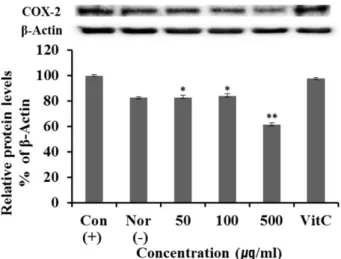
Western blot을 통한 iNOS 및 COX-2 단백질 발현 억제 효
과 측정염증유발인자인
iNOS, COX-2
단백질발현억제효과를측정하기위하여
western blot
을이용하여실험하였다.
이때 세포의여러조건에서도그발현정도의차이가거의없는house keeping gene
인β-actin
을positive control
로사용하 였다.
그결과Fig. 3, 4
과같이LPS
에의해증가된iNOS
와COX-2
단백질발현양이감소된것을확인할수있었으며,
대조군인
Vit. C
와비교하였을때대조군Vit. C
보다섬바디나물추출물에서
iNOS
와COX-2
단백질발현이더억제되 었음을확인할수있었다.
Fig. 2. Effect of Dystaenia takeshimana extract on production of nitric oxide in RAW 264.7 cell. Effect of 80% ethanol extracts of DT on NO production in LPS-induced RAW 264.7 cells. RAW 264.7 cells (1 × 10
5cells) were treated with DT 80% ethanol extract and LPS (1 µg/ml) for 24 h. NO production was deter- mined in culture supernatant by griess reagent. Con: control, in Raw 264.7 cells treated with LPS, Nor: normal, in Raw 264.7 cells not treated with LPS. Results are means ± S.D. of triplicate data.
Fig. 3. iNOS protein expression rate of extract from Dystaenia takeshimana on macrophage cell (Raw264.7). After Raw 264.7 cells (1 × 10
6cells) were started in serum free medium for 1 h the cells were treated 50, 100 and 500 µg/ml of extract from Dystae- nia takeshimana for 24 h. Con: control, in Raw 264.7 cells treated with LPS, Nor: normal, in Raw 264.7 cells not treated with LPS.
Results are means ± S.D. of triplicate data (Significant as com- pared to control. *p < 0.05, **p < 0.01).
Fig. 4. COX-2 protein expression rate of extract from Dystaenia takeshimana on macrophage cell (Raw264.7). After Raw264.7 cells (1 × 10
6cells) were started in serum free medium for 1 h the cells were treated 50, 100 and 500 µg/ml of extract from Dystae- nia takeshimana for 24 h. Con: control, in Raw 264.7 cells treated with LPS, Nor: normal, in Raw 264.7 cells not treated with LPS.
Results are means ± S.D. of triplicate data (Significant as com-
pared to control. *p < 0.05, **p < 0.01).
RT- PCR
을 통한 iNOS 및 COX-2 mRNA 발현 억제 효과 측정염증유발인자인
iNOS, COX-2 mRNA
발현억제효과를측정하기위하여
RT-PCR
을이용하여실험하였다.
이때세포의 여러 조건에서도 그 발현 정도의 차이가 거의 없는
house keeping gene
인GAPDH
을positive control
로사용 하였다.
그결과Fig. 5, 6
과같이LPS
에의해증가된iNOS
와
COX-2 mRNA
발현양이감소된것을확인할수있었으며
,
대조군인Vit. C
와비교하였을때대조군Vit. C
보다섬 바디나물추출물에서iNOS
와COX-2 mRNA
발현이유사하 거나억제되었음을확인할수있었다.
iNOS
억제활성을가진천연물질탐색을위한과거연구에서섬바디나물추출물은
LPS
로유도된NO
의생성을억 제한바있으며[7, 10], Kim
등은섬바디나물뿌리메탄올추 출물의헥산분획물과에틸아세테이트분획물에서분리한 쿠마린 및 플라보노이드가COX-2
와5-lipoxygenase (5-
LOX)
이중억제활성을가지며,
섬바디나물의소염작용은eicosanoid
의생성억제를통해부분적으로일어날수있다고보고한바있다
[8].
이러한사실을종합하였을때섬바디나물은항염증소재로서의가능성이있다고판단된다
.
요 약
본연구에서는섬바디나물의항염증효과를알아보기위 하여
LPS
로염증을유도한Raw 264.7
세포에대한섬바디나물
80%
에탄올추출물의효과를살펴보았다.
섬바디나물추출물을
LPS
로유도된Raw 264.7
대식세포에서전염증성 인자(iNOS, COX-2)
들을생성하여측정하였다.
섬바디나물 추출물의대식세포에서의세포독성측정을MTT
를수행하 였다.
섬바디 나물 추출물의 세포 독성을 측정한 결과, 1,000
μg/ml
의농도에서100%
이상의세포생존율을보였 다.
섬바디 나물 추출물의50, 100, 500
μg/ml
농도에서iNOS
와COX-2
의단백질발현억제효과를측정하기위해western blot
을통해측정하였고,
양성대조군으로는 β-actin
을사용하였다.
그결과,
섬바디나물추출물을western blot
을통해측정한
iNOS, COX-2
의단백질발현억제 효과는Fig. 5. iNOS mRNA expression rate of extract from Dystaenia takeshimana on macrophage cell (Raw264.7). After Raw264.7 cells (1 × 10
6cells) were started in serum free medium for 1 h the cells were treated 50, 100 and 500 µg/ml of extract from Dystaenia takeshimana for 24 h. Con: control, in Raw 264.7 cells treated with LPS, Nor: normal, in Raw 264.7 cells not treated with LPS. Results are means ± S.D. of triplicate data (Significant as compared to control. *p < 0.05, **p < 0.01).
Fig. 6. COX-2 mRNA expression rate of extract from Dystaenia takeshimana on macrophage cell (Raw264.7). After Raw264.7 cells (1 × 10
6cells) were started in serum free medium for 1 h the cells were treated 50, 100 and 500 µg/ml of extract from Dystaenia takeshimana for 24 h. Con: control, in Raw 264.7 cells treated with LPS, Nor: normal, in Raw 264.7 cells not treated with LPS. Results are means ± S.D. of triplicate data (Significant as compared to control. *p < 0.05, **p < 0.01).
Table 1. Sequence of the primers used for PCR.
Gene Primer Sequence (5’ → 3’)
GAPDH Sense TGA AGG TCG GTG TGA ACG GAT TTG GC Anti-sense CAT GTA GGC CAT GAG GTC CAC CAC COX-2 Sense GGA GAG ACT ATC AAG ATA GT
Anti-sense ATG GTC AGT AGA CTT TTA CA
iNOS Sense AAT GGC AAC ATC AGG TCG GCC ATC ACT
Anti-sense GCT GTG TGT CAC AGA AGT CTC GAA CTC
500
μg/ml
농도에서각각56%, 61.6%
로감소하였다.
섬바디 나물추출물의50, 100, 500
μg/ml
농도에서iNOS, COX-2
의
mRNA
의발현억제효과를측정하기위해RT-PCR
을통해측정하였으며