대한임상검사학회지 : 38권 제3호, 158-165, 2006
rpoB 유전자의 PCR-RFLP를 이용한 Mycobacterium 균종 동정의 유용성
미육군 121 병원 진단검사의학과1, 서울보건대학 임상병리과2
유 경 래1〮․박 정 오2
Identification of Mycobacterium species by rpoB Gene PCR-RFLP
Kyong-Nae Yu1 and Chung-Ho Park2
Department Pathology 121st General Hospital U.S. Army, Seoul 96205-0017, Korea1 Department of Biomedical Laboratory Science, Seoul Health College, Sungnam 461-713, Korea2
Although Mycobacterium tuberculosis complex strains remain responsible for the majority of diseases caused by mycobacterial infections worldwide, the increase in HIV infections has allowed for the emergence of other non-tuberculous mycobacteria as clinically significant pathogens. However, Mycobacterium species has a long period of incubation, and requires serious biochemical tests such as niacin, catalase, and nitrate test that are often tedious. The development of rapid and accurate diagnostics can aid in the early diagnosis of disease caused by Mycobacterium. The current DNA amplification and hybridization methods that have been developed target several genes for the detection of mycobacterial species such as hps65, 16S rDNA, rpoB, and dnaj. These methods produce rapid and accurate results. In this study, PCR-restriction fragment length polymorphism analysis(PCR-RFLP) based on the region of the rpoB gene was used to verify the identification of non-tuburculosis Mycobacterium species. A total of 8 mycobacterial reference strains and 13 clinical isolates were digested with restriction enzymes such as MspⅠ in this study. The results of using this process clearly demonstrated that all 13 specimens were identified by rpoB gene PRA method. The PCR-RFLP method based on the rpoB gene is a simple, rapid, and accurate test for the identification of Mycobacterium.
Key words : PCR-restriction fragment length polymorphism analysis rpoB gene, MspⅠ
1)
I. 서 론
Mycobacterium 속 균종 중에서 결핵균은 전 세계적으 로 심각한 보건상의 문제를 야기하는 병원성이 강한 균 종이다(김 등, 2002; Fitzgetrald 등. 2003). 1980년대 이후
교신저자 : 유경래, (우)96205-0017 미육군 121 병원 진단검사의학과
Tel : 011-9004-2502 E-mail : [email protected]
부터는 비결핵 Mycobacteria 감염증이 계속해서 증가되 고 있다(Guthertz 등, 1998; Ruiz 등, 2002). 이들은 항산 성, 호기성으로 일부 균주를 제외하고는 매우 느리게 성 장하는 균종들로써 약 70여 종의 다른 종으로 구성되어 있으며 이중 30여 종이 인간에게 각종 심각한 질환을 야 기한다고 보고 되고 있다(Cloud 등, 2002; Collins 등, 2003).
전통적인 방법은 균주의 성장속도, 색조, 균주 형태, 현미 경적 형태 관찰 및 생화학적인 검사를 시행한다(Shinners
등, 1999). Niacin, catalase, nitrate 등을 이용한 생화학적 인 방법은 균분리 이후 4~6주의 시간이 소요된다 (Collins 등, 1990). 그러나 결핵균의 신속한 동정법이 개 발됨에 따라 mycobacteria를 동정하는 전통적인 방법은 그 효용이 감소되고 있다. 현재 결핵균을 동정하는 모든 검사실은 신속한 동정법을 이용하도록 권고되고 있다. 신 속한 동정법으로는 DNA probe 이용, PCR, DNA sequencing, 그리고 chromatography와 같이 균의 구성성 분을 물리화학적 방법으로 직접 검출하는 법이 있다(김 등, 2002; Bannantine 등, 2002; Huard 등, 2003).
BACTEC 12B system은 p-nitroacetyl-p- aminohydroxy- propiophenone(NAP)을 이용하여 Mycobacterium tuber- culosis(MTB) complex와 nontubercuosis Mycobacterium (NTM)를 5~6일 만에 감별할 수 있다(Alcaide 등, 2003).
결핵균의 신속한 동정법이 개발됨에 따라 전통적인 동정 법은 그 효용이 감소되고 있다. 현재 결핵균을 동정하는 모든 검사실은 신속한 동정법을 이용하도록 권고되고 있 다. BACTEC 12B system은 NAP을 이용하여 TB complex와 NTM를 5~6일 만에 감별할 수 있다. 그러나 일부 NTM도 NAP에 의하여 억제될 수 있으므로 probe나 생화학적인 방법으로 확인하여야 한다.
최근 DNA 증폭기술의 발달로 개발된 분자유전 검사 법은, Mycobacterium 균속 내 여러 부위의 특이적인 특정 유전자를 이용하여 보다 빠르고 정확한 진단을 위한 유 전자 증폭 및 probe hybridization, 염기서열분석 등과 같 은 여러 검사방법이 시도되고 있다(Kim 등, 1997). 이와 같은 진단법에 이용되는 표적유전자(target gene)에는 hsp65, 16S rRNA, 그리고 dnaj 등 여러 유전자들이 사용 되고 있다(Lee 등, 1998; Kim 등, 1999; Lee 등, 2000;
Braccolo 등, 2003). 하지만 이들의 방법은 결과가 정확하 고 반복적이라는 장점이 있지만 고난이도의 기술과 여러 가지의 효소처리로 분석이 쉽지 않은 단점들도 있다.
II. 재료 및 방법
1. 대상
2005년 6월부터 8월 사이에 삼광의료재단 검사실에 의 뢰된 임상 검체로부터 분리 배양된 mycobacteria 13주와 한국결핵연구원(KIT)으로부터 분양한 표준균주 7주 (Table 1), 총 20주에 대하여 분석하였다. 13주의 임상분
Species Strain Source
M. avium ATCCa 25291 KITb
M. chelonea ATCC 35749 KIT
M. godonae ATCC 14470 KIT
M. kansasii type Ⅰ Pasteur institute KIT
M. smegmatis ATCC 19420 KIT
M. celatum type Ⅰ ATCC 51130 KIT M. absessus Pasteur institute YUMCc
M. tuberculosis ATCC 27294 KIT
aATCC, American type culture collection
bKIT, Korea institute of tuberculosis
cYUMC, Yonsei university college of medicine
Table 1. Reference strains of mycobacterial species used in this study
리주는 생화학적 검사로 균 동정을 확인하였다. 각 균주 는 전통적인 감별 방법인 Niacin strip test와 Gen-Probe test을 실행하여 결핵균의 여부를 재평가하였으며, 불일치 균주에 대해서는 PCR을 통한 확인 실험을 실시하였다.
2. 핵산추출과 중합효소연쇄반응
PCR 증폭을 위한 유전자 준비는 가열파쇄 유전자 추 출법을 사용하여 DNA를 추출하였다. Lowenstein-Jensen medium에서 배양된 균주의 집락을 증류수 400 μL와 screw-cap microcentrifuge tube에 넣어 5분간 boiling 하여 균체를 파쇄 하였다. 균파쇄액은 12000 rpm으로 5분간 원심분리하고 DNA가 함유된 상청액 100 μL를 상청액을 PCR에 사용하였다.
마이코박테리아 균종들의 rpoB 유전자를 증폭하기위 한 프라이머는 rpoB 유전자 염기서열(GenBank accession No. P47766) 내의 첫 번째 과변부위(V1)와 두 번째 상보 적 부위(conserved region2; C2) 중 대장균(Escherichia coil)의 유전적 정보를 바탕으로 V1의 171 bp와 C2의 189 bp 부위에 해당하는 부분을 표적으로 하여 제작하였다. 한 쌍의 프라이머는 5'-TCAAGGAGAAGCCGTACGA-3'(RPO5') 와 5'-GGATGTTGATCAGGTCTGC-3'(RPO3')로 유전자 내 902번째 염기에서부터 1261번째 염기까지 360 bp를 증폭하였다. PCR 반응은 10 pmol의 각각의 primer, 2 mM MgCl2, 200 mM deoxynucleoside triphosphate, 1 U 의 DyNAzyme Ⅰ DNA ploymerase(FINNZYMESY, Espoo, Finland)가 들어있는 DNA PCR Premix(M&D, Wonju,
Korea)를 이용하여 genomic DNA 10 μL와 멸균증류수 40 μL를 혼합하여 최종 부피가 50 μL가 되도록 혼합물을 만들었다.
첫 번째 pre-denatruation 과정은 94℃에서 5분실시 후에, denaturation 94℃ 20초, annealing 58℃ 20초, elongation 72℃ 30초로 35회 반복하였으며, 최종 elongation 72℃에 서 10분간 실시하였다. Thermocycler는 model 2700 PCR system(Aplied Biosystem, Fostercity, CA)가 사용되었다.
각 PCR 과정에서 positive, negative control, PCR size maker(M&D, Wonju, Korea)를 항상 사용되었다. 양성 대 조로는 M. bovis 표준균주를 PCR mixture에 사용하였고, 음성대조로 증류수를 사용하였다. 증폭산물은 2% agarose gel 위에서 전기영동을 한 후에 ethidium bromide(EtBr) 염색을 통해 확인하였다.
3. Restriction fragment length polymorphism
마이코박테리움 속 분류를 위하여 위 PCR 증폭산물을 MspⅠ(Boehringer Manheim biochemicals, Mannheim, Germany)을 이용하여 소화하고 RFLP 분석을 실시하였 다. 증폭된 PCR 산물 15 μL(genomic DNA 1.5 μg)에 MspⅠ(10 U/μL) 0.5 μL, 10x MspⅠ buffer 2 μL, 멸균된 증류수 2.5 μL를 넣어 20 μL의 혼합물을 잘 섞어주고, 12,000 rpm에서 3~5초간 원심 분리하고 37℃ 항온수조에 서 2시간 반응하였다. 2 μL의 loading buffer(0.25%
bromophenol blue, 40% sucrose)를 각 검체에 첨가한 후, PCR-RFLP DNA size marker(M&D, wonju, Korea)와 함께 4% metaphore agarose gel(FMC Bioproducts, Rockland, Maine)에 잠적한 후에 100 V에서 60~75분간 전기영동 탱크에 얼음을 담아 전기영동 하였다.
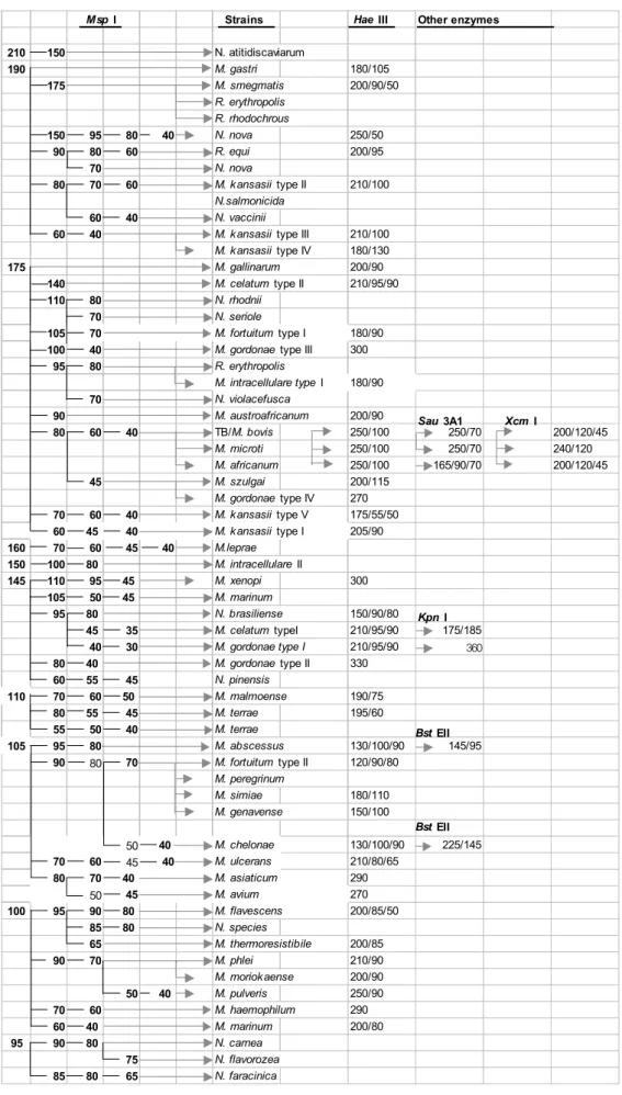
Gel을 EtBr 염색한 후, UV transilluminator로 분절편들의 결과를 확인하였다. 결과판독은 Mycobacteria- Nocardia 동 정 알고리즘(Fig. 4)을 참고하여 균을 동정하였다.
III. 결 과
1. rpoB 유전자 증폭 및 제한효소 분절법을 이용한 표준균주 동정
마이코박테리움 동정에 있어서 보다 신속, 정확, 간편 한 방법을 개발하기 위하여 여러 표적유전자를 이용한
유전자 증폭법이 연구되어 지고 있다. 여기서 이용한 표 적유전자 rpoB gene은 유전자 염기서열 분석을 통하여 PCR-RFLP 법으로 마이코박테리움 동정에 적합한 유전 자 구간으로 확인되었다.
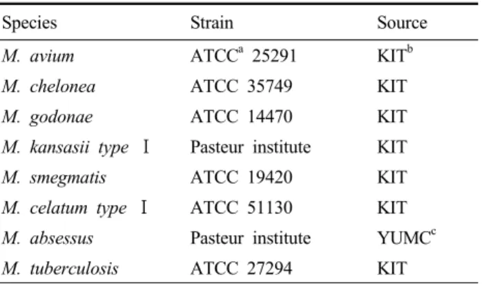
rpoB gene PCR을 통한 Mycobacterium의 표준균주에 대한 동정 가능 여부를 확인하기 위하여 대표적인 표준 균주 8주(Table 1)에 대하여 DNA 과변부위의 증폭으로 얻어진 산물에서의 다형태적 특성을 기초로 유전자 증폭 효소 분절 분석법을 사용하였다(Fig. 1).
600 bp 500 bp 400 bp
300 bp 200 bp
M 1 2 3 4 5 6 7 N P
Fig. 1. Results of PCR amplification of the rpoB gene using mycobacterial reference strains. PCR products were run on a 2% agarose gel. Lanes; M, DNA size marker; 1, M. avium; 2.
M. chelonea; 3, M. godonae; 4, M. kansasii Type I; 5, M.
smegmatis; 6, M. celatum; 7, M. abscessus; N, negative control;
P, positive control M. tuberculosis.
증폭된 유전자를 효소 분절 분석을 위하여 사용된 Msp
Ⅰ은 이미 M. tuberculosis, M. leprae, and M. smegmatis 의 rpoB gene 염기서열 분석정보를 통하여 선정되었다.
Msp Ⅰ는 DNA 염기서열 중 5'-C CGG-3', 3'-GGC C-5'에 서 forward sequence의 C와 C사이와 상보적인 reverse sequence의 C와 C사이를 sticky형태로 분절시키는 효과 가 있다. 효소 처리를 통한 RFLP 분석법에 의하여 표준 균주 8주에 대하여 동정한 결과 4% metaphore agarose gel 위에 Mycobacterium. avium은 PCR product가 105, 80, 50, 45-bp로 각각 분절되어 M. avium만의 고유한 분 절편을 형성하여 동정이 가능하였다(Fig. 2, Table 2).
rpoB gene으로 항산균 동정의 특이성을 확인한 후 삼광 의료재단의 임상검사실에서 임상검체로부터 분리, 배양, 동정된 임상표준균주 13주에 대해서도 PCR- restriction fragment length ploymorphism analysis (PCR- RFLP) 특 이도와 정확도를 빠르고 간편하게 Mycobacterium species 를 동정할 수 있었다
Reference strain of
Mycobacterium sp. Strain DNA fragment size(bp) Species identified by PCR-RFLP algorithm
M. avium ATCC 25291 105, 80, 50, 45 M. avium
M. chelonae ATCC 35749 105, 95, 80, 50, 40 M. chelonae
M. gordonae ATCC 14470 175, 80, 45 M. gordonae
M. kansasii type Ⅰ Pasteur institute 175, 60, 45, 40 M. kansasii type Ⅰ
M. smegmatis ATCC 19420 200, 90 M. smegmatis
M. celatum type Ⅰ ATCC 51130 145, 95, 45 M. celatum type Ⅰ
M. abscessus Pasteur institute 105, 95, 80 M. abscessus
M. tuberculosis ATCC 27294 175, 80, 60, 40 M. tuberculosis
Table 2. PCR-RFLP profiles obtained from reference strains of mycobacteria used in this study
M 1 2 3 N M 4 5 6 7 P M
3 50 b p
2 50 b p
2 00 b p
1 75 b p 1 50 b p 1 25 b p
1 05 b p 9 5 b p 8 0 b p 6 0 b p
5 0 b p 4 5 b p 4 0 b p
Fig. 2. Results of PCR-RFLP analysis of mycobacterial reference strains. A set of PCR primers, RPO5' and RPO3' was used. Amplified DNAs were digested with MspⅠand run on a 4% metaphore agarose gel. Lanes:
M, PCR-RFLP size marker; 1, M. avium; 2. M. chelonea; 3, M. gordonae; N, negative control; 5, M. kansasii Type I; 6, M. smegmatis; 7, M. celatum; 8, M. abscessus; P, positive control, M. tuberculosis
이와 같은 결과는 단일 효소 MspⅠ을 이용한 PCR-RFLP 법으로 임상검사실에서 분리되어지는 거의 모든 항산균을 분리 동정 할 수 있는 것을 확인하였다(Fig. 3, Table 3).
Ⅳ. 고 찰
Mycobacteria의 배양과 동정은 성장이 매우 느리고 각 균명을 정확히 동정하는 데 많은 시간과 노력이 소요되 어, mycobacteria 감염증 치료에 적절한 검사정보를 제공 하지 못하고 있다. 특히 최근에 증가되는 AIDS 환자와 비결핵성 mycobacteria 감염증과의 연관성이 밝혀짐에 따 라(Guthertz 등, 1998), Mycobacterium의 균속 동정에 더 욱더 많은 연구가 필요되고 있다. 이러한 이유로 보다 신 속하고 정확한 방법으로 mycobacteria를 동정하고자하는 연구가 진행되어 여러 가지 검사법이 소개되었다. 제한효
소와 DNA probe를 이용한 방법(Collins 등, 1990), 각 균 종에 특이적으로 반응하는 mycobacteriophage 기법 (Alcaide 등, 2003), DNA sequencing 법(Cloud 등, 2002), 그리고 PCR-RFLP 법(Kim 등, 1999; 김 등, 2002) 등이 소개되었으며, 이 중 DNA sequencing 법은 표준검사법으 로 정착하고 있고 PCR-RFLP 법은 임상검사로 정착하고 있다. 최근에는 real-time PCR 법(Broccolo 등, 2003)이 연구되고 있어 보다 쉽고 정확하게 mycobacteria 균속을 동정할 수 있게 될 것이다. 분자유전학적인 방법으로 mycobacteria 균속의 동정을 하기 위해서는 각 균종 간 변이가 심한 유전자를 선택하는 것이 중요하다. 전통적으 로는 16S rDNA 유전자가 많이 사용되었다(Cloud 등, 2002) 그리고 rpoB 유전자(Kim 등, 1999) 등이 시도되었 으며 PCR-RFLP 분석을 위해서는 rpoB 유전자가 가장 많 이 이용되고 있다(Lee 등, 2000; 김 등, 2002). RFLP 분석 에서 제한효소의 선택은 검사의 특이도를 결정하는 가장
M 9 10 11 M 12 13 14 15 M
350 bp
250 bp
200bp 175 bp 150bp 125 bp
105 bp 95 bp 80 bp
60bp
50 bp
45 bp
40 bp M 1 2 3 4 M 5 6 N 7 8 M
350 bp 250 bp
200bp 175 bp 150bp 125 bp
105 bp 95 bp 80 bp
60bp 50 bp 45 bp 40 bp
(B)
Fig. 3. PCR-RFLP analysis using rpoB gene for species identification of clinical isolates of mycobacteria. (A). Clincal isolates of mycobacteria were used for PCR amplification using primers RPO5' and RPO3'. PCR products were run on a 2% agarose gel. M, DNA size marker; Lane M, M&D PCR DNA size marker, lanes 1-14, clinical isolates of mycobacteria, lane 15, negative control. (B), PCR-RFLP analysis of PCR products shown in (A). MspⅠdigestion of PCR products were run on a 4% metaphore agarose gel. Lane M, M&D PCR-RFLP DNA size marker, lanes 1-14, clinical isolates of mycobacteria, lane 15. negative control.
600 500 400 300 200
M 1 2 3 4 5 6 7 8 9 10 11 12 13 14 15 (A)
Species identified by biochemical test DNA fragment size(bp) Species identified by PCR-RFLP algorithm
M. ulcerans 105, 70, 60, 42, 40 M. ulcerans
M. kansasii typeⅡ 195, 80, 70, 60 M. kansasii typeⅡ
M. malmoense 110, 75, 70, 60, 45 M. malmoense
M. cleatum typeⅡ 175, 140, 42 M. cleatum typeⅡ
M. intracellulare typeⅡ 150, 100, 80 M. intracellulare typeⅡ
M. gordonae Ⅳ 175, 80, 45 M. gordonae Ⅳ
M. tuberculosis 175, 80, 60, 40 M. tuberculosis
M. avium 105, 80, 50, 45 M. avium
M. godonae type Ⅲ 160, 95, 45 M. godonae type Ⅲ
M. celatum type Ⅰ 150, 95, 47, 42 M. celatum type Ⅰ
M. gordonae typeⅡ 150, 80, 45 M. gordonae type Ⅱ
M. abscessus 190, 120, 42, 40 M. abscessus
M. avium 105, 80, 50, 45 M. avium
M. tuberculosis 175, 80, 60, 40 M. tuberculosis
Table 3. PCR-RFLP profiles obtained from clinical isolates of mycobacteria used in this study
Msp I Strains Hae III Other enzymes
210 150 N. atitidiscaviarum
190 M. gastri 180/105
175 M. smegmatis 200/90/50
R. erythropolis R. rhodochrous
150 95 80 40 N. nova 250/50
90 80 60 R. equi 200/95
70 N. nova
80 70 60 M. kansasii type II 210/100
N.salmonicida
60 40 N. vaccinii
60 40 M. kansasii type III 210/100
M. kansasii type IV 180/130
175 M. gallinarum 200/90
140 M. celatum type II 210/95/90
110 80 N. rhodnii
70 N. seriole
105 70 M. fortuitum type I 180/90
100 40 M. gordonae type III 300
95 80 R. erythropolis
M. intracellulare type I 180/90
70 N. violacefusca
90 M. austroafricanum 200/90
80 60 40 TB/M. bovis 250/100 250/70 200/120/45
M. microti 250/100 250/70 240/120
M. africanum 250/100 165/90/70 200/120/45
45 M. szulgai 200/115
M. gordonae type IV 270
70 60 40 M. kansasii type V 175/55/50
60 45 40 M. kansasii type I 205/90
160 70 60 45 40 M.leprae
150 100 80 M. intracellulare II
145 110 95 45 M. xenopi 300
105 50 45 M. marinum
95 80 N. brasiliense 150/90/80
45 35 M. celatum typeI 210/95/90 175/185
40 30 M. gordonae type I 210/95/90 360
80 40 M. gordonae type II 330
60 55 45 N. pinensis
110 70 60 50 M. malmoense 190/75
80 55 45 M. terrae 195/60
55 50 40 M. terrae
105 95 80 M. abscessus 130/100/90 145/95
90 80 70 M. fortuitum type II 120/90/80
M. peregrinum
M. simiae 180/110
M. genavense 150/100
50 40 M. chelonae 130/100/90 225/145
70 60 45 40 M. ulcerans 210/80/65
80 70 40 M. asiaticum 290
50 45 M. avium 270
100 95 90 80 M. flavescens 200/85/50
85 80 N. species
65 M. thermoresistibile 200/85
90 70 M. phlei 210/90
M. moriokaense 200/90
50 40 M. pulveris 250/90
70 60 M. haemophilum 290
60 40 M. marinum 200/80
95 90 80 N. carnea
75 N. flavorozea
85 80 65 N. faracinica
Bst EII Kpn I
Sau 3A1 Xcm I
Bst EII
Fig. 4. The algorithm used the species identification of mycobacteria in this study(Lee, 등, 2000)
중요한 변수이다. rpoB 유전자의 각 균종 간 변이가 많은 부위에 Msp I 제한효소 자리가 분포하여 Mycobacterium 균종 간 차이를 잘 표현하는 것으로 알려졌다(Lee 등, 2000). MspⅠ는 DNA 염기서열 중 5'-C/CGG-3' 부위의 C 와 C사이를 sticky 형태로 분절시킨다. 본 연구에서 임상 검체에서 가장 흔하게 분리되는 8종의 Mycobacterium 표 준균주는 상호 교차반응이나 해석상의 어려움이 없이 정 확하게 감별되었다. 또한 13주의 임상균주도 동일한 성적 을 보였다. Kim 등(1999), Lee 등(2000), 그리고 김 등 (2002)의 결과와 큰 차이가 없었으며 현재 임상검사실에 서 결핵균 동정진단에 가장 널리 사용되고 있는 방법은 Gen-Probe로 전통적인 방법인 niacin 시험보다 빠르고 정확하다고 평가되고 있다. 그러나 비용이 많이 들고 비 결핵균성 mycobacteria의 감별에는 적용하기가 어려워 M. tuberculosis와 NTM과의 감별검사로만 사용되고 있는 실정이다. 따라서 여러 검사실에서 NTM의 감별동정을 위해 검사를 참고검사실(reference laboratory)로 의뢰하는 실정이다.
rpoB 유전자를 대상으로 하는 PCR-RFLP 방법은 최근 Kit system으로 개발되면서 검체 및 시약조작의 편이성, 결과의 정확성 및 재현성을 높여서 임상검사실에서 분리 된 결핵균 집락을 이용하여 쉽게 이용할 수 있도록 하였 다. 이 방법은 앞으로 임상검사실에서도 NTM 동정에 쉽 게 적용될 것으로 기대된다. 검체로부터 직접 PCR-RFLP 를 적용하여 mycobacteria를 동정하는 방법을 적용하면, 상재균으로 인하여 해석상 문제가 생기는 검체를 제외하 고는, 매우 유용한 적용방법이 될 것으로도 기대한다.
참 고 문 헌
1. Alcaide F, Gali N, Dominguez J, Berlanga P, Blanco S, Orus P, Martin R. Usefulenss of a New Myco- bacteriophage-Based Technique for Rapid Diagnosis of Pulmonary Tuberculosis. J Clin MIcrobiol 41:2867-2871, 2003.
2. Bannantine JP, Baechler E, Qing Z, LingLing Li, Kapur V. Genome Scale Comparison of Myco- bacterium avium subsp. paratuberculosis with Mycobacterium avium subsp. avium Reveals Potential Diagnostic Sequence. J Clin MIcrobiol 40:1303-1310, 2002.
3. Broccolo F, Scarpellini P, Locatelli G, Zingale A, Brambilla AM, Cichero P, Sechi L A, Lazzarin A, Lusso P, Maltati MS. Rapid diagnosis of Mycobacterial Infections and Quantitation of Mycobacterium tuberculosis Load by Two Real-Time Calibrated PCR assay. J Clin MIcrobiol 41:4565-4572, 2003.
4. Cloud JL, Neal H, Rosenberry R, Turenne CY, Jama M, Hillyard DR, Carroll KC. Identification of Mycobacterium spp. by using a Commercial 16S Ribosomal DNA Sequencing Kit and Additional Sequencing Libraries. J Clin MIcrobiol 40:400-406.
2002.
5. Collins DM, Gabric DM, de Lisle GW. Identification of two groups of Mycobacterium paratuberculosis strains by restriction endounclease and DNA hybridization J Clin MIcrobiol 28:1519-1596, 1990.
6. Collins DM, Zoete MD, Cavaignac SM.
Mycobacterium avium subsp. paratuberculosis Strains from Cattle and Sheep can be distinguished by a PCR test based on a Novel DNA Sequence difference. J Clin. MIcrobiol 40:4760-4762, 2002.
7. Fitzgetrald SD, Zwick LS, Berry DE, Church SV, Kaneene JB, Reed WM. Experiential inoculation of pigeons(Columba livia) with Mycobacterium bovis.
Avian disease 47:470-474, 2003.
8. Guthertz LS, Damasker B, Bottone EJ, Ford EG, Midura TF, Janda JM. Mycobacterium avium and Mycobacterium intracellulare infection in patients with and without AIDS. J Infect Dis 160:1037-1041, 1998.
9. Huard RC, Lazzarini LC de O, Butler WR, Sooligen DV, Ho JL. PCR-Based method to differentiate subspecies of the Mycobacterium tuberculosis Complex on the Basis of Genomic Deletions. J Clin MIcrobiol 41:1637-1650, 2003.
10. Kim BJ, Lee SH, Lyu MA, Kim SJ, Bai GH, Kim SJ, Chae GT, Kim EC, Cha CY, Kook YH.
Identification of Mycobacterial Species by comparative sequence analysis of the RNA Polymerase gene(rpoB). J Clin MIcrobiol. 37:1714-1720, 1999.
11. Kim BJ, Lee SH, Lee MA, Lyu SJ, Kim GH, Bai SJ, Kim GT, Chae EC, Kim CY, Cha, KooK YH.
Identification of mycobacterial species by comparative sequence analysis of the RNA polymerase gene (rpoB). J Clin MIcrobiol 37:1724-1720, 1990.
12. Lee, HY, Park HJ, Cho SN, Bai GH, Kim SJ. Species Identification of Mycobacteria by PCR-Restriction Fragment Length Polymorphism of the rpoB Gene. J Clin MIcrobiol 38:2966-2971, 2000.
13. Lee H, Cho SH, Bang HE, Lee JH, Bae GH, Kim SJ, Kim JD. Molecular analysis of rifampin-resistant Mycobacterium tuberculosis isolated from Korea by polymerase chain reaction-single strand conformation polymorphism sequence analysis. Int J Tuber lung dis 2:585-589, 1998
14. Kim bj, Kim SY, Park BH, Lyu MA, Park IK, Bai GH, KIm SJ, Cha CY, Kook YH. Mutations in the rpoB gene of Mycobacterium tuberculosis that interfere with PCR-single-strand conformation polymorphism
analysis for rifampin susceptibility testing. J Clin Microbiology 35(2):492-494, 1997.
15. Ruiz. P, gutierrez J, Zerolo J. Cassl M. Geno Type Mycobacterium Assay for identification of Myco- bacterial Species Isolated from Human Clinical Samples by Using Liquid Medium. J Clin MIcrobiol 40:3076-3078, 2002.
16. Shinners D, Yeager H Jr. Nontuberculous myco- bacterial infection; clinical syndromes and diagnosis;
overview, In Scholossberg D(ed.). Tuberculosis and nontuberculous mycobacterial infections, 4th ed., p341-350, W.B. Sanuders Co. Philadelphia, Pa, 1999.
17. 김범준, 변경희, 배길한, 김상재, 이근화, 황응수, 차 창룡, 국윤호. DT1-DT6 PCR에 의한 국내 분리 Mycobacterium avium complex의 감별. Korean J Bacteriol Virol 32:33-38, 2002.