Evaluation of Combined Use of BacT/ALERT 3D Liquid Culture System and PCR-RFLP for Detection and Identification of
Mycobacteria from Bronchial Specimens
Hae-Sun Chung, Chang-Seok Ki, Jang Ho Lee, Nam Yong Lee Department of Laboratory Medicine and Genetics, Samsung Medical Center,
Sungkyunkwan University School of Medicine, Seoul, Korea Background: We evaluated BacT/ALERT 3D liquid
culture system (bioMérieux, USA) and PCR-restric- tion fragment length polymorphism (RFLP) for recov- ery and direct identification of mycobacteria, and the results were compared with a conventional culture system using an egg-based solid medium.
Methods: A total of 3,037 bronchial specimens (2,309 bronchial washing fluids and 728 bronchoalveolar lavages) were collected. Decontaminated specimens were inoculated to both BacT/ALERT MP liquid me- dia and Ogawa solid media (3%, Shinyang, Korea).
Recovery rate and detection time were compared be- tween the two systems. Liquid media from positive cultures were centrifuged and the pellets were tested for direct identification of mycobacteria by PCR-RFLP using Myco-ID (M&D Inc., Korea).
Results: A total of 518 isolates, including 215 M. tu- berculosis (MTB) and 303 non-tuberculosis mycobac- teria (NTM), were recovered. The liquid media de- tected 492 isolates (16.2%), including 195 MTB and
297 NTM), whereas the solid media detected 416 isolates (13.7%), including 187 MTB and 229 NTM (P<0.001); 102 isolates (28 MTB and 74 NTM) were recovered only by the liquid media, while 26 (20 MTB and 6 NTM) isolates were recovered only by the solid media. The mean time to detection was 18.1 days by the liquid media and 29.3 days by the solid media (P<0.001). The overall time to species identification from inoculation was 21.8 days. Direct PCR-RFLP from the liquid media identified 39.1% of MTB, 6.3% of M. avium, 19.05 of M. abscessus, and 12.6% of M. intracellulare respectively.
Conclusion: Combined use of a liquid culture system and PCR-RFLP improved the recovery rate and shor- tened the detection time. However, solid media is still necessary to maximize the diagnostic efficiency.
(Korean J Clin Microbiol 2009;12:37-42)
Key Words: Mycobacteria, Culture, Liquid media, PCR- RFLP, Bronchial specimen
37
Received 5 August, 2008, Revised 12 December, 2008 Accepted 25 February, 2009
Correspondence: Nam Yong Lee, Department of Laboratory Medicine and Genetics, Samsung Medical Center, Sungkyunkwan University School of Medicine, 50 Ilwon-dong, Gangnam-gu, Seoul 135-710, Korea.
(Tel) 82-2-3410-2706, (Fax) 82-2-3410-2719, (E-mail) micro.lee@
samsung.com
서 론
마이코박테리아에는 결핵균(Mycobacterium tuberculosis, MTB), 나병균(M. leprae)과 이들을 제외한 비결핵 항산균(nontubercu- lous mycobacteria, NTM) 등이 포함된다. MTB에 의한 폐결핵 과 NTM에 의한 NTM 폐질환은 대표적인 폐 마이코박테리움 감염(Mycobacterial pulmonary infection)으로써, 전 세계적으로 중요한 질환이며 많은 관심의 대상이다[1-3]. 특히 우리나라는 폐결핵의 유병률이 높고, 최근에는 NTM 폐질환의 중요성도 강조되고 있다[4].
폐 마이코박테리움 감염의 효과적인 관리에 있어서 신속하고 정확한 진단은 필수적이다. 그러나 기존의 전통적인 검사방법 인 항산균 염색과 배양 검사는 한계가 있었다. 항산균 염색은 저렴하고 간편하며 검사 결과를 빨리 얻을 수 있는 장점이 있 으나, MTB와 NTM을 구별할 수 없고 민감도와 특이도가 낮은 단점이 있다. 배양 검사의 경우, 가장 확실한 방법이며 표준 진 단법(gold standard) 방법이지만 오랜 시간이 필요하다는 단점 이 있다. 배양 배지는 크게 고체 배지와 액체 배지로 구분되는 데, 특히 고체 배지의 경우 검출 시간이 길고 액체 배지에 비해 민감도가 낮다. 그에 비해 액체 배지는 검출 시간이 빠르고 검 출률이 높으며, 장비를 이용하여 더 편리하게 우수한 성과를 얻을 수 있다. 그러나 액체 배지는 오염률이 높고, 배양 양성 시 MTB와 NTM을 구별할 수 없으며, 비용이 높다는 등의 단 점도 있다. 현재 시판되어 이용되고 있는 액체 배지 배양 장비 로는 BACTEC MGIT 960 system (Becton Dickinson, Sparks, MD, USA), VersaTRECK (ESP culture system Ⅱ; Trek
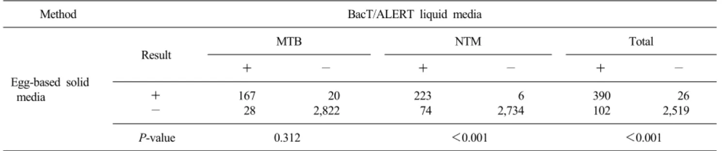
Table 1. Isolations of mycobacteria from 3,037 specimens
Method BacT/ALERT liquid media
Egg-based solid media
Result MTB NTM Total
+ − + − + −
+ 167 20 223 6 390 26
− 28 2,822 74 2,734 102 2,519
P-value 0.312 <0.001 <0.001
Abbreviations: MTB, M. tuberculosis; NTM, nontuberculosis mycobacteria.
Diagnostics, Inc., Westlake, OH, USA), BacT/ALERT 3D liquid culture system (bioMérieux, Durham, NC, USA) 등이 있다[5,6].
마이코박테리아 배양 시 Center for Disease Control (CDC), Clinical and Laboratory Standards Institute (CLSI), College of American Pathologists (CAP) 등에서는 액체 배지와 고체 배지 를 포함하여 두 개 이상의 배지를 사용하도록 권고하고 있다 [1,7-9]. 그러나 국내에서는 아직 액체 배지의 사용이 제한되어 있는 실정이다[10].
본 연구에서는 기관지 검체에서 액체 배지 배양 자동화 장비 BacT/ALERT 3D liquid culture system과 중합효소연쇄반응-제 한절편길이다형성(PCR-restriction fragment length polymor- phism, PCR-RFLP)을 이용하여 마이코박테리아의 검출 및 동 정을 시행하고, 기존의 고체 배지 배양과 비교하였다.
재료 및 방법 1. 재료
2006년 4월부터 2007년 9월까지 결핵균 도말 및 배양 검사 가 의뢰된 총 3,037건의 기관지 검체(기관지 세척액 2,309예, 기관지폐포 세척액 728예)를 대상으로 하였다.
2. 검체 전처리
50 mL의 원추형 시험관에 검체와 동량의 N-acetyl-L-cys- teine (NALC)-2% NaOH 용액을 넣고 15분간 실온에 방치 후 멸균 증류수로 50 mL까지 채워 냉장 원심분리기에서 3,000 g 에서 15분간 원침하였다. 상청액을 제거하고 남은 침전물로 염 색과 배양을 실시하였다.
3. 항산균 염색 및 결과 판독
전처리가 끝난 검체를 유리 슬라이드에 도말한 후 형광 염색 법으로 항산균 염색을 실시하였다. 형광 염색 결과 CDC의 기 준에 의한 “±” 이상의 슬라이드는 Ziehl-Neelsen 염색법으로 확 인하여 CDC 판정법에 따라 보고하였다.
4. 마이코박테리아 배양
전처리된 검체를 고체 배지(3% Ogawa; Shinyang Chemical Co., Seoul, Korea)에 접종하여 37oC에서 배양하였고, 1주에 한 번씩 8주 동안 균 집락을 관찰하였다. 또한 동시에 Middlebrook 7H9 broth base가 포함되어 있는 BacT/ALERT MP 액체 배지 (bioMérieux, Durham, NC, USA)에 접종하여 BacT/ALERT 3D liquid culture system에 배양하였다. BacT/ALERT 3D liquid culture system은 매 10분 마다 마이코박테리아가 성장하면 생 성되는 CO2를 pH 변화로 감지하여 정해진 알고리즘에 따라 지 속적으로 마이코박테리아 성장 유무를 자동으로 검색한다. 장 비에서 양성 신호가 발생되면 배양병을 꺼내어 배양액을 원심 분리하여 침사를 얻어 항산균 염색을 실시하고 균 동정을 실시 하였다. BacT/ALERT MP 액체 배지에서 양성 신호가 발생되 었지만, 이후 배양액 항산균 염색이나 균종 동정에서 음성 결 과가 나온 경우는 위양성으로 판단하였다.
5. 마이코박테리아의 균종 동정
배양 양성인 BacT/ALERT MP 액체 배지 병에서 배양액을 취해 100oC 끓는 물에 20분간 담가 놓은 후 원심분리하여 추출 된 DNA로 직접 Myco-ID (M&D, Seoul, Korea)를 이용하여 PCR-RFLP 방법으로 마이코박테리아의 균종 수준까지 동정하 였다. Myco-ID는 rpoB 유전자의 다형성을 이용한 것으로, rpoB 유전자의 일정 부위를 다량 증폭한 후 제한효소 처리를 하여 절편의 크기에 따라 키트에 포함된 ‘마이코박테리아 및 노카디아 동정 알고리즘’표를 기준으로 균을 동정한다.
6. 통계 처리
두 배양 방법 간의 비교는 SPSS 프로그램(Window version 11.5)을 이용하여, 검출률의 차이는 McNemar’s test 검정을 하 였고, 검출 시간의 차이는 Wilcoxon signed rank test 검정을 시 행하였다. 통계적 유의 수준은 P값 0.05 이하로 하였다.
Table 2. Time (days) to detect mycobacteria
BacT/ALERT Egg-based
Method P-value
liquid media solid media
MTB 24.6 30.8 <0.001
NTM 12.8 28.0 <0.001
Total 18.1 29.3 <0.001
Abbreviations: See Table 1.
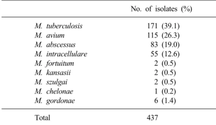
Table 3. Isolation frequency of mycobacteria from bronchial spe- cimen
No. of isolates (%)
M. tuberculosis 171 (39.1)
M. avium 115 (26.3)
M. abscessus 83 (19.0)
M. intracellulare 55 (12.6)
M. fortuitum 2 (0.5)
M. kansasii 2 (0.5)
M. szulgai 2 (0.5)
M. chelonae 1 (0.2)
M. gordonae 6 (1.4)
Total 437
결 과 1. 검출률 비교(Table 1)
총 3,037개의 검체 중 518검체(17.1%)에서 항산균이 배양되 었다. 이 중 MTB는 215건(7.1%), NTM은 303건(9.9%)이었다.
BacT/ALERT MP 액체 배지에서는 492건(16.2%; 195 MTB, 297 NTM)이 검출되었고, 고체 배지에서는 416건(13.7%; 187 MTB, 229 NTM)이 검출되었다. BacT/ALERT MP 액체 배지와 고체 배지의 검출률 차이가 MTB에서는 유의하지 않았으나 (P=0.312), 마이코박테리아 전체와 NTM에서는 유의한 차이가 있었다(P<0.001).
BacT/ALERT MP 액체 배지에서만 검출된 경우는 102건 (3.4%; 28 MTB, 74 NTM)이었고, 고체 배지에서만 검출된 경 우는 26건(0.9%; 20 MTB, 6 NTM)이었다.
BacT/ALERT MP 액체 배지에서 위양성인 경우는 총 3,037 개의 검체 중에 66건(2.2%)이었으며, 고체 배지가 오염된 경우 는 62건(2.0%)이었다.
2. 검출 시간 비교(Table 2)
마이코박테리아를 검출하는 데 소요된 평균 시간은 BacT/
ALERT MP 액체 배지에서는 18.1일, 고체 배지에서는 29.3일 로 유의한 차이가 있었다(P<0.001). 이러한 검출 시간의 차이
는 MTB, NTM 모두에서 유의하게 나타났다. BacT/ALERT MP 액체 배지에 접종 후 양성 배양병에서 직접 PCR-RFLP를 이용하여 균을 동정하기까지 소요된 총 평균 시간은 21.8일이 었다.
3. 마이코박테리아 균종 동정 결과(Table 3)
BacT/ALERT MP 액체 배지 양성 중 총 468개의 배양병에서 추출한 DNA로 직접 PCR-RFLP를 시행하여 총 437건의 마이코 박테리아를 동정하였다. 22건(5.0%)은 증폭 산물을 얻는 데 실 패하였으며, 9건(2.1%)은 동정 알고리즘에 의해 동정이 되지 않 았다. 동정된 마이코박테리아 중 MTB가 가장 흔했고(39.1%), 그 다음으로 M. avium (26.3%), M. abscessus (19.0%), M. intra- cellulare (12.6%) 순으로 흔하게 동정되었다. 다른 균종은 1%
미만으로 동정되었다. 전체 동정된 마이코박테리아 중 NTM의 비율은 60.9%였다. 2가지 이상의 마이코박테리아가 배양된 경 우는 1건이었다.
고 찰
본 연구 결과, BacT/ALERT MP 액체 배지에서는 양성 검체 의 95.0%가 검출되었고, 고체 배지에서는 양성 검체의 84.6%
만 검출되어, BacT/ALERT MP 액체 배지의 마이코박테리아 검출률이 고체 배지보다 우수하였다. 그 차이는 MTB (90.1 vs.
87.0%)에서는 유의하지 않았으나(P=0.312), 마이코박테리아 전체(95.0 vs. 84.6%)와 NTM (98.0 vs. 75.6%)에서는 유의하게 BacT/ALERT MP 액체 배지가 고체 배지보다 검출률이 높았다(P
<0.001). Gil-Setas 등의 연구[11]에서는 MB/BacT 배지(Organon Teknika, Turnhout, Belgium)에서는 90.1%, Lowenstein-Jensen (L-J) 배지(Becton Dickinson, Cockeysville, MD, USA)에서는 77.5%가 검출되었다고 하였고, Mirovic과 Lepsanovic[12]은 MB/BacT 배지(Organon Teknika, Durham, NC, USA)에서는 93.2%, L-J 배지에서는 67.3%로 보고한 바 있다. 또한, Ogawa 배지와 액체 배지를 비교한 Abe 등의 연구[13]에서 3% Ogawa 배지에서의 검출률은 75.6%로 액체 배지 배양 장비인 BACTEC 에서의 93.0%보다 유의하게 낮았다. 이상의 연구들에서 액체 배지가 계란기초의 고체 배지인 L-J 배지와 Ogawa 배지보다 우수한 검출률을 보이는 것을 확인할 수 있고, 그 외에도 여러 논문들에서 일관되게 액체 배지가 고체 배지에 비해서 마이코 박테리아의 검출에 유리하다고 보고되고 있다[14-17]. 하지만 액체 배지와 고체 배지를 병용 시 검출률이 높아진다는 점이 보고된 바 있으며[11,14,17,18], 본 연구에서도 고체 배지에서 만 검출된 경우가 총 양성 검체 중 5.0%, 특히 MTB 양성 검체 중 9.3% 있어, 고체 배지 배양도 배양의 민감도를 높이기 위해 필요할 것으로 판단된다. 이는 CDC, CLSI, CAP 등에서 권고 하는 바와도 일치한다[1,7-9].
BacT/ALERT MP 액체 배지에서의 위양성은 다른 세균의 성 장 등으로 인한 pH의 변화에 의한 것으로, 본 연구에서는 2.2%
의 위양성률을 보였다. 위양성인 경우, 마이코박테리아에 대한 검사 이외의 세균에 대한 검사를 시행하지 않아, 위양성 중 오 염에 의한 것을 정확히 구분할 수는 없었지만, 상당 부분이 오 염에 의한 위양성일 것으로 생각된다. 기존의 액체 배지에 대 한 연구에서 오염률이 1.6∼9%로 다양하게 보고되고 있는데, 일반적으로 고체 배지보다 높은 것으로 알려져 있다[11-16]. 한 편, 일부 연구에서 오염률과 구별하여 위양성률을 0∼0.95%로 보고하였다[11,14,16]. 본 연구에서 오염률을 포함한 전체 위양 성률은 기존에 알려진 것보다 낮은 수준이며, 고체 배지의 오 염률과 크게 차이가 없는 것으로 나타났다.
마이코박테리아가 검출된 시간에 있어서 BacT/ALERT MP 액체 배지가 고체 배지보다 유의하게 빨라(18.1 vs. 29.3일) 결 과 보고를 10일 이상 단축할 수 있었다. 이 차이는 MTB (24.6 vs. 30.8일)와 NTM (12.8 vs. 28.0일) 모두에서 유의하였으며 (P<0.001), 특히 NTM에서 매우 단축되었다. BacT/ALERT 3D system의 경우 기기에 의한 양성 신호 검출의 예민도가 높고, 매 10분마다 지속적으로 균의 성장을 감시 및 보고하는 방법으 로 운용되기 때문에 1주일에 한 번 빈도로 균 집락 유무를 육 안으로 확인하는 고체 배지에 비해 균의 성장을 검출하는 데 소요되는 시간이 단축될 수 있었다. 기존의 액체 배지와 고체 배지 비교 연구들에서도 액체 배지 사용 시 검출 시간이 단축 됨이 보고되었다[11-16]. 하지만 본 연구에서 균을 동정하기까 지 소요된 총 평균 시간은 21.8일로, CDC에서 권고하는 동정 소요 기간인 14일에는 미치지 못하였다. 이는 대상 검체가 기 관지 검체 중 기관지 세척액과 기관지폐포 세척액이어서 균이 희석되었을 가능성에 의한 것일 수 있다. 또한 낮은 오염률이 검출 시간의 연장과 관계가 있다는 보고가 있어[16], 본 연구의 결과를 뒷받침한다.
마이코박테리아 균종 동정 결과 MTB가 단일 균종으로는 가 장 흔하게 분리되었으며(39.1%), 그 외 흔하게 분리된 NTM은 M. avium (26.3%), M. abscessus (19.0%), M. intracellulare (12.6%)이었다. NTM의 균종 비율은 지역적인 차이가 있는 것 으로 알려져 있으며[19], 본 연구에서 발견된 흔한 NTM 균종 은 다른 국내 연구와 비슷한 결과였다[19-24]. 한편, 전체 NTM 의 비율은 60.9%로 다소 높은 비율이었는데, 그 요인으로 대상 검체가 기관지경을 시행하여 채취한 기관지 검체라는 점, 본원 에 내원한 NTM 폐질환 환자의 비율이 타 병의원에 비해 높다 는 점 등을 생각해 볼 수 있다.
본 연구 결과에서 확인하였듯이, 액체 배지가 검출률이 높고 검출 시간이 빠르다는 장점이 있으나, 반면 몇 가지 단점도 지 적되고 있다. 그 중 액체 배지에서 양성 시, 육안으로 균종이 구별되는 고체 배지에서와는 달리, MTB와 NTM의 감별이 안 된다는 점이 있다. 하지만 이러한 단점은 본 연구에서 시행한
PCR-RFLP나 다른 MTB를 검출할 수 있는 분자유전학적 방법 을 동원하면 극복할 수 있을 것이다. 또 액체 배지 사용의 중요 한 단점이 비용 측면에서 불리하다는 점이며, 이 점 때문에 국 내에서의 액체 배지 사용이 제한되어 있는 실정이다.
국내에서는 마이코박테리아의 액체 배지 배양에 대한 연구 로는 BACTEC MGIT 960 system에 대한 평가가 주로 이루어 져 있고[25-28], BacT/ALERT MP 액체 배지에 대한 평가는 부 족한 실정이다[29]. 한편, 마이코박테리아 균 동정을 위한 PCR-RFLP 방법에 대한 연구는 보고된 바 있으나[30], 양성 액 체 배지를 검체로 한 연구도 부족하다. 따라서, 본 연구가 국내 에서 BacT/ALERT 3D liquid culture system과 PCR-RFLP의 병 용에 대해 평가한 첫 번째 연구이며, 유용한 정보를 제공할 것 으로 생각된다.
그러나 이 연구에서는 몇 가지 제한점이 있었는데, 그 중 임 상적 의의에 대한 고려가 배제된 점을 들 수 있다. 특히, NTM 의 경우 NTM 폐질환의 유무는 임상 양상, 영상 소견 등 다양 한 임상 소견을 바탕으로 판단되며, 검사실에서의 NTM의 동 정이 중요한 의미를 가지지만, 절대적인 지표는 아니다[31]. 따 라서, 앞으로 분리된 NTM의 임상적 의의에 대한 연구가 이루 어져야 할 것이다. 또한, 본 연구에서 시행한 방법을 실제 검사 실에 모두 적용할 수 있을지에 대해서 인력이나 재정적인 문제 에 대한 고려가 필요할 것이다.
결론적으로, 마이코박테리아 배양 시 BacT/ALERT 3D sys- tem 액체 배지를 사용함으로써 검출률을 높일 수 있었으나, 검 출률을 최대화하기 위해서는 고체 배지와의 병용이 권고된다.
또한, 액체 배지 배양과 PCR-RFLP의 병용은 검출률을 높일 뿐 아니라, 마이코박테리아 검출 시간과 균종 동정까지의 시간을 단축할 수 있어, 폐결핵 및 NTM 폐질환 환자의 조기 진단 및 치료에 많은 도움이 될 것으로 생각한다.
참 고 문 헌
1. Shinnick TM, Iademarco MF, Ridderhof JC. National plan for reliable tuberculosis laboratory services using a systems approach.
Recommendations from CDC and the Association of Public Health Laboratories Task Force on Tuberculosis Laboratory Services.
MMWR Recomm Rep 2005;54:1-12.
2. Griffith DE, Aksamit T, Brown-Elliott BA, Catanzaro A, Daley C, Gordin F, et al. An official ATS/IDSA statement: diagnosis, treat- ment, and prevention of nontuberculous mycobacterial diseases.
Am J Respir Crit Care Med 2007;175:367-416.
3. Global tuberculosis control: surveillance, planning, financing. WHO report 2007. Geneva, World Health Organization, (WHO/HTM/
TB/2007.376).
4. Hwang EJ. Evaluation of a tuberculosis control program at community health centers. J Korean Acad Public Health Nurs 2007;21:241-51.
5. Della-Latta P. Mycobacteriology and Antimycobacterial Suscep- tibility Testing. In: Isengerg HD, ed. Clinical Microbiology Proce-
dures Handbook. 2nd ed, Washington, D.C.; ASM Press, 2004:
71-8.
6. Pfyffer GE. Mycobacterium: General Characteristics, Laboratory Detection, and Staining Procedures. In: Murray PR, ed. Manual of Clinical Microbiology. 9th ed, Washington, D.C.; ASM Press, 2007:
543-72.
7. WHO. New WHO Policies: Liquid culture backgroud document.
http://www.who.int/tb/dots/laboratory/policy/en/index.html [Online]
(last visited on 15 July 2008).
8. Clinical and Laboratory Standards Institute. Laboratory detection and identification of mycobacteria; proposed guideline. CLSI docu- ment M48-P. Wayne, PA; CLSI, 2007.
9. College of American Pathologists. CAP Microbiology Checklists.
http://www.cap.org [Online] (last visited on 15 July 2008).
10. Chang CL, Park TS, Kim MN, Lee NY, Lee HJ, Suh JT. Survey on changes in mycobacterial testing practicies in Korean laboratories.
Korean J Cin Microbiol 2001;4:108-14.
11. Gil-Setas A, Torroba L, Fernandez JL, Martinez-Artola V, Olite J.
Evaluation of the MB/BacT system compared with Middlebrook 7H11 and Lowenstein-Jensen media for detection and recovery of mycobacteria from clinical specimens. Clin Microbiol Infect 2004;10:224-8.
12. Mirovic V and Lepsanovic Z. Evaluation of the MB/BacT system for recovery of mycobacteria from clinical specimens in compa- rison to Lowenstein-Jensen medium. Clin Microbiol Infect 2002;8:
709-14.
13. Abe C, Hosojima S, Fukasawa Y, Kazumi Y, Takahashi M, Hirano K, et al. Comparison of MB-Check, BACTEC, and egg-based media for recovery of mycobacteria. J Clin Microbiol 1992;30:
878-81.
14. Alcaide F, Benitez MA, Escribà JM, Martin R. Evaluation of the BACTEC MGIT 960 and the MB/BacT systems for recovery of mycobacteria from clinical specimens and for species identification by DNA AccuProbe. J Clin Microbiol 2000;38:398-401.
15. Harris G, Rayner A, Blair J, Watt B. Comparison of three isolation systems for the culture of mycobacteria from respiratory and non-respiratory samples. J Clin Pathol 2000;53:615-8.
16. Piersimoni C, Scarparo C, Callegaro A, Tosi CP, Nista D, Bornigia S, et al. Comparison of MB/Bact alert 3D system with radiometric BACTEC system and Lowenstein-Jensen medium for recovery and identification of mycobacteria from clinical specimens: a multicenter study. J Clin Microbiol 2001;39:651-7.
17. Adler H, Straub C, Frei R. Comparison of BacT/ALERT 3D, Lowenstein-Jensen medium and Middlebrook 7H10/7H11 biplate for recovering mycobacteria from clinical specimens. Eur J Clin Microbiol Infect Dis 2005;24:499-500.
18. Crump JA, Tanner DC, Mirrett S, McKnight CM, Reller LB.
Controlled comparison of BACTEC 13A, MYCO/F LYTIC, BacT/ALERT MB, and ISOLATOR 10 systems for detection of mycobacteremia. J Clin Microbiol 2003;41:1987-90.
19. Koh WJ, Kwon OJ, Jeon K, Kim TS, Lee KS, Park YK, et al.
Clinical significance of nontuberculous mycobacteria isolated from respiratory specimens in Korea. Chest 2006;129:341-8.
20. Lee JY, Choi HJ, Lee H, Joung EY, Huh JW, Oh YM, et al.
Recovery rate and charascteristics of nontuberculous mycobacterial isolates in a university hospital in Korea. Tuberc Respir Dis 2005;58:385-91.
21. Koh WJ, Kwon OJ, Ham HS, Suh GY, Chung MP, Kim HJ, et al.
Clinical significance of nontuberculous mycobacteria isolated from respiratory specimens. Korean J Med 2003;65:10-21.
22. Koh WJ, Kwon OJ, Lee KS. Diagnosis and treatment of nontu- berculous mycobacterial pulmonary diseases: a Korean perspective.
J Korean Med Sci 2005;20:913-25.
23. Lew WJ, Ahn DI, Yoon YJ, Cho JS, Kwon DW, Kim SJ, et al.
Clinical experience on mycobacterial diseases other than tubercu- losis. Tuberc Respir Dis 1992;39:425-32.
24. Bai GH, Park KS, Kim SJ. Clinically isolated mycobacteria other than mycobacterium tuberculosis from 1980 to 1990 in Korea. J Korean Soc Microbiol 1993;28:1-5.
25. Kim YS, Jo YH, Lee HJ, Suh JT, Lee YJ. Comparison of MGIT (Mycobacteria Growth Indicator Tube) with Ogawa media for recovery of mycobacteria. Korean J Cin Microbiol 2001;4:58-61.
26. Choi YM. Evaluation of MGIT 960 system for recovery of mycobacteria from body fluids. Korean J Cin Microbiol 2003;
6:69-73.
27. Choi YM and Lee MH. Evaluation of BACTEC MGIT 960 system for recovery of mycobacteria. Korean J Clin Pathol 2000;20:56-61.
28. Lee JY, Kim JP, Shin JH, Suh SP, Ryang DW. Detection of Mycobacterium tuberculosis using BACTEC Mycobacteria Growth Indicator Tube (MGIT) 960 system-Comparision with BACTEC 460 TB system and Ogawa media. Korean J Lab Med 2000;20:
384-91.
29. Lee KE, Park DS, Lee YJ, Cho JH. Effects on detection rate and turnaround time by changes in the mycobacterial culture and identification methos. Korean J Lab Med 2005;25:46-52.
30. Park CM, Heo SR, Park KU, Song J, Lee JH, Lee CT, et al.
Isolation of nontuberculous mycobacteria using polymerase chain reaction-restriction fragment length polymorphism. Korean J Lab Med 2006;26:161-7.
31. Yang HY, Lee HJ, Park SY, Lee KK, Suh JT. Comparison of in-house polymerase chain reaction assay with conventional techniques for the detection of mycobacterium tuberculosis. Korean J Lab Med 2006;26:174-8.
=국문초록=
기관지 검체에서의 마이코박테리아 검출 및 동정을 위한 BacT/ALERT 3D System과 PCR-RFLP 병용
성균관대학교 의과대학 삼성서울병원 진단검사의학과 정혜선, 기창석, 이장호, 이남용
배경: 본 연구에서는 기관지 검체에서 액체 배지 배양 자동화 장비 BacT/ALERT 3D liquid culture system (bioMérieux, USA)과 중합효소연쇄반응-제한절편길이다형성(PCR-restriction fragment length polymorphism, PCR-RFLP)을 이용하여 마 이코박테리아의 검출 및 동정을 시행하고, 기존의 고체 배지 배양과 비교하였다.
방법: 2006년 4월부터 2007년 9월까지, 총 3,037건의 기관지 검체(기관지 세척액 2,309예, 기관지폐포 세척액 728예)를 대상으로, BacT/ALERT MP 액체 배지(bioMérieux, USA)와 고체 배지(3% Ogawa; Shinyang, Korea)에 동시에 접종하여, 두 방법 간의 검출률과 검출 시간의 차이를 비교하였다. BacT/ALERT MP 액체 배지에서 양성이면, Myco-ID (M&D Inc., Korea)를 이용하여 PCR-RFLP 방법으로 균종을 동정하였다.
결과: 총 3,037개의 검체 중 518검체(17.1%)에서 항산균이 배양되었으며, Mycobacterium tuberculosis (MTB) 215건, non- tuberculous mycobacteria (NTM) 303건이었다. 액체 배지에서는 492건(16.2%; 195 MTB, 297 NTM), 고체 배지에서는 416건 (13.7%; 187 MTB, 229 NTM)이 검출되었다(P<0.001). 액체 배지에서만 검출된 경우는 102건(28 MTB, 74 NTM)이었고, 고체 배지에서만 검출된 경우는 26건(20 MTB, 6 NTM)이었다. 마이코박테리아를 검출하는 데 소요된 평균 시간은 액체 배지가 고체 배지보다 빨랐다(18.1 vs 29.3일; P<0.001). 액체 배지에 접종 후 균을 동정하기까지 소요된 총 평균 시간은 21.8일이었다. 양성 액체 배지로 직접 PCR-RFLP를 시행한 결과, MTB (39.1%), M. avium (26.3%), M. abscessus (19.0%), M. intracellulare (12.6%) 등이 동정되었다.
결론: 액체배지 배양과 PCR-RFLP의 병용은 검출률을 높이고 검출 시간을 단축시킬 수 있었으며, 검출률을 최대화하기 위해 고체 배지와의 병용이 권고된다. [대한임상미생물학회지 2009;12:37-42]
교신저자 : 이남용, 135-710, 서울시 강남구 일원동 50번지 삼성서울병원 진단검사의학과
Tel: 02-3410-2706, Fax: 02-3410-2719 E-mail: [email protected]