0095-1137/10/$12.00 doi:10.1128/JCM.02167-09
Copyright © 2010, American Society for Microbiology. All Rights Reserved.
Genetic Diversity of Mycobacterium tuberculosis Isolates from a
Tertiary Care Tuberculosis Hospital in South Korea
䌤
Isdore Chola Shamputa,
1Jongseok Lee,
2Caroline Allix-Be
´guec,
3Eun-Jin Cho,
2Ji-im Lee,
2Vignesh Rajan,
1Eun Gae Lee,
4Jin Hong Min,
5Matthew W. Carroll,
1Lisa C. Goldfeder,
1Jin Hee Kim,
5Hyung Seok Kang,
5Soohee Hwang,
5Seok-Yong Eum,
2Seung Kyu Park,
5Hyeyoung Lee,
6Philip Supply,
3,7,8Sang-Nae Cho,
4Laura E. Via,
1and Clifton E. Barry III
1*
Tuberculosis Research Section, Laboratory of Clinical Infectious Disease, National Institute of Allergy and Infectious Diseases, National Institutes of Health, Bethesda, Maryland1; International Tuberculosis Research Center, Masan, Republic of Korea2;
Genoscreen, Lille, France3; Department of Microbiology, Yonsei University College of Medicine, Seoul, Republic of Korea4;
National Masan Tuberculosis Hospital, Masan, Republic of Korea5; Department of Biomedical Laboratory Sciences,
College of Health Sciences, Yonsei University, Wonju, Republic of Korea6; INSERMU629, Lille, France7; and
Institut Pasteur de Lille, Lille, France8
Received 5 November 2009/Returned for modification 30 November 2009/Accepted 7 December 2009
Tuberculosis (TB) remains an immense public health problem in the Republic of Korea despite a more than fivefold decrease in the prevalence of the disease over the last 3 decades. The rise in drug-resistant TB has compounded the situation. We analyzed 208 clinical isolates of M. tuberculosis from the National Masan Tuberculosis Hospital by spoligotyping, IS6110 restriction fragment length polymorphism (RFLP), and 24-locus-based mycobacterial interspersed repetitive unit-variable number tandem repeat (MIRU-VNTR) typing to assess the diversity and transmission dynamics of the tubercle bacilli in the Republic of Korea. The majority of the isolates (97.1%) belonged to the Beijing genotype. Cluster analysis by MIRU-VNTR yielded a low clustering rate of 22.3%, with most of the clusters comprising isolates with diverse drug resistance patterns. The discriminatory capacity of the typing methods was high for RFLP and MIRU-VNTR (allelic diversity [h]ⴝ 0.99) but low for spoligotyping (hⴝ 0.31). Although analysis of 19 MIRU-VNTR loci was needed to achieve maximum discrimination, an informative set of 8 loci (960, 1955, 2163b, 2165, 2996, 3192, 4052, and 4348) (hⴝ 0.98) that was able to differentiate most of the closely related strains was identified. These findings suggest that 24-locus-based MIRU-VNTR typing is a likely suitable alternative to RFLP to differentiate clinical isolates in this setting, which is dominated by M. tuberculosis Beijing strains. Within the study limits, our results also suggest that the problem of drug-resistant TB in the Republic of Korea may be largely due to acquired resistance as opposed to transmission.
Tuberculosis (TB) is still a major public health problem in many parts of the world despite multiple efforts to combat it. In the Republic of Korea, the prevalence of TB has decreased substantially in the last 3 decades, from 668 per 100,000 pop-ulation in 1965 to an estimated 123 per 100,000 poppop-ulation in 2006 (15, 45), partly due to improved economic and living standards (33). However, the advent of drug-resistant
Myco-bacterium tuberculosis isolates, including multidrug resistance
(MDR), i.e., resistance to at least isoniazid (INH) and rifampin (RIF), and extensive drug resistance (XDR; resistance to at least INH, RIF, a fluoroquinolone, and an injectable drug) have exacerbated the problem. In contrast to the general de-cline in TB cases, there has been a steady increase in drug resistance, including MDR and XDR (45).
DNA fingerprinting has contributed significantly to the un-derstanding of the epidemiology and control of TB by provid-ing information on transmission dynamics (19, 21, 38), deter-mining the importance of reactivation versus exogenous reinfection (10, 34), investigating/confirming outbreaks (29), and confirmation of laboratory cross contamination (2). The
most widely applied and current standard method for compar-ing the genetic relatedness of M. tuberculosis strains is IS6110-based restriction fragment length polymorphism (RFLP) (41). However, the method is labor intensive, slow, requires weeks for culturing the isolates necessary for the large amount of chromosomal DNA needed, and has inherent limitations in the interpretation of complex banding patterns. It is also less dis-criminatory among M. tuberculosis isolates with low numbers of elements (IS6110 copy number,⬍5) (5).
In the last decade, several PCR-based methods have been developed to discriminate among strains, including spoligotyp-ing and mycobacterial interspersed repetitive unit-variable number tandem repeat typing (MIRU-VNTR). These meth-ods are considerably faster to perform and interpret, require small amounts of DNA, and can easily be digitalized and shared among laboratories. In addition, there is now a freely accessible web-based program for analyzing data generated by the above-mentioned methods (3). Spoligotyping relies on analyzing the polymorphism of 43 unique DNA sequences comprising identical 36-bp fragments that are repeated in the direct repeat region of the mycobacterial genome (18). MIRU-VNTR is based on analysis of tandemly repeated sequences of multiple loci that are amplified using primers flanking regions of each locus, followed by size determination of the resulting PCR products, which indicates the numbers of the targeted
* Corresponding author. Mailing address: Bldg 33, Rm. 2W2OD, 33 North Drive, NIAID, NIH, Bethesda, MD 20892. Phone: (301) 435-7509. Fax: (301) 480-5705. E-mail: [email protected].
䌤Published ahead of print on 16 December 2009.
MIRU-VNTR copies. A set of loci has been standardized (34) and favorably evaluated (4, 23, 28, 39) for discriminating clin-ical isolates of M. tuberculosis. To date, studies of the applica-tion of DNA typing methods in differentiating clinical isolates of M. tuberculosis in the Republic of Korea are rare or old, and the usefulness of the recommended MIRU-VNTR loci has not been evaluated.
This study aimed at exploring the genetic diversity of M.
tuberculosis isolates from Korean subjects while assessing the
utility of 24-locus MIRU-VNTR in typing these clinical iso-lates. In addition, we sought to evaluate the clonal heteroge-neity of the K family, a sublineage of the Beijing genotype, whose members are commonly isolated from TB patients in the Republic of Korea (21, 30).
MATERIALS AND METHODS
Samples and study population.A total of 208 M. tuberculosis isolates from
samples collected from new TB cases and retreatment cases of subjects who were enrolled in a prospective longitudinal cohort study (ClinicalTrials.gov identifier, NCT00341601) at the National Masan Tuberculosis Hospital (NMTH) in the Republic of Korea from May 2005 to December 2006 were included in this study. The majority of TB patients presenting at the NMTH are referred from other public and private health facilities countrywide, while some are self-referred.
The subjects were at least 20 years old; had clinical signs and symptoms suggestive of TB; and were sputum smear positive for acid-fast bacilli, HIV seronegative, and not pregnant. Demographic and epidemiologic data were collected after provision of informed consent. Clinical data were obtained from the subjects’ medical records. This study was approved by the Institutional Review Boards of both the National Institute of Allergy and Infectious Diseases, National Institutes of Health (United States), and the NMTH (Republic of Korea). All subjects provided informed consent for the collection and study of their isolates.
Cultures.All sputum samples were digested and decontaminated using the
N-acetyl-l-cysteine-sodium hydroxide method (20) and were examined for the presence of acid-fast bacilli using the Ziehl-Neelsen method. These isolates were cultured in an MB/BacT liquid culture system (bioMe´rieux) and on Ogawa slants (ShinYang Chemicals, Republic of Korea). The cultures were incubated at 37°C in ambient air for up to 8 weeks. Primary cultures were identified using classical methods (22).
Drug susceptibility testing.Drug susceptibility testing (DST) was performed
for all M. tuberculosis primary isolates as described elsewhere (7), using the proportion method on Lo¨wenstein-Jensen (L-J) medium with drugs at the fol-lowing final concentrations: isoniazid (0.2g/ml), rifampin (40 g/ml), etham-butol (2g/ml), streptomycin (10 g/ml), ofloxacin (2 g/ml), kanamycin (40 g/ml), ethionamide (40 g/ml), cycloserine (30 g/ml), and p-aminosalicylic acid (1g/ml). DST for pyrazinamide was determined by using the pyrazinami-dase assay (44).
DNA extraction.Chromosomal DNA was extracted by boiling a suspension of
mycobacteria scraped from L-J slants in 400l of 10 mM Tris-HCl and 1 mM EDTA (pH 8.0) buffer for 10 min or purified by the standardized method (42).
DNA fingerprinting. Spoligotyping was performed as previously described
(18). Spoligotype families were assigned as described elsewhere (12), as were MIRU-VNTR patterns, according to the MIRU-VNTRplus database (3). Stan-dardized RFLP fingerprinting was performed according to the method of van Embden and colleagues (41), and standardized 24-locus MIRU-VNTR typing was done as described by Supply et al. (37).
Analysis of genotyping data.BioNumerics software version 4.6 from Applied
Maths, St. Marten-Latem, Belgium, and MIRU-VNTRplus were used to analyze genotyping data. Dendrograms were generated by using the unweighted pair group method with arithmetic averages and the Dice or the categorical coeffi-cient. For each genotyping method, a cluster was defined as two or more patterns with identical DNA fingerprints. The allelic diversity (h) at a given MIRU-VNTR locus was calculated as h⫽ [n/(n ⫺ 1)] ⫻ 1 ⫺ ⌺ xi2, where xiis the
frequency of the ith allele at the locus and n is the number of isolates. The clustering rate was defined as (nc⫺ c)/n, where ncis the total number of clustered cases, c is the number of clusters, and n is the total number of cases in the sample.
Quality control.To prevent cross contamination during decontamination, the
culturing of samples, DNA extraction, and sample preparation for PCR and
DNA fingerprinting were performed in laminar flow cabinets. To assess the reproducibility of the MIRU-VNTR analysis, each 96-well plate contained 4 blindly coded and 4 uncoded duplicate samples. The H37Rv M. tuberculosis strain (spoligotyping and MIRU-VNTR) and water were used in each experi-ment as positive and negative controls, respectively. M. tuberculosis strain mt14323 was included as an external reference for RFLP, and the expected pattern was consistently obtained. We did not detect any reagent contamination, as all of the negative controls remained negative on amplification (spoligotyping and MIRU-VNTR) and the correct number of MIRU-VNTR repeats and no double alleles at any locus were detected from the positive controls. In addition, all duplicate and coded samples were correctly identified by a complete match of the 24-locus genotypes.
RESULTS
Description of isolates.A total of 208 M. tuberculosis com-plex isolates from 181 male and 27 female TB subjects with a median age of 43 (range 21 to 90 yrs) were included in the study. Seventy-eight of the subjects were new cases, while the remaining 130 subjects were retreatment cases. Complete DST results for the isolates used herein and some demographic data on the subjects were included in a previous report (17). Briefly, isolates from 62 (29.8%) subjects were pansusceptible, 29 (13.9%) monoresistant, 34 (16.3%) polyresistant (non-MDR), 63 (30.3%) MDR, and 20 (9.6%) XDR. All 208 isolates were genotyped by spoligotyping and by 24-locus MIRU-VNTR, while a subset of 154 of the 208 isolates with sufficient DNA was additionally subjected to RFLP typing. M. tuberculosis isolates in this subset were also used in cluster analysis to compare the ability of the three methods to differentiate among isolates.
Spoligotyping. A total of four spoligotypes were obtained, including 202 (97.1%) members of the Beijing genotype, 4 (1.9%) of the Uganda genotype, and 1 (0.5%) each that be-longed to the Cameroon and Ural genotypes, respectively. Among the 202 Beijing genotype strains, the classical Beijing genotype spoligotype pattern, consisting of the absence of the first 34 spacer oligonucleotides and the presence of spacers 35 to 43, was observed among 164 isolates. The remaining 38 Beijing-like strains lacked one or more spacers that are present in the classical Beijing spoligotype pattern.
MIRU-VNTR. The full set of results for all 24 loci was obtained for 198 of 208 isolates. For 10 of the isolates, no PCR amplicon was obtained at one or more loci. An occasional lack of PCR amplification of some loci has been reported in previ-ous studies (16, 24, 37). This might be explained by chromo-somal deletion, nucleotide polymorphisms in the sequences complementary to PCR primers (1), or insufficient DNA qual-ity or amount (especially suggested by amplification failure in multiple, independent PCRs and loci). Four isolates had dou-ble alleles at one locus, suggestive of the presence of clonal variants in the sample, while two isolates were identified as a mixture of two independent strains as defined by the presence of double alleles at two or more loci (35, 37). The cases with no or double alleles at only one locus were treated as missing data at the respective loci, whereas the mixed infection cases were excluded from cluster analysis. These observations remained the same even after repeated testing with freshly prepared materials.
Altogether, MIRU-VNTR cluster analysis grouped 71 (34.5%) isolates in 25 clusters of 2 to 8 representatives, result-ing in a low clusterresult-ing rate of 22.3%. The largest cluster
con-tained 8 isolates; other clusters were composed of between 2 and 5 strains, whereas the remaining 135 strains had unique genotypes. Sixty percent (12/20) of the XDR and 34.9% (22/ 63) of the MDR strains were in MIRU-VNTR clusters. Among these, one cluster contained 3 XDR strains exclusively and 3 other clusters were composed of 2 MDR strains each. Data were not available to assess possible epidemiologic links be-tween/among the subjects in the above clusters prior to hospi-tal admission. However, two subjects in one cluster shared the same hospital room and at least two subjects each in three other clusters shared rooms. There were also 13 (21.0%) pan-susceptible strains in 5 clusters, in many cases being found together with drug-resistant isolates (data not shown). We did not have sufficient data to determine whether most of the MDR and XDR cases were retreatment cases or not.
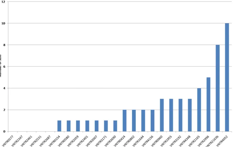
Determination of minimal set of MIRU-VNTR loci.In order to identify the most variable MIRU-VNTR loci among the isolates in our study, an allelic diversity index was calculated for each locus from the 162 distinct profiles obtained (Table 1). Because of the close genetic relationships of most isolates (almost all belonging to the Beijing genotype), we performed redundancy analysis by looking at single-locus variants (SLVs) (Fig. 1) for an in-depth determination of the minimal set of loci necessary for maximal discrimination. Eight loci (960, 1955, 2163b, 2165, 2996, 3192, 4052, and 4348) involved in 3 to 10 SLVs were able to differentiate most of the isolates in our sample. Stepwise addition of an auxiliary set of four loci with 2 SLVs and 7 loci with one SLV provided marginal improve-ment. No SLVs were observed for only five loci (loci 577, 2347, 2461, 2561, and 2687).
Comparison and congruence of RFLP and MIRU-VNTR data.All the isolates included in the analysis had a high IS6110 copy number, varying between 8 and 18 elements, and
dis-played typical Beijing banding patterns. When an IS6110-RFLP-based dendrogram was generated, three large and a few smaller groups were observed. A careful examination of the banding patterns revealed that isolates in two of the three large groups closely resembled the K family, a sublineage of the Beijing genotype which is commonly isolated from TB patients in the Republic of Korea (21, 30). The third large group had banding patterns typical of Beijing genotype isolates from China (43).
Interestingly, this grouping based on IS6110-RFLP analysis was congruent with the grouping obtained using a minimum spanning tree based on MIRU-VNTR data (Fig. 2). This con-gruence demonstrates the consistency of the broad phyloge-netic groupings of the M. tuberculosis isolates in our sample using either genotyping method and thus relevantly defines the clonal populations within the K family. Isolates belonging to one branch of the K family are predominant in the city of Masan, in contrast to the other branch, which has been isolated from many parts of the country (S. N. Cho, T. S. Song, and H. Y. Lee, unpublished data). The former branch has there-fore been designated Masan and the latter the Beijing-Korea branch, respectively. Based on this classification, 138 out of the 154 isolates were classified into one of the three broad groups. Among these, 41.3% (57/138) of the isolates belonged to the Beijing-China branch and 32.6% (45/138) to the Beijing-Korea branch, while 26.1% (36/138) of the isolates were designated the Beijing-Masan branch. We did not ob-serve a differential distribution of resistant (non-MDR), MDR, or XDR strains among the 3 branches.
Within technical limits inherent to analysis of complex band-ing patterns such as are typical of Beijband-ing strains, RFLP grouped 41 isolates into 16 clusters with 113 unique patterns, resulting in a strain-clustering rate of 16.2%. Seven of these
TABLE 1. Allelic diversity of M. tuberculosis isolates from the Republic of Korea
MIRU-VNTR locus
No. of isolates with indicated MIRU allele Allelic
diversity index 0 1 2 3 4 5 6 7 8 9 10 11 2687 162 0.00 2531 160 2 0.01 2461 1 159 1 0.03 0154 2 159 1 0.03 0580 2 158 2 0.03 0577 1 157 2 1 0.04 3171 1 5 156 0.06 2347 7 3 152 0.09 2165 1 9 148 0.09 2059 6 152 3 0.11 2401 10 1 148 2 0.10 1644 6 148 6 1 0.16 0960 7 4 144 4 0.14 3007 1 1 145 14 0.16 4348 2 16 136 4 3 0.23 3690 1 17 116 12 9 3 3 0.43 3192 1 6 30 112 10 1 1 0.48 2996 20 5 5 9 109 8 4 1 0.49 0802 2 51 99 8 2 0.52 1955 5 10 73 69 2 2 1 0.58 4156 5 31 85 39 1 0.61 0424 5 47 53 52 2 0.68 4052 3 2 2 3 10 18 69 46 3 1 0.70 2163b 2 8 4 13 66 42 17 4 2 0.73
clusters were completely concordant with MIRU-VNTR (ac-counting for the missing data in one locus of one isolate in cluster 4), and 2 other clusters had some isolates with identical and variant MIRU-VNTR patterns, respectively (clusters 10
and 16). The MIRU-VNTR patterns of four isolates in differ-ent RFLP clusters differed at only one locus, while the other seven RFLP clusters of two isolates each were not matched by MIRU-VNTR data (Fig. 3). The MIRU-VNTR patterns of
FIG. 1. Single-locus variation analysis of 24-locus MIRU-VNTR of M. tuberculosis isolates from the Republic of Korea. Numbers on the x axis designate MIRU-VNTR loci according to their positions (in kilobase pairs) on the H37Rv chromosome.
FIG. 2. Minimum spanning tree of M. tuberculosis Beijing isolates from the Republic of Korea, generated by using the 24-locus MIRU-VNTR typing data in the MIRU-VNTRplus database (3). Circles show the various sublineage clonal complexes identified by the 24 MIRU-VNTR loci among the M. tuberculosis isolates analyzed. The size of each circle is proportional to the number of MIRU-VNTR types belonging to a particular complex. The color code for red, blue, and green is indicated by the labels in the figure; white circles indicate genotypes that have uncertain sublineage assignments but are related to the respective branch.
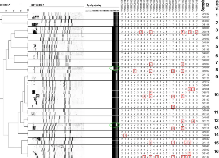
four isolates within different RFLP clusters differed at only one locus (corresponding to the SLVs), while in 9 other cases, the isolates within RFLP clusters differed by 2 to 7 loci. Spoligo-typing independently confirmed the splitting of 2 of the RFLP clusters by two or more MIRU-VNTR loci in two instances (clusters 8 and 10).
Conversely, 5 of the 18 MIRU-VNTR clusters were also identical by RFLP clustering (clusters 6, 9 to 11, and 18). One cluster had identical RFLP and MIRU-VNTR patterns, but one of the two isolates lacked one spacer (spacer 40) in spo-ligotyping (cluster 3). Twelve MIRU-VNTR clusters were par-tially or totally split by RFLP. However, 11 of these clusters were split by minor differences of between one and three IS6110 bands. Only in cluster 16 did one isolate have four extra IS6110 bands in comparison to a common profile shared by four other isolates. Not surprisingly, the combination of MIRU-VNTR, RFLP, and spoligotyping techniques provided the highest apparent discriminatory power and grouped 19 isolates in 8 clusters (Fig. 4).
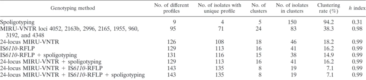
The discriminatory capacity of the different methods used alone or in combination was high (⬎0.98) in all cases except when spoligotyping was considered alone (h⫽ 0.31) (Table 2).
The resolution of MIRU-VNTR alone was only slightly less than that of RFLP (18.2% versus 16.2%, respectively) but was identical when used in conjunction with spoligotyping.
DISCUSSION
Our results demonstrate that this Korean collection of M.
tuberculosis isolates is very homogeneous, as only four
spoli-gotype families were obtained for the 208 isolates, with nearly all (97%) the isolates belonging to the single Beijing family. The predominance of the Beijing family in East Asia is well documented (26). The proportions of Beijing family isolates in previous reports have ranged from 18.5% to 72% (21, 30), with a previous maximum of up to 81.9% reported in China (13). Thus, to date, this study from the Republic of Korea represents the most extreme proportion of Beijing family isolates to be reported. This could be explained partly by the fact that these isolates were obtained from hospitalized subjects in a referral setting, compared to the more general population-based sam-ples used in other studies (30). The high proportion of isolates belonging to the Beijing family reported herein coupled with the fact that at least 39% of the samples analyzed were either
FIG. 3. Comparison between RFLP clusters, MIRU-VNTR patterns, and spoligotyping of M. tuberculosis isolates from the Republic of Korea. Polymorphic MIRU-VNTR loci and spoligotyping spacers within each RFLP cluster are shown in red boxes and green circles, respectively.
MDR (30.3%) or XDR (9.6%) confirms previous reports sug-gesting an association between the Beijing family and drug resistance (11, 25). These findings suggest that our study sub-jects could have been selectively infected with M. tuberculosis strains that are more prone to the establishment of chronic infections and are more difficult to treat.
Although the samples analyzed were not necessarily repre-sentative of the bulk of the extant strains in Korea, the low clustering rate (22% by MIRU-VNTR typing) and high level of
drug resistance of these strains suggest that the problem of drug-resistant TB in Korea may be largely due to acquisition of drug resistance rather than transmission. This low strain clustering rate is most probably a maximum value, at least for this study population sample. Both MIRU-VNTR and RFLP showed a high diversity index (h⫽ 0.99), cluster number con-cordance (18 versus 16, respectively), and concon-cordance in unique genotype number (113 versus 108) in our sample. About half of the RFLP clusters in our study were split by
FIG. 4. Comparison between MIRU-VNTR clusters and RFLP of M. tuberculosis isolates from the Republic of Korea. The variant RFLP bands and spoligotyping spacers among different M. tuberculosis isolates within each MIRU-VNTR cluster are shown in red and green circles, respectively.
TABLE 2. Discriminatory capacities of the three different typing methods, based on different sets of MIRU-VNTR loci, alone or in combination
Genotyping method No. of different profiles
No. of isolates with unique profile No. of clusters No. of isolates in clusters Clustering rate (%) h index Spoligotyping 9 4 5 150 94.2 0.31 MIRU-VNTR loci 4052, 2163b, 2996, 2165, 1955, 960, 3192, and 4348 95 71 24 83 38.3 0.98 24-locus MIRU-VNTR 126 108 18 46 18.2 0.99 IS6110-RFLP 129 113 16 41 16.2 0.99 IS6110-RFLP⫹ spoligotyping 131 116 15 38 14.9 0.99
24-locus MIRU-VNTR⫹ spoligotyping 129 113 16 41 16.2 0.99
24-locus MIRU-VNTR⫹ IS6110-RFLP 143 135 8 19 7.1 0.99
MIRU-VNTR by 1 to 7 loci. Taking into account the high degree of clonal stability of these MIRU-VNTR loci and evi-dence available from well-defined epidemiologic studies (28, 32, 37, 40), differences of two or more loci and even, to a lesser extent, of a single locus remain strongly predictive of absence of a direct transmission link (i.e., of infection by independent strains). Although sufficient information was not available to investigate epidemiologic links among isolates within these RFLP clusters, the observed splitting may therefore represent epidemiologically meaningful differences of strains with iden-tical RFLP fingerprints that share only remote common clonal ancestors.
Although data were not available to systematically assess possible epidemiologic links among the patients in the MIRU-VNTR clusters prior to their admission to the hospital, two patients in one of these clusters shared the same hospital room and at least two patients in each of three other clusters shared rooms. Moreover, all but one of the differences observed in the MIRU-VNTR clusters that were split by RFLP were limited to 1 to 3 IS6110 bands, which may represent microevolutionary changes within the same strain transmission chain (6, 8, 9, 27, 36). These data suggest a high likelihood of nosocomial trans-mission of strains among in-patients in this hospital.
Generally, more than three RFLP band differences between patterns has been considered sufficient to unambiguously de-fine different strains (36). However, even after standardization, evaluation of slight RFLP changes is sometimes subject to error, especially among the complex banding patterns observed in Beijing strains. This difficulty may be overlooked in compar-isons of discriminatory power with that of other genotyping methods. Nonetheless, in apparent contrast to the results of other studies (14, 16), our observations suggest that 24-locus-based MIRU-VNTR typing can compare favorably with RFLP for study of TB transmission, even in a setting largely domi-nated by M. tuberculosis Beijing strains. The 24-locus MIRU-VNTR typing was able to broadly classify the K family of the Beijing genotype into Beijing-Korea and Beijing-Masan lin-eages that were in agreement with the RFLP analysis, although specific allelic signatures that could unambiguously discrimi-nate these two subgroups within the K family were not tected in individual loci. Moreover, this study successfully de-fined a reduced set of loci (960, 1955, 2163b, 2165, 2996, 3192, 4052, and 4348) with a discriminatory power close to those of both RFLP and 24-locus MIRU-VNTR (allelic diversity, 0.98 versus 0.99), which could be reliably used to discriminate most isolates in our sample. We believe that this reduced MIRU-VNTR locus set can be used to discriminate isolates from our study setting and, probably, from the whole Republic of Korea and will therefore facilitate additional epidemiological inves-tigations involving larger numbers of samples. Three of these loci (960, 2996, and 3192) were also shown to be moderately to highly discriminatory (h⬎ 0.6) in a recent study that analyzed a predominantly non-Beijing sample in which three other loci (1644, 3007, and 802) that we found to have low discrimination were reported to have moderate discriminatory power (h ⬎ 0.5) (46).
The low rate of mixed infection observed in this study is comparable to the findings of previous reports (31, 36), albeit from other settings, and may be because most of the subjects studied were retreatment cases where a single dominant strain
would have established the infection. Korea has a relatively low TB incidence rate that makes superinfection less likely than in other settings.
In summary, this study has determined that MIRU-VNTR typing is a useful alternative to RFLP typing of M. tuberculosis isolates, and we have identified a reduced set of MIRU-VNTR loci that can be applied for reliable strain differentiation. Our results also suggest that the bulk of drug-resistant TB in Korea could be due to acquired drug resistance as opposed to trans-mission of drug-resistant strains but that occasional transmis-sion of MDR and XDR strains may occur, particularly in the tertiary hospital setting.
ACKNOWLEDGMENTS
We sincerely thank the many subjects and the doctors and nurses of the National Masan Tuberculosis Hospital that have been willing to sacrifice their time and energy to contribute to this study.
Funding for this work was provided (in part) by the Intramural Research Program of the National Institutes of Health, National In-stitute of Allergy and Infectious Disease, and (in part) by continuing support from the Ministry for Health, Welfare, and Family Affairs of the Republic of Korea. P.S. is a Researcher of the Centre National de la Recherche´ Scientifique.
REFERENCES
1. Affolabi, D., G. Anyo, F. Faïhun, N. Sanoussi, I. C. Shamputa, L. Rigouts, L.
Kestens, S. Anagonou, and F. Portaels.2009. First molecular
epidemiolog-ical study of Mycobacterium tuberculosis in Benin. Int. J. Tuberc. Lung Dis.
13:317–322.
2. Allix, C., P. Supply, and M. Fauville-Dufaux. 2004. Utility of fast mycobac-terial interspersed repetitive unit-variable number tandem repeat genotyping in clinical mycobacteriological analysis. Clin. Infect. Dis. 39:783–789. 3. Allix-Be´guec, C., D. Harmsen, T. Weniger, P. Supply, and S. Niemann.2008.
Evaluation and strategy for use of MIRU-VNTRplus, a multifunctional database for online analysis of genotyping data and phylogenetic identifica-tion of Mycobacterium tuberculosis complex isolates. J. Clin. Microbiol. 46: 2692–2699.
4. Allix-Be´guec, C., M. Fauville-Dufaux, and P. Supply.2008. Three-year pop-ulation-based evaluation of standardized mycobacterial interspersed repeti-tive-unit–variable-number tandem-repeat typing of Mycobacterium tubercu-losis. J. Clin. Microbiol. 46:1398–1406.
5. Barlow, R. E., D. M. Gascoyne-Binzi, S. H. Gillespie, A. Dickens, S. Qamer,
and P. M. Hawkey.2001. Comparison of variable number tandem repeat and
IS6110-restriction fragment length polymorphism analyses for discrimination of high- and low-copy-number IS6110 Mycobacterium tuberculosis isolates. J. Clin. Microbiol. 39:2453–2457.
6. Braden, C. R., G. P. Morlock, C. L. Woodley, K. R. Johnson, A. C. Colombel,
M. D. Cave, Z. Yang, S. E. Valway, I. M. Onorato, and J. T. Crawford.2001.
Simultaneous infection with multiple strains of Mycobacterium tuberculosis. Clin. Infect. Dis. 33:e42–e47.
7. Canetti, G., W. Fox, A. Khomenko, H. T. Mahler, N. K. Menon, D. A.
Mitchison, N. Rist, and N. A. Smelev.1969. Advances in techniques of
testing mycobacterial drug sensitivity, and the use of sensitivity tests in tuberculosis control programmes. Bull. World Health Organ. 41:21–43. 8. Cave, M. D., K. D. Eisenach, G. Templeton, M. Salfinger, G. Mazurek, J. H.
Bates, and J. T. Crawford.1994. Stability of DNA fingerprinting patterns
produced with IS6110 in strains of Mycobacterium tuberculosis. J. Clin. Mi-crobiol. 32:262–266.
9. de Boer, A. S., K. Kremer, M. W. Borgdorff, P. E. de Haas, H. F. Heersma,
and D. van Soolingen.2000. Genetic heterogeneity in Mycobacterium
tuber-culosis isolates reflected in IS6110 restriction fragment length polymorphism patterns as low-intensity bands. J. Clin. Microbiol. 38:4478–4484. 10. Dobler, C. C., G. B. Marks, S. E. Simpson, and A. B. Crawford. 2008.
Recurrence of tuberculosis at a Sydney chest clinic between 1994 and 2006: reactivation or reinfection? Med. J. Aust. 188:153–155.
11. Drobniewski, F., Y. Balabanova, V. Nikolayevsky, M. Ruddy, S. Kuznetzov,
S. Zakharova, A. Melentyev, and I. Fedorin.2005. Drug-resistant
tubercu-losis, clinical virulence, and the dominance of the Beijing strain family in Russia. JAMA 293:2726–2731.
12. Filliol, I., J. R. Driscoll, D. Van Soolingen, B. N. Kreiswirth, K. Kremer, G.
Vale´tudie, D. D. Anh, R. Barlow, D. Banerjee, P. J. Bifani, K. Brudey, A.
Cataldi, R. C. Cooksey, D. V. Cousins, J. W. Dale, O. A. Dellagostin, F. Drobniewski, G. Engelmann, S. Ferdinand, D. Gascoyne-Binzi, M. Gordon,
M. C. Gutierrez, W. H. Haas, H. Heersma, G. Ka¨llenius, E.
Mostro¨m, I. Mokrousov, V. Narbonne, O. Narvskaya, A. Nastasi, S. N. Niobe-Eyangoh, J. W. Pape, V. Rasolofo-Razanamparany, M. Ridell, M. L. Rossetti, F. Stauffer, P. N. Suffys, H. Takiff, J. Texier-Maugein, V. Vincent,
J. H. De Waard, C. Sola, and N. Rastogi. 2002. Global distribution of
Mycobacterium tuberculosis spoligotypes. Emerg. Infect. Dis. 8:1347–1349. 13. Han, H., F. Wang, Y. Xiao, Y. Ren, Y. Chao, A. Guo, and L. Ye. 2007. Utility
of mycobacterial interspersed repetitive unit typing for differentiating Myco-bacterium tuberculosis isolates in Wuhan, China. J. Med. Microbiol. 56:1219– 1223.
14. Hanekom, M., G. D. van der Spuy, N. C. Gey van Pittius, C. R. McEvoy,
K. G. Hoek, S. L. Ndabambi, A. M. Jordaan, T. C. Victor, P. D. van Helden,
and R. M. Warren.2008. Discordance between mycobacterial interspersed
repetitive-unit–variable-number tandem-repeat typing and IS6110 restric-tion fragment length polymorphism genotyping for analysis of Mycobacte-rium tuberculosis Beijing strains in a setting of high incidence of tuberculosis. J. Clin. Microbiol. 46:3338–3345.
15. Hong, Y. P., S. J. Kim, W. J. Lew, E. K. Lee, and Y. C. Han. 1998. The seventh nationwide tuberculosis prevalence survey in Korea, 1995. Int. J. Tuberc. Lung Dis. 2:27–36.
16. Iwamoto, T., S. Yoshida, K. Suzuki, M. Tomita, R. Fujiyama, N. Tanaka, Y.
Kawakami, and M. Ito.2007. Hypervariable loci that enhance the
discrim-inatory ability of newly proposed 15-loci and 24-loci variable-number tandem repeat typing method on Mycobacterium tuberculosis strains predominated by the Beijing family. FEMS Microbiol. Lett. 270:67–74.
17. Jeon, C. Y., S. H. Hwang, J. H. Min, D. R. Prevots, L. C. Goldfeder, H. Lee,
S. Y. Eum, D. S. Jeon, H. S. Kang, J. H. Kim, B. J. Kim, D. Y. Kim, S. M.
Holland, S. K. Park, S. N. Cho, C. E. Barry III, and L. E. Via.2008.
Extensively drug-resistant tuberculosis in South Korea: risk factors and treat-ment outcomes among patients at a tertiary referral hospital. Clin. Infect. Dis. 46:42–49.
18. Kamerbeek, J., L. Schouls, A. Kolk, M. van Agterveld, D. van Soolingen, S.
Kuijper, A. Bunschoten, H. Molhuizen, R. Shaw, M. Goyal, and J. van
Embden.1997. Simultaneous detection and strain differentiation of
Myco-bacterium tuberculosis for diagnosis and epidemiology. J. Clin. Microbiol.
35:907–914.
19. Kan, B., I. Berggren, S. Ghebremichael, R. Bennet, J. Bruchfeld, E.
Chrys-santhou, G. Ka¨llenius, R. Petersson, B. Petrini, V. Romanus, S. Sylvan, and
M. Kalin.2008. Extensive transmission of an isoniazid-resistant strain of
Mycobacterium tuberculosis in Sweden. Int. J. Tuberc. Lung Dis. 12:199–204. 20. Kent, P. T., and G. P. Kubica. 1985. Public health mycobacteriology: a guide for the level III laboratory. U.S. Department of Health and Human Services, Centers for Disease Control, Atlanta, GA.
21. Kim, S. J., G. H. Bai, H. Lee, H. J. Kim, W. J. Lew, Y. K. Park, and Y. Kim. 2001. Transmission of Mycobacterium tuberculosis among high school stu-dents in Korea. Int. J. Tuberc. Lung Dis. 5:824–830.
22. Le´vy-Fre´bault, V. V., and F. Portaels.1992. Proposed minimal standards for the genus Mycobacterium and for description of new slowly growing Myco-bacterium species. Int. J. Syst. Bacteriol. 42:315–323.
23. Maes, M., K. Kremer, D. van Soolingen, H. Takiff, and J. H. de Waard. 2008. 24-locus MIRU-VNTR genotyping is a useful tool to study the molecular epidemiology of tuberculosis among Warao Amerindians in Venezuela. Tu-berculosis (Edinb.) 88:490–494.
24. Mokrousov, I., O. Narvskaya, A. Vyazovaya, J. Millet, T. Otten, B.
Vish-nevsky, and N. Rastogi.2008. Mycobacterium tuberculosis Beijing genotype in
Russia: in search of informative variable-number tandem-repeat loci. J. Clin. Microbiol. 46:3576–3584.
25. Mokrousov, I., T. Otten, T. Zozio, E. Turkin, V. Nazemtseva, A. Sheremet, B.
Vishnevsky, O. Narvskaya, and N. Rastogi.2009. At Baltic crossroads: a
molecular snapshot of Mycobacterium tuberculosis population diversity in Kaliningrad, Russia. FEMS Immunol. Med. Microbiol. 55:13–22. 26. Murase, Y., S. Mitarai, I. Sugawara, S. Kato, and S. Maeda. 2008. Promising
loci of variable numbers of tandem repeats for typing Beijing family Myco-bacterium tuberculosis. J. Med. Microbiol. 57:873–880.
27. Niemann, S., E. Richter, and S. Rusch-Gerdes. 1999. Stability of Mycobac-terium tuberculosis IS6110 restriction fragment length polymorphism patterns and spoligotypes determined by analyzing serial isolates from patients with drug-resistant tuberculosis. J. Clin. Microbiol. 37:409–412.
28. Oelemann, M. C., R. Diel, V. Vatin, W. Haas, S. Ru¨sch-Gerdes, C. Locht, S.
Niemann, and P. Supply.2007. Assessment of an optimized mycobacterial
interspersed repetitive-unit–variable-number tandem-repeat typing system combined with spoligotyping for population-based molecular epidemiology studies of tuberculosis. J. Clin. Microbiol. 45:691–697.
29. Paranjothy, S., M. Eisenhut, M. Lilley, S. Bracebridge, I. Abubakar, R.
Mulla, K. Lack, D. Chalkley, J. Howard, S. Thomas, and M. McEvoy.2008.
Extensive transmission of Mycobacterium tuberculosis from 9 year old child with pulmonary tuberculosis and negative sputum smear. BMJ 337:a1184. 30. Park, Y. K., G. H. Bai, and S. J. Kim. 2000. Restriction fragment length
polymorphism analysis of Mycobacterium tuberculosis isolated from countries in the Western Pacific region. J. Clin. Microbiol. 38:191–197.
31. Richardson, M., N. M. Carroll, E. Engelke, G. D. Van Der Spuy, F. Salker,
Z. Munch, R. P. Gie, R. M. Warren, N. Beyers, and P. D. Van Helden.2002.
Multiple Mycobacterium tuberculosis strains in early cultures from patients in a high-incidence community setting. J. Clin. Microbiol. 40:2750–2754. 32. Savine, E., W. M. Warren, G. D. van der Spuy, N. Beyers, P. D. van Helden,
C. Locht, and P. Supply.2002. Stability of variable-number tandem repeats
of mycobacterial interspersed repetitive units from 12 loci in serial isolates of Mycobacterium tuberculosis. J. Clin. Microbiol. 40:4561–4566.
33. Seung, K. J., G. H. Bai, S. J. Kim, W. J. Lew, S. K. Park, and J. Y. Kim. 2003. The treatment of tuberculosis in South Korea. Int. J. Tuberc. Lung Dis.
7:912–919.
34. Shamputa, I. C., A. Van Deun, M. A. Salim, M. A. Hossain, K. Fissette, P. de
Rijk, L. Rigouts, and F. Portaels.2007. Endogenous reactivation and true
treatment failure as causes of recurrent tuberculosis in a high incidence setting with a low HIV infection. Trop. Med. Int. Health 12:700–708. 35. Shamputa, I. C., L. Jugheli, N. Sadradze, E. Willery, F. Portaels, P. Supply,
and L. Rigouts.2006. Mixed infection and clonal representativeness of a
single sputum sample in tuberculosis patients from a penitentiary hospital in Georgia. Respir. Res. 7:99.
36. Shamputa, I. C., L. Rigouts, L. A. Eyongeta, N. A. El Aila, A. van Deun, A. H.
Salim, E. Willery, C. Locht, P. Supply, and F. Portaels.2004. Genotypic and
phenotypic heterogeneity among Mycobacterium tuberculosis isolates from pulmonary tuberculosis patients. J. Clin. Microbiol. 42:5528–5536. 37. Supply, P., C. Allix, S. Lesjean, M. Cardoso-Oelemann, S. Ru¨sch-Gerdes, E.
Willery, E. Savine, P. de Haas, H. van Deutekom, S. Roring, P. Bifani, N. Kurepina, B. Kreiswirth, C. Sola, N. Rastogi, V. Vatin, M. C. Gutierrez, M. Fauville, S. Niemann, R. Skuce, K. Kremer, L. Locht, and D. van Soolingen.
2006. Proposal for standardization of optimized mycobacterial interspersed repetitive unit–variable-number tandem repeat typing of Mycobacterium tu-berculosis. J. Clin. Microbiol. 44:4498–4510.
38. Tostmann, A., S. V. Kik, N. A. Kalisvaart, M. M. Sebek, S. Verver, M. J.
Boeree, and D. van Soolingen.2008. Tuberculosis transmission by patients
with smear-negative pulmonary tuberculosis in a large cohort in the Neth-erlands. Clin. Infect. Dis. 47:1135–1142.
39. Valcheva, V., I. Mokrousov, O. Narvskaya, N. Rastogi, and N. Markova. 2008. Utility of new 24-locus variable-number tandem-repeat typing for discriminating Mycobacterium tuberculosis clinical isolates collected in Bul-garia. J. Clin. Microbiol. 46:3005–3011.
40. van Deutekom, H., P. Supply, P. E. de Haas, E. Willery, S. P. Hoijng, C.
Locht, R. A. Coutinho, and D. van Soolingen.2005. Molecular typing of
Mycobacterium tuberculosis by mycobacterial interspersed repetitive unit– variable-number tandem repeat analysis, a more accurate method for iden-tifying epidemiological links between patients with tuberculosis. J. Clin. Microbiol. 43:4473–4479.
41. van Embden, J. D., M. D. Cave, J. T. Crawford, J. W. Dale, K. D. Eisenach,
B. Gicquel, P. Hermans, C. Martin, R. McAdam, T. M. Shinnick, and P. M.
Small.1993. Strain identification of Mycobacterium tuberculosis by DNA
fingerprinting: recommendations for a standardized methodology. J. Clin. Microbiol. 31:406–409.
42. van Soolingen, D., P. W. Hermans, P. E. de Haas, D. R. Soll, and J. D. van
Embden.1991. Occurrence and stability of insertion sequences in
Mycobac-terium tuberculosis complex strains: evaluation of an insertion sequence-dependent DNA polymorphism as a tool in the epidemiology of tuberculosis. J. Clin. Microbiol. 29:2578–2586.
43. van Soolingen, D., L. Qian, P. E. de Haas, J. T. Douglas, H. Traore, F.
Portaels, H. Z. Qing, D. Enkhsaikan, P. Nymadawa, and J. D. van Embden.
1995. Predominance of a single genotype of Mycobacterium tuberculosis in countries of East Asia. J. Clin. Microbiol. 33:3234–3238.
44. Wayne, L. G. 1974. Simple pyrazinamidase and urease tests for routine identification of mycobacteria. Am. Rev. Respir. Dis. 109:147–151. 45. World Health Organization. 2008. Tuberculosis control in the Western Pacific
region: 2008 Report. WHO Regional Office for the Western Pacific, Manilla, Phillipines. http://www.wpro.who.int/publications/PUB_9789290613855.htm. 46. Yun, K. W., E. J. Song, G. E. Choi, I. K. Hwang, E. Y. Lee, and C. L. Chang.
2009. Strain typing of Mycobacterium tuberculosis isolates from Korea by mycobacterial interspersed repetitive units-variable number of tandem re-peats. Korean J. Lab. Med. 29:314–319.