Received November 15, 2012, Revised June 13, 2013, Accepted for publication July 9, 2013
Corresponding author: Kyu Joong Ahn, Department of Dermatology, Konkuk University School of Medicine, 120 Neungdong-ro, Gwangjin- gu, Seoul 143-729, Korea. Tel: 82-2-2030-5171, Fax: 82-2-2030-5179, E-mail: [email protected]
This is an Open Access article distributed under the terms of the Creative Commons Attribution Non-Commercial License (http://
creativecommons.org/licenses/by-nc/3.0) which permits unrestricted non-commercial use, distribution, and reproduction in any medium, provided the original work is properly cited.
ORIGINAL ARTICLE
Identification of Dermatophytes by Polymerase Chain Reaction-Restriction Fragment Length Polymorphism Analysis of Metalloproteinase-1
Ho Jung Jung1, Soo Young Kim1, Jae Wook Jung1, Hyun Jung Park1, Yang Won Lee1,2, Yong Beom Choe1,2, Kyu Joong Ahn1,2
1Department of Dermatology, Konkuk University School of Medicine,
2Research Institute of Medical Science, Konkuk University, Seoul, Korea
Background: Transgenic research on metalloproteinase-1 is an emerging field in the area of plant molecular biology. The new method reported here can similarly be applied in fungal molecular biology to identify different dermatophytes. Our method is more accurate than traditional methods such as molecular analyses. Objective: To identify Trichophyton rubrum, T. mentagrophytes var. mentagrophytes, T. tonsu- rans, T. mentagrophytes var. interdigitale, Microsporum canis and M. gypseum, by using the restriction fragment length polymorphism (RFLP) analysis and polymerase chain reaction (PCR) to detect polymorphisms in the metallopro- teinase-1 gene (MEP1). Methods: From each fungal strain, we isolated genomic DNA and performed PCR to amplify the region coding for metalloproteinase-1. Primers for the meta- lloproteinase-1 gene were designed based on the sequence in NCBI GenBank. Subsequently, we purified the amplified PCR product and performed RFLP analysis. After restriction enzyme digestion, BsrDI (NEB, England), the samples were subjected to electrophoresis. Four different patterns of DNA fragments were observed among 6 fungal species. Results:
The DNA fragments for T. mentagrophytes var. menta- grophytes, T. mentagrophytes var. interdigitale and T. ton- surans showed similar patterns on electrophoresis and were
not distinguishable, whereas T. rubrum, M. canis, and M.
gypseum showed different patterns. Conclusion: To our knowledge, it is the first study to introduce the analysis of the nucleotide sequence of metalloproteinase-1 enzyme to study differentiation in dermatophytes. Based on our results, more accurate differentiation and subtyping of T. rubrum and T. mentagrophytes var. interdigitale might be possible. This might contribute to better understanding of the epide- miology and pathogenesis of dermatophyte. (Ann Dermatol 26(3) 338∼342, 2014)
-Keywords-
Metalloproteinases, Microsporum, Polymerase chain re- action, Restriction fragment length polymorphism, Trich- ophyton
INTRODUCTION
Dermatophytes are pathogenic, keratinophilic fungi that invade the stratum corneum of epidermis, hair, fingernails, and toenails of animals. Throughout the world, 43 species of dermatophyte fungi have been identified. Among these, 20 are known to infect humans and other animals.
Trichophyton mentagrophytes var. mentagrophytes, T.
mentagrophytes var. interdigitale, T. tonsurans, T. ver- rucosum, T. schönleinii, T. rubrum, Microsporum canis, M. gypseum, and Epidermophyton floccosum have been reported in South Korea. Of the nine species, T. rubrum is the most abundant and accounts for 85% to 90% of all reported cases of infection. Accurate differentiation of dermatophyte subspecies of T. rubrum, T. tonsurans, and T. mentagrophytes var. interdigitale is, therefore, of
Species Strain
Trichophyton rubrum ATCC28188
T. mentagrophytes var. mentagrophytes CBS113880 T. mentagrophytes var. interdigitale IFM48155
T. tonsurans CBS109036
Microsporum canis IFM45829
M. gypseum IFM5292
Table 1. Standard strains of dermatophytes used in our study clinical significance.
Dermatophytes can be differentiated by the variation in the colors and shapes of colonies when observed under the microscope and in culture. Nevertheless, differen- tiation of dermatophytes by these methods remains difficult. Therefore, other molecular techniques need to be considered. A variety of methods, including mitochondrial DNA restriction fragment length polymorphism (RFLP) pattern and chitin synthetase 1 nucleotide sequence ana- lysis, have been used for simple, fast, and accurate identification of dermatophytes. Recently, the nucleotide sequences of internal transcribed spacer (ITS) regions that represent organism diversity have been analyzed.
However, the ITS sequences of T. mentagrophytes, T.
tonsurans, T. rubrum and M. gypseum are very similar, making differentiation using this method difficult. More- over, this method requires the utilization of a variety of restriction enzymes1-4.
It has recently been reported that proteins from various organisms are damaged upon the activation of proteolytic enzymes, such as metalloproteinase-1, upon exposure to ultraviolet light or high temperatures. It was found that during the recovery of the damaged DNA sequences, slight alterations occur in the sequences. These alterations often result in speciation. Recently, transgenic research using the proteolytic enzyme metalloproteinase-1 has gained popularity in the area of plant molecular biology.
This method can also be applied to the study diffe- rentiation in dermatophytes and may prove to be more accurate than other known molecular methods.
In this study, common pathogenic species of derma- tophytes, T. rubrum, T. mentagrophytes var. mentagro- phytes, T. tonsurans, T. mentagrophytes var. interdigitale, M. canis and M. gypseum were isolated and identified using the polymerase chain reaction (PCR)-RFLP tech- nique. The gene coding for metalloproteinase-1 (MEP1) was analyzed for polymorphisms upon restriction digestion with BsrDI.
MATERIALS AND METHODS
Bacterial species
The standard species of T. rubrum, T. mentagrophytes var.
mentagrophytes, T. mentagrophytes var. interdigitale, T.
tonsurans, M. canis and M. gypseum were spread evenly on Mycosel agar culture medium (Papaic digest of soybean meal 10 g, dextrose 10 g, cycloheximide 0.4 g, chloramphenicol 0.05 g and bactoagar 15.5 g), sterilized at 121oC for 15 minutes, and incubated at 34oC for 14 days (Table 1).
Methods
1) DNA extraction
To collect the samples with roots, a 0.5×0.5 cm×1 to 2 mm block of Mycosel agar with colonies was placed in a 1.5-ml eppendorf tube (e-tube) and centrifuged at 13,570
×g at 4oC for 10 minutes. The samples were resuspended in 200 μl of Lysis buffer (100 mM Tris-HCl pH 9.5, 1 M KCl and 10 mM EDTA), pulverized with a plastic pestle, and heated at 50oC for 30 minutes. Lysis buffer (200 μl) was added to the samples and they were centrifuged at 13,570×g at 4oC for 2 minutes. The floating upper layer of the resulting supernatant was transferred to a 1.5-ml e-tube and treated with 50 ng of proteinase K for 16 hours at 55oC. The samples were then incubated at 100oC for 30 minutes to inactivate the proteinase K. A mixture of phenol : chloroform : isoamyl alcohol (25 : 24 : 1, v/v; 400 μl) was added to the sample and centrifuged at 15,920
×g at 4oC for 15 minutes. The supernatant was then transferred to a new tube. Isopropanol, the same volume as the supernatant, was added to the tube and the samples were incubated at -80oC for 1 hour. The tubes were then spun at 15,920×g at 4oC for 20 minutes. Seventy percent ice-cold ethanol was added to the samples, followed by centrifugation at 15,920×g at 4oC for 5 minutes. The sam- ples were then dried and the DNA concentration was determined using a NanoDrop spectrophotometer (ND- 1000; NanoDrop Technologies, Wilmington, DE, USA) 2) Metalloproteinase-1 primer
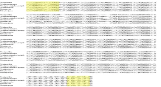
For the PCR, the forward (5'-GACGGTTCTTTGGCTTTG- 3') and reverse (5'-ACTTACGACCGTGGGTGTA-3') pri- mers were designed based on the nucleotide sequence of metalloproteinase-1 deposited in GenBank Genebank (Na- tional Center for Biotechnology Information, Bethesda, MD, USA) (Fig. 1).
3) Polymerase chain reaction amplification
This study adopted colony PCR analysis to amplify the
Fig. 1. Nucleotide sequence align- ment of metalloproteinase-1 genes of various dermatophytes. Complemen- ts of the sequences highlighted in the yellow boxes were used as pri- mers for the amplification of metall- oproteinase-1.
Species BsrDI
Trichophyton rubrum A
T. mentagrophytes var. mentagrophytes B*
T. mentagrophytes var. interdigitale B*
T. tonsurans B*
Microsporum canis C
M. gypseum D
Patterns were designated arbitrarily as A, B, C and D. A: 351/216 base pair (bp), B: 321/216/34 bp, 320/216/34 bp, 325/216/34 bp, C: 321 bp, D: 428/321/31 bp. *Considered to be identical patterns because differences of 1∼5 base pairs were not identified by electrophoresis.
Table 2. Restriction fragments length polymorphism patterns according to species
metalloproteinase-1 gene from the extracted genomic DNA. Each PCR mixture contained 10-μl 10× reaction buffer; 100-μmol/L (each) dATP, dCTP, dGTP, and dTTP;
2.5 U Taq polymerase; 30 pmol of each primer; and 5-μl DNA template solution. Ultrapure water was added to increase the volume of the reaction mixture to 100 μl.
PCR amplification was performed in a Veriti 96-Well Fast Thermal Cycler (Life Technologies, New York, NY, USA).
Each reaction mixture was preheated to 94oC for 5 minutes. PCR amplification was performed for 35 cycles under the following conditions: 94oC for 1 minute; 52.8oC for 30 seconds; 72oC for 1 minute and 72oC for 7 minutes.
The products were separated by electrophoresis on a 3%
agarose gel. A single band of about 0.74 kilo base pairs was detected by ultraviolet (UV) visualization.
4) Purification of amplified DNA
Bands of the appropriate size were excised from the agarose gel. DNA extraction from the excised gel parts was performed using LaboPassTM Gel (Cosmo, Seoul, Korea) and PCR Clean-up kit (Cosmo) according to the manufacturer’s instructions.
5) Detection of restriction fragment length polymor- phism in the metalloproteinase-1 regions
Ten micrograms of purified PCR sample was digested at 65oC for 16 hours with 10 U of restriction endonuclease BsrDI (New England Biolabs, Ipswich, MA, USA). Dige- sted fragments were separated by electrophoresis on a 3%
agarose gel.
RESULTS
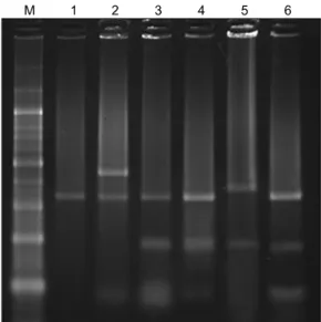
We performed RFLP analysis for six isolated strains of
dermatophytes (T. rubrum, T. mentagrophytes var. menta- grophytes, T. mentagrophytes var. interdigitale, T. ton- surans, M. canis, and M. gypseum). From the DNA frag- ments obtained after BsrDI restriction digestion, we were able to distinguish four different patterns (Table 2). The DNA fragment sizes in each pattern were as follows: T.
rubrum, 351/216 base pairs (bp); T. mentagrophytes var.
mentagrophytes, 321/216/34 bp; T. mentagrophytes var.
interdigitale, 320/216/34 bp; T. tonsurans, 325/216/34 bp;
M. canis, 321 bp; and M. gypseum, 428/321/31 bp. Frag- ments from T. mentagrophytes var. mentagrophytes, T.
mentagrophytes var. interdigitale, and T. tonsurans showed an identical pattern because the differences between them were of the order of 1 to 5 bp, which cannot be dis- tinguished by electrophoresis. In addition, T. rubrum, M.
canis, and M. gypseum showed their own three distinct patterns (Fig. 2).
Fig. 2. Restriction fragment length polymorphism patterns obtained upon DNA digestion with BsrDI. M: 100-base pair (bp) marker, 1: Microsporumcanis (IFM45829), 2: M. gypseum (IFM- 5292), 3: Trichophyton mentagrophytes var. interdigitale (IFM- 48155), 4: T. mentagrophytes var. mentagrophytes (CBS113880), 5: T. rubrum (ATCC28188), 6: T. tonsurans (CBS109036).
DISCUSSION
Dermatophytes are a major cause of frequent fungal infections. In in vitro conditions, morphology of derma- tophytes can easily change depending on the culture conditions5. Classification of dermatophytes based on their phenotypes is not systematic. Since the 1980s, molecular classification techniques have been developed to categorize these fungi more accurately6-8.
While mitochondrial DNA RFLP analyses (using restriction endonucleases) as well as nuclear ribosomal RNA gene analyses (using nucleic acid sequences) have been at- tempted9, some species exist that cannot be distinguished using such methods. Since the mid-1990s, analysis of diverse ITS sequences began. Summerbell et al.10 per- formed the ITS region analysis of T. rubrum and similar strains. They found that T. rubrum, T. raubitschekii, T.
fischeri, and T. kanei share similar sequences, and that T.
soudanense and T. megninii have nearly identical se- quences. Furthermore, several researchers performed an ITS sequence analysis using BsYiI, HinfI, DdeI, and MvaI, and they were able to differentiate 13 strains, including T.
rubrum, M. canis, and T. mentagrophytes11-14.
Dermatophytes are known to have evolved from a single ancestor that co-evolved with mammals for 50 million years. Mitochondrial DNA, which differs from the existing genetic information, is translated to RNA, which is then transcribed into amino acids. Eventually, the reconstructed protein differs from the existing protein, resulting in the
development of a new species. Metalloproteinase-1 is an essential factor in the differentiation process of these orga- nisms.
Due to the above process of differentiation, the metallo- proteinase-1 sequence in each dermatophyte strain is different. Therefore, it has been possible to classify the dermatophyte strains by molecular techniques using metalloproteinase-1 sequence analysis.
In this study, T. rubrum, the main causative agent of superficial fungal infections, was easily differentiated from T. mentagrophytes var. mentagrophytes, T. mentagro- phytes var. interdigitale, T. tonsurans, M. canis, and M.
gypseum. Moreover, we were able to differentiate M.
canis and M. gypseum from the other four species. On the other hand, we could not differentiate T. mentagrophytes var. mentagrophytes, T. mentagrophytes var. interdigitale, and T. tonsurans. These species could be differentiated by the observation of colony morphology in culture and by microscopy. ITS-1 and ITS-2 sequence analysis using 3 to 4 restriction enzymes for identifying dermatophytes has been reported previously15. To our knowledge, this is the first report to use metalloproteinase-1 sequence analysis for the differentiation of dermatophytes. Various types of proteases such as metalloproteinase-1, metalloproteinase- 2, metalloproteinase-9, and metalloproteinase-12 have been previously reported. The use of metalloproteinase DNA sequences for molecular analysis of dermatophytes might lead to improvements in the understanding of their epidemiology and pathogenesis. Furthermore, our current method of categorizing dermatophytes can contribute to more efficient genotyping analysis.
ACKNOWLEDGMENT
This work was supported by Konkuk University.
REFERENCES
1. Okeke CN, Tsuboi R, Kawai M, Hiruma M, Ogawa H.
Isolation of an intron-containing partial sequence of the gene encoding dermatophyte actin (ACT) and detection of a fragment of the transcript by reverse transcription-nested PCR as a means of assessing the viability of dermatophytes in skin scales. J Clin Microbiol 2001;39:101-106.
2. Gräser Y, el Fari M, Presber W, Sterry W, Tietz HJ. Identi- fication of common dermatophytes (Trichophyton, Micro- sporum, Epidermophyton) using polymerase chain reactions.
Br J Dermatol 1998;138:576-582.
3. Harmsen D, Schwinn A, Bröcker EB, Frosch M. Molecular differentiation of dermatophyte fungi. Mycoses 1999;42:
67-70.
4. Turenne CY, Sanche SE, Hoban DJ, Karlowsky JA, Kabani
AM. Rapid identification of fungi by using the ITS2 genetic region and an automated fluorescent capillary electropho- resis system. J Clin Microbiol 1999;37:1846-1851.
5. Summerbell R, Kane J. Laboratory handbook of dermato- phytes: a clinical guide and laboratory handbook of dermatophytes and other filamentous fungi from skin, hair, and nails. Belmont (CA): Star Publishing, 1997:103-107.
6. Liu D, Pearce L, Lilley G, Coloe S, Baird R, Pedersen J. PCR identification of dermatophyte fungi Trichophyton rubrum, T. soudanense and T. gourvilii. J Med Microbiol 2002;51:
117-122.
7. Liu D, Coloe S, Baird R, Pedersen J. Application of PCR to the identification of dermatophyte fungi. J Med Microbiol 2000;49:493-497.
8. Gräser Y, Kuijpers AF, Presber W, De Hoog GS. Molecular taxonomy of Trichophyton mentagrophytes and T. ton- surans. Med Mycol 1999;37:315-330.
9. Jackson CJ, Barton RC, Evans EG. Species identification and strain differentiation of dermatophyte fungi by analysis of ribosomal-DNA intergenic spacer regions. J Clin Microbiol 1999;37:931-936.
10. Summerbell RC, Haugland RA, Li A, Gupta AK. rRNA gene
internal transcribed spacer 1 and 2 sequences of asexual, anthropophilic dermatophytes related to Trichophyton rub- rum. J Clin Microbiol 1999;37:4005-4011.
11. Makimura K, Tamura Y, Mochizuki T, Hasegawa A, Tajiri Y, Hanazawa R, et al. Phylogenetic classification and species identification of dermatophyte strains based on DNA sequ- ences of nuclear ribosomal internal transcribed spacer 1 regions. J Clin Microbiol 1999;37:920-924.
12. Kwon-chung KJ, Bennett JE. Medical mycology. Philadel- phia: Lea and Febiger, 1992:301-311.
13. Liu D, Coloe S, Baird R, Pedersen J. PCR identification of Trichophyton mentagrophytes var. interdigitale and T. men- tagrophytes var. mentagrophytes dermatophytes with a ran- dom primer. J Med Microbiol 1997;46:1043-1046.
14. Kim JA. Molecular biological approaches to the study of dermatophytes. Korean J Med Mycol 2002;7:1-5.
15. Shin JH, Sung JH, Park SJ, Kim JA, Lee JH, Lee DY, et al.
Species identification and strain differentiation of derma- tophyte fungi using polymerase chain reaction amplification and restriction enzyme analysis. J Am Acad Dermatol 2003;
48:857-865.