ISSN: 2233-601X (Print) ISSN: 2093-6516 (Online)
Received: July 25, 2016, Revised: September 29, 2016, Accepted: October 17, 2016, Published online: June 5, 2017
Corresponding author: Deok Heon Lee, Department of Thoracic and Cardiovascular Surgery, Kyungpook National University Hospital, Kyungpook National University School of Medicine, 130 Dongdeok-ro, Jung-gu, Daegu 41944, Korea
(Tel) 82-53-200-5665 (Fax) 82-53-426-4765 (E-mail) [email protected]
© The Korean Society for Thoracic and Cardiovascular Surgery. 2017. All right reserved.
This is an open access article distributed under the terms of the Creative Commons Attribution Non-Commercial License (http://creativecommons.org/
licenses/by-nc/4.0) which permits unrestricted non-commercial use, distribution, and reproduction in any medium, provided the original work is properly cited.
Mid-Term Outcomes of Single-Port versus Conventional Three-Port Video-Assisted Thoracoscopic Surgery
for Primary Spontaneous Pneumothorax
Hanna Jung, M.D., Tak Hyuk Oh, M.D., Joon Yong Cho, M.D., Deok Heon Lee, M.D.
Department of Thoracic and Cardiovascular Surgery, Kyungpook National University Hospital, Kyungpook National University School of Medicine
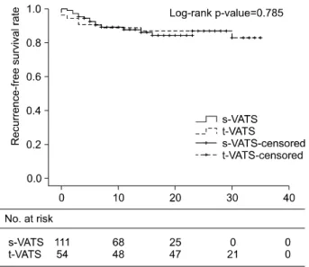
Background: The benefits of video-assisted thoracoscopic surgery (VATS) have been demonstrated over the past decades; as a result, VATS has become the gold-standard treatment for primary spontaneous pneumo- thorax (PSP). Due to improvements in surgical technique and equipment, single-port VATS (s-VATS) is emerg- ing as an alternative approach to conventional three-port VATS (t-VATS). The aim of this study was to eval- uate s-VATS as a treatment for PSP by comparing operative outcomes and recurrence rates for s-VATS ver- sus t-VATS. Methods: Between March 2013 and December 2015, VATS for PSP was performed in 146 pa- tients in Kyungpook National University Hospital. We retrospectively reviewed the medical records of these patients. Results: The mean follow-up duration was 13.4±6.5 months in the s-VATS group and 28.7±3.9 months in the t-VATS group. Operative time (p<0.001), the number of staples used for the operation (p=0.001), duration of drainage (p=0.001), and duration of the postoperative stay (p<0.001) were sig- nificantly lower in the s-VATS group than in the t-VATS group. There was no difference in the overall re- currence-free survival rate between the s-VATS and t-VATS groups. Conclusion: No significant differences in operative outcomes and recurrence rates were found between s-VATS and t-VATS for PSP. Therefore, we cautiously suggest that s-VATS may be an appropriate alternative to t-VATS in the treatment of PSP.
Key words: 1. Video-assisted thoracic surgery 2. Pneumothorax
3. Recurrence
Introduction
Since first introduced, the benefits of video-as- sisted thoracoscopic surgery (VATS) have been clear- l y demonstrated; as a resul t, VATS has become the gold-standard treatment for primary spontaneous pneumothorax (PSP), as it is a quick, safe, and effec- tive procedure, with comparable recurrence to open thoracotomy [1]. Due to improvements in surgical technique and equipment, single-port VATS (s-VATS)
is emerging as an alternative approach to conven- tional three-port VATS (t-VATS) [2-4]. PSP is not a critical disease, but may become problematic due to recurrence. With the s-VATS approach, since all the instruments are manipulated through just one small hole, limitations in the visual field and surgical pro- cedure are inevitabl e [5]. There are stil l doubts about the degree to which it is actually possible to fully explore the thoracic cavity to find blebs or bul- lae, and about whether this technique allows for the
https://doi.org/10.5090/kjtcs.2017.50.3.184
precise wedge resection of the targeted blebs or bullae. Thus, despite all the benefits of s-VATS, the most important point to consider is recurrence after s-VATS. The aim of this study was to evaluate s-VATS as a treatment for PSP by comparing oper- ative outcomes and recurrence between s-VATS and t-VATS [6].
Methods
1) Study population
Between March 2013 and December 2015, VATS for PSP was performed in 146 patients at Kyungpook National University Hospital. We retrospectively re- viewed the medical records of these patients. One case was counted as a single operation on one side (right or l eft). A total of 165 cases of VATS for PSP were performed. Fifty-one patients underwent 54 t-VATS procedures between March 2013 and April 2014. In May 2014, s-VATS became the routine approach at our institution for treating PSP in cases requiring surgical access; 101 patients underwent 111 s-VATS procedures between May 2014 and December 2015.
The eligibility criteria for this study included ipsi- lateral recurrent pneumothorax, a previous history of contralateral pneumothorax, persistent air leakage for more than 3 days, visible blebs or a collapsed lung on radiologic examinations, and age under 30 years.
The exclusion criteria were a history of any lung dis- ease, a previous history of ipsilateral thoracic sur- gery, and two-port VATS and t-VATS cases performed after s-VATS was adopted. This study was approved by the institutional review board of Kyungpook National University Hospital (IRB no. 2016-04-013).
2) Surgical technique
The operations were all performed by a single surgeon. All patients scheduled for VATS underwent high-resolution computed tomography to locate bul- l ae and bl ebs. Al l operations were performed with the patient in the l ateral decubitus position under general anesthesia with a double-lumen endotracheal tube allowing one-lung ventilation.
PSP patients who required closed thoracostomy to relieve symptoms routinely had a 12F trocar catheter (Argyle, suture rib trocar catheter; Covidien, Mansfield, MA, USA) inserted through the fifth intercostal space (ICS) at the mid-axillary line to use as a single port
after lengthening the skin incision to 20 mm.
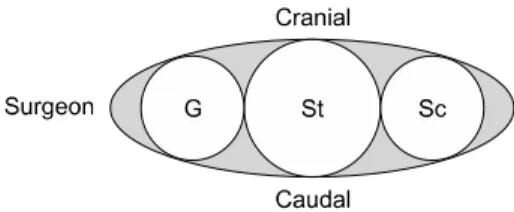
The s-VATS approach was performed with a 20-mm skin incision on the mid-axillary line of the fifth ICS. A 5-mm 30
ovideo thoracoscope and roticulator endog- rasp (Covidien) were used. We al so empl oyed a wound protector (T-port; Yuwon Meditech, Wonju, Korea) to retract the soft tissue and intercostal muscle and to allow maximum exposure of the incision site.
All instruments were then inserted through the single incision. In most cases, the thoracoscope was placed at the right side of the incision, al though sometimes the orientation of the instruments along the incision was changed. We did not require a single-incision lap- aroscopic surgery port [5] or an additional invasive anchoring suture (tower crane technique) [7].
For the t-VATS approach, we used inverted tri- angl e positioning. A 10-mm 30
ovideo thoracoscope was inserted through the seventh ICS mid-axillary line. In addition, 10-mm working ports were inserted through the fifth ICS anterior axillary line and the sixth ICS posterior axillary line.
After inserting the thoracoscope, the entire lung was carefully inspected throughout the lobes, with particular attention paid to the apex and the superior segment of the lower lobe. When it was possible to identify definite blebs or bullae, complete wedge re- section of these l esions was performed using an en- doscopic stapler (Endo GIA articulating stapler;
Covidien). We reinforced the stapler lines by cover- ing them with absorbable cellulose mash (Surgicel;
Ethicon, Somerville, NJ, USA) with fibrin glue (Green pl ast; Green Cross Corp., Yongin, Korea); we did not perform mechanical pleural abrasion. After bleeding control was performed, we placed a 20F or 24F thoracic catheter at the apex of the pleural space.
3) Postoperative management
Postoperative pain control started in the recovery room with intravenous patient-controlled analgesia.
As soon as the patient arrived at their room, neg-
ative suction (−20 cm H
2O) was applied. If no air
leakage was present on postoperative day 1, we
changed to natural water-sealed drainage [8]. The
thoracic catheter was removed when air leakage was
no longer present and when the lung was observed
to expand well on plain chest films. Patients were
discharged on the day of or after removal of the
thoracic catheter.
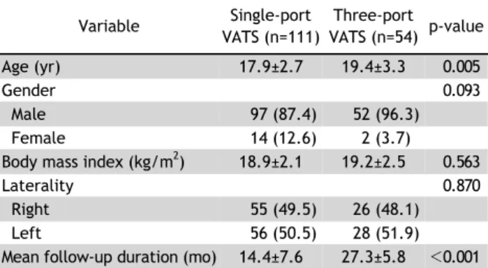
Table 1. Patient characteristics
Variable Single-port VATS (n=111)
Three-port VATS (n=54) p-value
Age (yr) 17.9±2.7 19.4±3.3 0.005
Gender 0.093
Male 97 (87.4) 52 (96.3)
Female 14 (12.6) 2 (3.7)
Body mass index (kg/m
2) 18.9±2.1 19.2±2.5 0.563
Laterality 0.870
Right 55 (49.5) 26 (48.1)
Left 56 (50.5) 28 (51.9)
Mean follow-up duration (mo) 14.4±7.6 27.3±5.8 <0.001 Values are presented as mean±standard deviation or number (%).
VATS, video-assisted thoracoscopic surgery.
Table 2. Operative outcomes
Variable Single-port VATS (n=111)
Three-port
VATS (n=54) p-value Operative time (min) 30.7±9.1 51.4±18.0 <0.001 No. of wedge resections 1.3±0.5 1.4±0.5 0.672
No. of staples 2.4±0.9 3.0±1.1 0.001
Drainage (day) 1.6±1.0 2.7±2.2 0.001
Postoperative stay (day) 2.3±1.3 3.6±2.4 <0.001 Values are presented as mean±standard deviation.
VATS, video-assisted thoracoscopic surgery.
Table 3. Recurrence
Variable Single-port VATS (n=111)
Three-port
VATS (n=54) p-value Overall recurrence 14 (12.6) 8 (14.8) 0.808
Intervention
a)8 (7.2) 7 (13.0) 0.255
Values are presented as number (%).
VATS, video-assisted thoracoscopic surgery.
a)