Introduction
Ovarian cancer is the second most common gynecologic malignancy, with the highest mortality rate [1]. Because it is difficult to detect ovarian cancer at an early stage, it is usu- ally not diagnosed until the advanced stage. As with most cancers, ovarian cancer has a high 5-year relative survival rate (92.6%) if detected early [2]. Ovarian cancer is surgically staged, and an optimal staging operation is required to de- termine the exact stage, treatment plan, and prognosis. The surgical methods for staging ovarian cancer include total hys- terectomy, bilateral salpingo-oophorectomy, pelvic and para- aortic lymphadenectomy, omentectomy, peritoneal biopsy,
Comparison of single-port laparoscopy and laparotomy in early ovarian cancer surgical staging
Kyu Hee Cho, MD 1,* , Yeon Ju Lee, MD 1,* , Kyung Jin Eoh, MD, PhD 2 , Yong Jae Lee, MD 1 , Jung-Yun Lee, MD, PhD 1 , Eun Ji Nam, MD, PhD 1 , Sunghoon Kim, MD, PhD 1 , Young Tae Kim, MD, PhD 1 , Sang Wun Kim, MD, PhD 1
Department of Obstetrics and Gynecology,
1Women’s Cancer Center, Yonsei Cancer Center and Institute of Women’s Life Medical Science, Severance Hospital, Yonsei University College of Medicine, Seoul,
2Yongin Severance Hospital, Yonsei University College of Medicine, Yongin, Korea
Objectives
The aims of this study were to assess the feasibility of single-port laparoscopic surgical staging (SPLS) in early ovarian cancer and to compare the surgical outcomes of SPLS with those of staging laparotomy.
Methods
Between January 2014 and December 2018, 40 patients underwent SPLS and 41 patients underwent staging laparotomy at Yonsei Cancer Center. The patients were diagnosed with International Federation of Gynecology and Obstetrics (FIGO) stage I ovarian cancer. Variables such as patient age, body mass index (BMI), tumor size, FIGO stage, and perioperative surgical outcomes and survival outcomes of SPLS and laparotomy were compared.
Results
The total operation time was similar between the 2 groups (SPLS: 201.4 vs. laparotomy: 203.0 minutes, P=0.806). The median tumor diameters in the SPLS and laparotomy groups were 11.0 (2.5–28 cm) and 15.4 (6–40 cm), respectively (P=0.001). The SPLS group had lower tumor spillage rate (5.0% vs. 19.5%, P=0.047), less intraoperative blood loss (102.0 vs. 371.5 mL, P<0.001), less postoperative pain, and shorter postoperative hospital stay (5 vs. 9.5 days, P<0.001). The intraoperative major complication rate was similar between groups (2.5% vs. 4.9%, P=0.571). There was no significant difference in progression-free survival between the 2 groups (P=0.945). There were no deaths in either group.
Conclusion
SPLS is feasible in early ovarian cancer and has better perioperative surgical outcomes, in some aspects, than staging laparotomy without compromising survival outcomes. SPLS could be performed in patients suspected to have early ovarian cancer.
Keywords: Minimal invasive surgery; Laparoscopic
surgery; Laparotomy; Cancer staging; Ovary Received: 2020.07.25. Accepted: 2020.09.18. Published: 2020.10.12.
Corresponding author: Sang Wun Kim, MD, PhD
Department of Obstetrics and Gynecology, Women’s Cancer Center, Yonsei Cancer Center and Institute of Women’s Life Medical Science, Yonsei University College of Medicine, 50-1 Yonsei-ro, Seodaemun-gu, Seoul 03722, Korea
E-mail: [email protected]
https://orcid.org/0000-0002-8342-8701
*These authors have equally contributed to this paper.
Jung-Yun Lee and Young Tae Kim have been an Editorial Board of Obstetrics &
Gynecology Science; however, they were not involved in the peer reviewer selection, evaluation, or decision process of this article. Otherwise, no other potential conflicts of interest relevant to this article was reported.
Articles published in Obstet Gynecol Sci are open-access, distributed under the terms of the Creative Commons Attribution Non-Commercial License (http://creativecommons.
org/licenses/by-nc/3.0/) which permits unrestricted non-commercial use, distribution, and reproduction in any medium, provided the original work is properly cited.
Copyright © 2021 Korean Society of Obstetrics and Gynecology https://doi.org/10.5468/ogs.20216
eISSN 2287-8580
and peritoneal washing for cytology [3,4].
Recently, many gynecologic surgeries have been performed using a minimally invasive approach compared with the previ- ous invasive approaches, with improved surgical outcomes such as better cosmetic results, less postoperative pain and in- traoperative blood loss, and shorter hospital stay. In particular, single-port laparoscopy using a single incision in the umbilicus maximizes the advantages of conventional laparoscopy [5-8].
There have been few studies on single-port laparoscopy for ovarian cancer staging [9,10]. Staging laparotomy is still the traditional method, and minimally invasive staging surgery is not widely available owing to limitations in exploring the full extent of the peritoneal surface, port-site metastasis, and a higher incidence of intraoperative tumor rupture [7,11,12].
The aims of this study were to introduce and evaluate the safety and feasibility of single-port laparoscopic surgical stag- ing (SPLS), and to compare its perioperative surgical and sur- vival outcomes with those of staging laparotomy in patients with early ovarian cancer.
Materials and methods
1. Data collection
We retrospectively reviewed the medical records of patients
who underwent staging surgery for suspected ovarian cancer between January 1, 2014 and December 31, 2018 at Yonsei Cancer Center, Severance Hospital, Yonsei University Col- lege of Medicine in Seoul, Korea. During the study period, a total of 1,126 patients were diagnosed with ovarian cancer and underwent surgical staging, of whom 725 patients un- derwent staging laparotomy and 401 patients underwent staging laparoscopy. In the staging laparoscopy group, 149 patients underwent SPLS. There were 40 patients in the SPLS group and 41 patients in the laparotomy group who were diagnosed with International Federation of Gynecology and Obstetrics (FIGO) stage I ovarian cancer after surgical stag- ing. Patients found to have metastatic cancers or borderline tumors in the final pathology were excluded. Only patients who underwent primary surgical staging at our hospital were included in this study (Fig. 1).
The collected data included patient age, body mass index (BMI), pelvic adhesion, tumor size confirmed with preopera- tive imaging, tumor histology and grade, preoperative clinical stage, FIGO stage, operation time, estimated blood loss, he- moglobin change, transfusion rate, harvested lymph nodes (LNs), length of hospital stay, intraoperative and postopera- tive complications, postoperative pain scores (immediately postoperation, postoperative day 1, and postoperative day 3), postoperative adjuvant chemotherapy, and recurrence rate.
Fig. 1. Flowchart of patient selection. SPLS, single-port laparoscopic surgical staging; FIGO, International Federation of Gynecology and Obstetrics.
Exclusion:
- No carcinoma, 33
- No primary ovarian cancer, 2 - FIGO stage >I, 56
- No primary staging surgery, 18
401 patients who underwent the staging laparoscopy
1,126 patients who underewent staging operation due to the suspected ovarian cancer
40 patients who diagnosed with FIGO stage I after the staging surgery 149 patients who underwent the
SPLS
725 patients who underwent the staging laparotomy
41 patients who diagnosed with FIGO stage I after the staging surgery
Exclusion:
- No carcinoma, 18
- No primary ovarian cancer, 3 - FIGO stage >I, 536
- No primary staging surgery, 127
Operative time was defined as the time from skin incision to completion of skin closure. Pain was evaluated using the nu- meric pain intensity scale (NPIS). Intraoperative complications were defined as adjacent organ or vessel injury, and wound complications were defined as wound discharge, dehiscence, and operative wound herniation. The number of cycles of postoperative adjuvant chemotherapy was compared. Fol- low-up duration was defined as the number of months from the operation date to the last follow-up date.
2. Surgical techniques
1) Single-port laparoscopic surgical staging
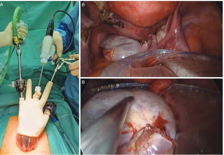
Under general anesthesia, the patient was placed in the lithotomy position and the skin was prepared in the usual manner. A uterine manipulator was used. As in our previous studies, after a 1.2–1.5 cm vertical intraumbilical skin incision with a 1.5–2.0 cm rectus fasciotomy was made, an Alexis
wound retractor (Applied Medical, Rancho Santa Margarita, CA, USA) was inserted through the umbilical incision and a surgical glove was combined with the Alexis wound retractor for a single-port entry system (Fig. 2A). Subsequently, the ab- dominal cavity was filled with CO
2gas and the patient was placed in the Trendelenburg position [13,14].
After the pelvic and abdominal cavities were thoroughly explored, peritoneal washing cytology samples were ob- tained first. Thereafter, each procedure (e.g., hysterectomy, salpingo-oophorectomy, lymphadenectomy, omentectomy, peritonectomy, and appendectomy) was performed using bipolar or monopolar electrocautery and LigaSure (Covidien Valleylab, Boulder, CO, USA) or the Thunderbeat system (Olympus Medical Systems Corp., Tokyo, Japan) [4].
To avoid spilling into the intraperitoneal cavity, ovarian tumors were removed using a laparoscopic tissue retrieval bag (LapBag; Sejong Medical Co., Ltd., Seoul, Korea) (Fig. 2B and C). In the case of large tumors (longest diameter ≥15 cm),
Fig. 2. Single-port laparoscopic surgical staging outside view (A) and safe tumor removal using a laparoscopic tissue retrieval bag (B, C).
A B
B
SW Kim’s technique was used to insert the tumors in a re- trieval bag [15], the bag was retracted through the umbili- cus, and the tumor was removed by aspirating and cutting it inside the bag. After hemostasis and irrigation, the Alexis wound retractor was removed and the umbilical fascia and subcutaneous layer were approximated with 1–0 and 2–0 Vicryl sutures (Ethicon, Piscataway, NJ, USA) [13,14].
2) Staging laparotomy
The preoperative preparation, surgical procedures, and postoperative management were essentially the same as for SPLS, except for a low midline or extended midline incision and nonuse of CO
2gas.
3. Statistical methods
Statistical analysis was performed using SPSS version 25 (IBM, Armonk, NY, USA). A P-value of <0.05 was considered statis- tically significant. Categorical variables are presented as fre- quencies and percentages, and the Pearson χ
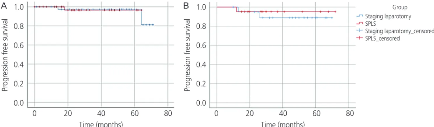
2test was used to evaluate differences between proportions. For continuous variables, medians and ranges of variables are presented, and the Mann-Whitney U test was performed. Kaplan-Meier estimates were used to evaluate differences in time to recur- rence (Fig. 3A and B), and propensity score matching was used to adjust for selection bias (Supplementary Table 1) [16-18].
Results
In total, 81 patients who underwent surgical staging for early
ovarian cancer were selected (40 SPLS and 41 laparotomy).
In the SPLS group, 1 patient (2.5%) was converted to staging laparotomy because of a large tumor and 3 patients (7.5%) required an additional port.
The patient characteristics are shown in Table 1. The me- dian age of patients in the SPLS group was younger than that of patients in the laparotomy group (46.4 [15–86] vs.
53.8 [19–61] years, P=0.005). There was no significant dif- ference in median BMI (24.0 [15.0–36.8] vs. 23.2 [16.7–32.9]
kg/m
2, P=0.461) or pelvic adhesion rate (50.0% vs. 39.0%, P=0.320). The median tumor size was 11 cm in the SPLS group and 15.4 cm in the laparotomy group (P=0.001). The most common tumor histology type in the SPLS group was mucinous adenocarcinoma (30.0%), followed by clear cell carcinoma (25.0%), sex cord-stromal tumor (12.5%), and endometrioid adenocarcinoma (10.0%). In the laparotomy group, mucinous (22.0%) and endometrioid (22.0%) ad- enocarcinomas were the most common histologic types, followed by serous adenocarcinoma (19.5%) and clear cell carcinoma (19.5%). The tumor grade was not significantly different between the 2 groups (SPLS group vs. laparotomy group, grade 1: 27.5% vs. 17.1%, grade 2: 20.0% vs.
34.1%, grade 3: 25.0% vs. 24.4%; P=0.464). With respect to the FIGO stage, stage IA was the most common stage in both groups (70.0% and 39.0% in the SPLS and laparotomy groups, respectively), and the SPLS group had more stage IA cases (P=0.010). FIGO stage IC1 constituted 5.0% and 19.5%, IC2 20.0% and 22.0%, and IC3 2.5% and 19.5% in the SPLS and laparotomy groups, respectively.
Of 149 patients who underwent SPLS for suspected ovar-
Fig. 3. Comparison of survival curves between single-port laparoscopic surgical staging (SPLS) and staging laparotomy. (A) Before pro- pensity score matching (SPLS, n=40 and staging laparotomy, n=41). (B) After propensity score matching (SPLS, n=21 and staging lapa- rotomy, n=21).
Group Staging laparotomy SPLS
Staging laparotomy_censored SPLS_censored
Pr ogr ession fr ee survival Pr ogr ession fr ee survival
1.0 0.8 0.6 0.4 0.2 0.0
1.0 0.8 0.6 0.4 0.2 0.0 0 20 40 60 80
Time (months)
0 20 40 60 80 Time (months)
A B
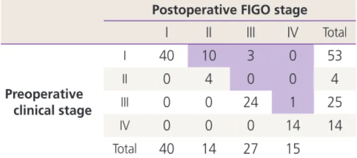
ian cancer, 96 patients were finally diagnosed with ovarian cancer after the surgery. Table 2 shows the preoperative clinical stage and postoperative FIGO stage of patients who were finally diagnosed with ovarian cancer. Fourteen patients (14.6%) were upstaged from preoperative clinical stage I to postoperative FIGO stage II to IV (FIGO stage II: 10 patients, stage III: 3 patients, and stage IV: 1 patient).
Furthermore, we performed propensity score matching to minimize the difference in clinical characteristics between the
two groups. The number of patients after propensity score matching decreased to 21 in each group, and the patient characteristics are summarized in Supplementary Table 1.
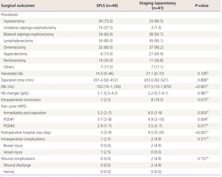
There was no significant difference between the 2 groups in terms of surgical procedures (Table 3). The number of harvested LNs (14.3 vs. 21.0, P=0.145), total operation time (201.4 vs. 203.0 minutes, P=0.806), and postoperative he- moglobin change (2.1 vs. 2.2 g/dL, P=0.981) did not differ between the SPLS and laparotomy groups. The estimated blood loss was smaller in the SPLS group (median: 102 mL, range: 10–1,100 mL) than in the laparotomy group (median:
371.5 mL, range: 10–1,870 mL) (P<0.001). Moreover, the intraoperative transfusion rate was lower in the SPLS group (1/40 [2.5%] vs. 8/41 [19.5%], P=0.015). The SPLS group showed lower postoperative pain score (immediately postop- eration: 5.2 [2–7] vs. 6.0 [2–9], P=0.003; postoperative day 1: 3.7 [2–8] vs. 4.9 [2–10], P = 0.004; postoperative day 3:
2.9 [1–7] vs. 3.5 [2–7], P=0.017) and shorter postoperative hospital stay (5 vs. 9.5 days, P<0.001). The intraoperative major complication rate was similar between groups (2.5%
vs. 4.9%, P=0.571). The SPLS group had 1 case of vascular injury, and the laparotomy group had 2 cases of bowel injury.
There were no wound complications such as wound dehis- cence or umbilical hernia in the SPLS group, whereas wound discharge was observed in 2 patients in the laparotomy group.
The adjuvant chemotherapy rate was lower in the SPLS group than in the laparotomy group (40.0% vs. 75.6%, P=0.001) (Table 4). The median number of chemotherapy cycles was similar in the two groups (5.1 vs. 5.3 cycles, P=0.426). The SPLS group showed a longer time to chemo- therapy after the surgery (17.3 vs. 15.3 days, P=0.031).
Table 2. Comparison of preoperative clinical stage and postop- erative surgical stage in patients who were diagnosed with ovarian cancer after single-port laparoscopic surgical staging (n=96)
Postoperative FIGO stage
I II III IV Total
Preoperative clinical stage
I 40 10 3 0 53
II 0 4 0 0 4
III 0 0 24 1 25
IV 0 0 0 14 14
Total 40 14 27 15
FIGO, 2014 International Federation of Gynecology and Obstetrics.
Table 1. Patient characteristics in the single-port laparoscopic surgical staging (SPLS) and staging laparotomy groups (n=81)
Characteristics SPLS (n=40) Staging lapa-
rotomy (n=41) P-value Age (yr) 46.4 (15–86) 53.8 (19–61) 0.005
a)BMI (kg/m
2) 24 (15.0–36.8) 23.2 (16.7–32.9) 0.461
a)Pelvic adhesion 20 (50) 16 (39.0) 0.320
b)Tumor size (cm) 11 (2.5–28) 15.4 (6–40) 0.001
a)FIGO stage 0.010
b)IA 28 (70.0) 16 (39.0)
IB 0 (0.0) 0 (0.0)
IC1 2 (5.0) 8 (19.5)
IC2 8 (20.0) 9 (22.0)
IC3 1 (2.5) 8 (19.5)
Unknown 1 (2.5) 0 (0.0)
Histology 0.111
b)Serous 3 (7.5) 8 (19.5)
Mucinous 12 (30.0) 9 (22.0)
Seromucinous 2 (5.0) 3 (7.3) Endometrioid 4 (10.0) 9 (22.0) Clear cell 10 (25.0) 8 (19.5) Malignant germ
cell 1 (2.5) 3 (7.3)
Sex cord-stromal 5 (12.5) 1 (2.4)
Others 3 (7.5) 0 (0.0)
Tumor grade 0.464
b)Grade 1 11 (27.5) 7 (17.1)
Grade 2 8 (20.0) 14 (34.1)
Grade 3 10 (25.0) 10 (24.4)
N/A 11 (27.5) 10 (24.4)
Data are presented as median (range) or number (%).
BMI, body mass index; N/A, not applicable; FIGO, 2014 International Federation of Gynecology and Obstetrics.
a)
Calculated using the Mann-Whitney nonparametric test;
b)Calcu-
lated using the χ
2parametric test.
Table 3. Surgical outcomes of single-port laparoscopic surgical staging (SPLS, n=40) and staging laparotomy (n=41)
Surgical outcomes SPLS (n=40) Staging laparotomy
(n=41) P-value
Procedures
Hysterectomy 30 (75.0) 33 (80.5)
Unilateral salpingo-oophorectomy 15 (37.5) 3 (7.3)
Bilateral salpingo-oophorectomy 24 (60.0) 38 (92.7)
Lymphadenectomy 34 (85.0) 39 (95.1)
Omentectomy 32 (80.0) 37 (90.2)
Appendectomy 6 (15.0) 27 (65.9)
Peritonectomy 14 (35.0) 11 (26.8)
Others 7 (17.5) 7 (17.1)
Harvested LNs 14.3 (0–46) 21.1 (0–72) 0.145
a)Operation time (min) 201.4 (92–412) 203.0 (92–521) 0.806
a)EBL (mL) 102 (10–1,100) 371.5 (10–1,870) <0.001
a)Hb changes (g/dL) 2.1 (0.3–4.2) 2.2 (0.7–4.1) 0.981
a)Intraoperative transfusion 1 (2.5) 8 (19.5) 0.015
b)Pain score (NPIS)
Immediately post-operation 5.2 (2–7) 6.0 (2–9) 0.003
a)POD#1 3.7 (2–8) 4.9 (2–10) 0.004
a)POD#3 2.9 (1–7) 3.5 (2–7) 0.017
a)Postoperative hospital stay (day) 5 (2–9) 9.5 (5–20) <0.001
a)Intraoperative complications 1 (2.5) 2 (4.9) 0.571
b)Bowel injury 0 (0.0) 2 (4.9)
Vessel injury 1 (2.5) 0 (0.0)
Wound complications 0 (0.0) 2 (4.9) 0.157
b)Wound discharge 0 (0.0) 2 (4.9)
Hernia 0 (0.0) 0 (0.0)
Data are presented as median (range) or number (%).
LN, lymph node; EBL, estimated blood loss; Hb, hemoglobin; NPIS, numeric pain intensity scale; POD, postoperative day.
a)
Calculated using the Mann-Whitney nonparametric test;
b)Calculated using the χ
2parametric test.
Table 4. Postoperative adjuvant chemotherapy rate in the 2 groups
Variables SPLS (n=40) Staging laparotomy
(n=41) P-value
Adjuvant chemotherapy 16 (40.0) 31 (75.6) 0.001
b)Cycles of chemotherapy 5.1 (3–6) 5.3 (1–8) 0.426
a)Time to chemotherapy (day) 17.3 (7–31) 15.3 (6–25) 0.031
a)Data are presented as median (range) or number (%).
SPLS, single-port laparoscopic surgical staging.
a)