• 검색 결과가 없습니다.

Usefulness of SUV Ratio for Differentiating Benign from Malignant Focal Thyroid Lesions Incidentally Detected by F-18 FDG PET/ CT: Comparison with SUV Analysis

N/A
N/A
Protected

Academic year: 2021

Share "Usefulness of SUV Ratio for Differentiating Benign from Malignant Focal Thyroid Lesions Incidentally Detected by F-18 FDG PET/ CT: Comparison with SUV Analysis"

Copied!
8
0
0

로드 중.... (전체 텍스트 보기)

전체 글

(1)

ARTICLE Vol. 2, No. 2, November 2009

Received February 20, 2009 / Accepted July 17, 2009

Correspondence: June-Key Chung, MD, PhD, Department of Nuclear Medicine, Seoul National University College of Medicine, 28, Yeongeon-dong, Jongno-gu, Seoul 110-744, Korea

Tel: 82-2-2072-3376, Fax: 82-2-766-9083, E-mail: jkchung@plaza.snu.ac.kr

Usefulness of SUV Ratio for Differentiating Benign from Malignant Focal Thyroid Lesions Incidentally Detected by F-18 FDG PET/ CT:

Comparison with SUV Analysis

Bom Sahn Kim, MD 1 , Won Jun Kang, MD 2 , So Won Oh, MD 3 , Dong Soo Lee,

MD 3,5,6 , June-Key Chung, MD 3,4,5 and Myung Chul Lee, MD 3

Department of Nuclear Medicine, Ewha Womans University College of Medicine

1

; and Division of Nuclear Medicine, Department of Radiology, Yonsei University College of Medicine

2

; and Department of Nuclear Medicine

3

; Cancer Research Institute

4

; Research Institute of Radiation Medicine

5

, Department of Molecular Medicine and Biopharmaceutical Science

6

, Seoul National University College of Medicine, Seoul, Korea

Background and Objectives: We investigated the usefulnesses of SUV and SUV ratios in terms of discriminating focal thyroid lesions incidentally detected by F-18 FDG PET/CT (FDG PET) in patients with various cancers.

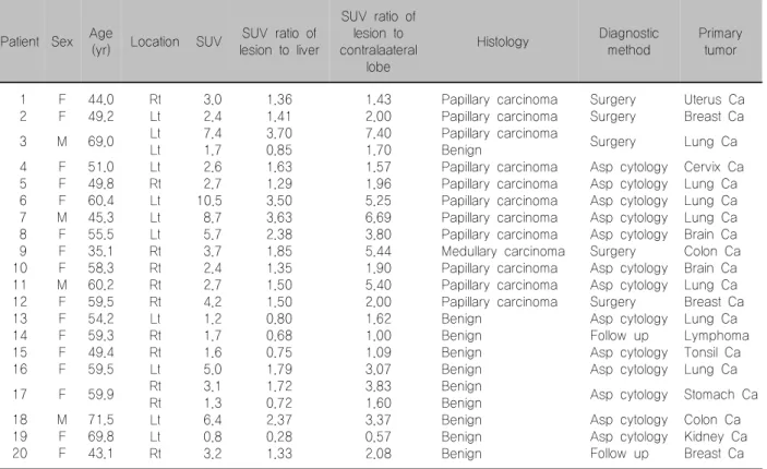
Materials and Methods: Among 2,635 patients, 20 patients with 22 thyroid incidentalomas (benign vs.

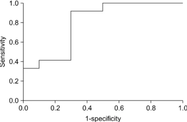
malignant=10 vs. 12) on FDG PET were assessed. Maximal SUV was measured by drawing a ROI on thyroid lesions, on contralateral thyroid lobes, and on the liver. SUV ratio was calculated as the thyroid lesions versus the contralateral lobes and liver. Results: A marginally significant difference in maximal SUV was found between malignant and benign nodules (4.67±2.78 vs. 2.59±1.84, p=0.05). However, more significant differences were found using SUV ratio to normal organs than that of SUV. Malignant nodules had a higher SUV ratio versus liver than benign nodules (2.09±0.96 vs. 1.13±0.65, p=0.012). The SUV ratio of lesions versus contralateral lobes were found to significantly predict the presence of malignancy (3.74±2.19 vs. 1.89±1.17, p=0.027). By ROC curve analysis, the best SUV value cut-off for the prediction of malignancy was 2.42 with a sensitivity of 91.7% and a specificity of 60.0% (AUC=0.758). Similarly, the best SUV ratio cut-off versus the liver was 1.34 with a sensitivity of 91.7% and a specificity of 70.0% (AUC=0.808). But, there was no significant difference between areas of the ROC curves (p=0.736). Conclusion: The SUV ratio had a tendency to discriminate benign from malignant thyroid incidentalomas better than the SUV method. Further investigations with larger number of patients are needed.

Key Words: FDG PET/CT, Thyroid incidentaloma, SUV, SUV ratio

Introduction

Thyroid incidentaloma is defined as a coinci- dentally detected thyroid lesion during image studies, such as, ultrasonography (US), computed tomography (CT), and magnetic resonance imaging (MRI) con- ducted for other purposes.

1)

According to one autopsy study, the incidence of thyroid nodules was over than 50% in patients without a history of a thyroid ab-

normality.

2)

Therefore, the detection of thyroid inci- dentaloma during radiology studies is not uncom- mon. In previous reports on US, the detection rate of thyroid incidentaloma was 19∼46%. However, most conventional imaging modalities are not efficient at discriminating malignancy.

3-5)

Recently, F-18 fluorodeoxyglucose positron emi-

ssion tomography (FDG PET) has been widely used

for the detection and staging of various cancers,

6)

and

frequently detects thyroid incidentalomas. Some in-

(2)

vestigators have reported that FDG PET detects focal abnormal thyroid lesions with increased FDG uptakes in 1.2∼4.3% of examinations.

7-10)

Usually, standard uptake values (SUVs) of FDG are used to determine whether thyroid lesions are malignant. However, the role of SUV in the differential diagnosis of thyroid nodules is controversial. Some articles have reported that SUV have substantial discriminatory power,

11-15)

whereas others have concluded that SUV is incapable of differentiating benign and malignant thyroid le- sions.

16,17)

It is possible that SUV ratio of thyroid nodules versus normal organs can resolve this problem. In the case of FDG PET, SUVs can be underestimated or over- estimated by several fold, due to procedural in- adequacies (leakage, study time inaccuracies) and individual features (overweighting, an elevated blood glucose level), and thus, the SUV ratios versus normal organs might overcome these shortcomings. Several trials using SUV ratio versus liver have been reported for gallbladder cancer and adrenal masses.

18-20)

How- ever, no report has yet been issued on the use of SUV ratio for determining the malignancies of thyroid nodules.

In the present study, we compared the abilities of SUV ratio and SUV in patients to diagnose malignancy in focal thyroid lesions incidentally detected during cancer staging by combined FDG PET/CT.

Materials and Methods

Patients

From November 2003 to May 2007, 2,635 patients with malignant tumors underwent FDG PET/CT for initial cancer staging at Seoul National University Hospital. Fifty-six of these 2,635 patients (2.1%) showed focal abnormal thyroid lesions by FDG PET/

CT. Thyroid incidentalomas in 20 patients (55.2±9.2 yr; F:M=14:6) were confirmed by pathology (n=18) or by clinical follow up over at least 5 months and by various radiologic examinations. Patients with hepatic disease and those receiving therapies (e.g., chemo- or radiotherapy) capable of interfering with the liver metabolism were excluded.

FDG PET/CT protocol

Before administering intravenous (i.v.) F-18 FDG (370∼555 MBq; 10∼15 mCi), patients were fasted for at least 6 hours to ensure a blood glucose level below 140 mg/dl. Drinking water was encouraged during the fasting period. Sixty minutes after administering F-18 FDG, whole body PET images were acquired using a conventional 3-dimensional F-18 FDG PET protocol using a Gemini PET/CT camera (Philips, Knoxville, TN, USA). The transaxial field of view (FOV) is 576 mm in diameter and the axial FOV is 180 mm. Low-dose CT non-contrast (120 kV, 50 mA) data was acquired from head to pelvis for attenuation correction and anatomic localization with natural shallow breathing. PET emission images were acquired for 2 min 30 seconds per bed (total 9 beds) in 3-D acquisition mode. Transaxial images were used for quantitative analysis. And the voxel dimension was 4×4×4 mm.

Image analysis

FDG PET/CT quantitative analysis was performed by visual interpretation and maximal SUV values, which were acquired using attenuation corrected images and calculated as decay-corrected activity (kBq) per milliliter of tissue volume per injected F-18 FDG activity (kBq) per body mass (g). FDG PET and noncontrast CT images were used to detect focal abnormal thyroid lesions (thyroid incidentaloma) in- cluded in either anatomically abnormal or metabolically abnormal lesions. Diffuse FDG uptake by the whole thyroid gland was excluded. Using non-contrast CT images, thyroid lesions with a non-normal atte- nuation were interpreted as focal thyroid lesions and their metabolisms were examined by FDG PET. The calcified statuses of thyroid lesions were also do- cumented during image analysis.

In this study, we measured the maximal SUVs of

lesions, contralateral thyroid lobes, and of liver by

drawing regions of interest (ROI) to central region of

right lobe, and then calculated SUV ratios versus the

liver and contralateral lobe.

(3)

Table 1. Patient characteristics

Patient Sex Age

(yr) Location SUV SUV ratio of lesion to liver

SUV ratio of lesion to contralaateral

lobe

Histology Diagnostic

method

Primary tumor

1 2 3 4 5 6 7 8 9 10 11 12 13 14 15 16 17 18 19 20

F F M F F F M F F F M F F F F F F M F F

44.0 49.2 69.0 51.0 49.8 60.4 45.3 55.5 35.1 58.3 60.2 59.5 54.2 59.3 49.4 59.5 59.9 71.5 69.8 43.1

Rt Lt Lt Lt Lt Rt Lt Lt Lt Rt Rt Rt Rt Lt Rt Rt Lt Rt Rt Lt Lt Rt

3.0 2.4 7.4 1.7 2.6 2.7 10.5 8.7 5.7 3.7 2.4 2.7 4.2 1.2 1.7 1.6 5.0 3.1 1.3 6.4 0.8 3.2

1.36 1.41 3.70 0.85 1.63 1.29 3.50 3.63 2.38 1.85 1.35 1.50 1.50 0.80 0.68 0.75 1.79 1.72 0.72 2.37 0.28 1.33

1.43 2.00 7.40 1.70 1.57 1.96 5.25 6.69 3.80 5.44 1.90 5.40 2.00 1.62 1.00 1.09 3.07 3.83 1.60 3.37 0.57 2.08

Papillary carcinoma Papillary carcinoma Papillary carcinoma Benign

Papillary carcinoma Papillary carcinoma Papillary carcinoma Papillary carcinoma Papillary carcinoma Medullary carcinoma Papillary carcinoma Papillary carcinoma Papillary carcinoma Benign

Benign Benign Benign Benign Benign Benign Benign Benign

Surgery Surgery Surgery Asp cytology Asp cytology Asp cytology Asp cytology Asp cytology Surgery Asp cytology Asp cytology Surgery Asp cytology Follow up Asp cytology Asp cytology Asp cytology Asp cytology Asp cytology Follow up

Uterus Ca Breast Ca Lung Ca Cervix Ca Lung Ca Lung Ca Lung Ca Brain Ca Colon Ca Brain Ca Lung Ca Breast Ca Lung Ca Lymphoma Tonsil Ca Lung Ca Stomach Ca Colon Ca Kidney Ca Breast Ca

Ca: cancer, Rt: right, Lt: left, Asp cytology: US-guided aspiration biopsy, Follow up: follow up of radiologic exam

Statistical analysis

Patients were allocated to malignant and benign groups based on final diagnoses and these allo- cations were compared with FDG PET/CT results.

Data are presented as means±SD and analyzed using the independent SPSS 12 sample test (SPSS INC., IL, USA). Receiver operating characteristic (ROC) curves were drawn to determine optimal SUV ratio cutoff values and optimal maximal SUV values in terms of differentiating malignant from benign thyroidal le- sions. Comparisons of area under independent ROC curves were done by MedCalc software (MedCalc Software, Mariakerke, Belgium). Statistical significance was accepted for p values of <0.05.

Results

In the 20 patients enrolled, primary cancers were located in lung (n=7), brain (n=2), breast (n=3), and in the gastrointestinal (n=3), urogenital (n=3), and other

regions (n=2). Twelve of the 20 thyroid lesions were histologically proven to be malignant and the re- maining 10 thyroid lesions to be benign (2 patients had 2 focal thyroid lesions). The histological types of the malignant thyroid lesions were papillary car- cinoma in 11 patients and medullary carcinoma in one (Table 1). Of the 10 benign lesions, 6 focal thyroid nodules were detected by non contrast CT but did not have an abnormal metabolism by FDG PET.

Lesion SUVs and SUV ratios versus liver and the contralateral lobe

A marginally significant difference was observed between the SUVs of malignant and benign nodules (4.67±2.78 vs. 2.59±1.84, p=0.05), but more sig- nificant differences were observed for SUV ratios.

Malignant nodules had higher SUV ratio versus liver

than benign nodules (2.09±0.96 vs. 1.13±0.65,

p=0.012), and higher SUV ratios versus contralateral

lobes (3.74±2.19 vs. 1.89± 1.17, p=0.027) (Fig. 1).

(4)

Fig. 1. Results of the independent sample test in patients with focal thyroid incidentaloma. Maximal SUV, SUV ratios of lesions versus liver, and SUV ratios of lesions versus contralateral thyroid lobes are presented as means±SD.

Fig. 2. ROC curve analysis: SUV ratio of lesion vs. liver.

ROC curve analysis of SUV ratios versus liver and contralateral lobes and of SUV values for the 22 focal thyroid incidentalomas

The significances of SUV ratios were greater than SUV values according to ROC analysis. The best SUV cut-off value was 2.42 with a sensitivity of 91.7% and a specificity of 60.0% (AUC=0.758). The best SUV ratio vs. liver cut-off value was 1.34 with a sensitivity of 91.7% and a specificity of 70.0% (AUC=0.808, Fig.

2), and similarly, the best SUV ratio vs. contralateral lobes cut-off value was 1.8 with a sensitivity of 83.3%

and a specificity of 60.0% (AUC=0.758) (Fig. 3, 4).

Comparison of areas under independent ROC curves didn’t have significantly difference between SUV and SUV ratios vs liver cut-off value (p=0.736) and con- tralateral lobes cut-off value (p=1.0).

Noncontrast CT results for the 22 focal thyroid incidentalomas

Of the 5 focal thyroid lesions showing calcification, 1 lesion was benign and 4 were malignant. But, there were no statistical significance between calcification and malignancy (p=0.212).

The Hounsfield unit values of focal benign thyroid lesions were not different from those of malignant lesions (54.4±71.6 vs. 71.6±18.7; p=0.083). How- ever, two lesions with very low attenuations (HU=11 and 24) were benign.

Discussion

The incidence of thyroid incidentaloma by FDG PET/CT in the general population is not known because of the high cost of imaging. However, several studies have reported prevalences in selected co- horts, such as, among cancer screening groups or cancer patients staged by FDG PET. In the present study, 2.1% of patients had thyroid incidentaloma by FDG PET/CT, which was performed for cancer staging, and this prevalence is similar to those re- ported previously.

7-10)

For example, Kang et al.

8)

reported a prevalence of 1.9% among patients that underwent FDG PET for cancer staging.

The role of SUV in the differential diagnosis of

thyroid nodules is controversial. Several reports have

concluded that SUV can significantly differentiate the

malignant and benign thyroid nodules,

7-15)

whereas

other have found that SUV is incapable of dif-

(5)

Fig. 4. Right thyroid incidentaloma F-18 FDG PET/CT and follow up CT images. A 59 year old female with lymphoma underwent F-18 FDG PET/CT for stage work up. The maximal SUV of the incidentaloma was 1.7, and the maximal SUVs vs. liver and the contralateral thyroid lobe were 2.5 and 1.7, respectively, giving SUV ratios of lesion vs. liver of 0.68 and of lesion vs. the contralateral lobe of 1.0. The thyroid nodule was confirmed to be benign by aspiration cytology.

Fig. 3. Left thyroid incidentaloma F-18 FDG PET/CT and follow up CT images. A 60 year old female with lung cancer underwent F-18 FDG PET/CT for stage work up. The maximal SUV of the incidentaloma was 10.5, and the maximal SUVs vs. liver and the contralateral thyroid lobe were 3.0 and 2.0, respectively, giving SUV ratios of lesion vs. liver of 3.50 and of lesion vs. the contralateral lobe of 5.25. Papillary thyroid cancer was confirmed by aspiration cytology.

ferentiating benign and malignant thyroid lesions.

16,17)

In the present study, maximal SUV values were marginally different for malignant and benign thyroid nodules (p=0.05).

These disagreements may be due to the over- lapping of glucose metabolisms in malignant and benign tumors or to the inaccuracies of SUV mea- surement. Usually, malignant tumors show higher glucose metabolism than benign lesions. In cases of thyroid, malignant thyroid tumors are slowly growing, and benign thyroid lesions have elevated metabolisms directed toward the production of endocrine hor- mones. This nature can make overlapping metabolism between malignant thyroid tumors and benign thyroid lesions. Nevertheless, malignant lesions are more metabolically active than their benign counterparts.

Several factors can affect the accuracy of SUV measurements, e.g., blood glucose level, imaging time after F-18 FDG injection, the amount of radioactivity actually delivered (i.e., amount of extravasated dose),

patient size, recovery coefficients, ROI partial volume effects, and pixel size.

21-26)

According to a previous study, elevated glucose levels can result in under- estimation of FDG uptake in target regions.

22)

In our patients, the mean blood glucose level was 90.0

±27.6 (range; 36∼134) mg/dl, and this was similar for those with malignant and benign nodules. Beaulieu et al.

23)

found that the rate of change SUV was linearly correlated with SUV measurements at different times after injection. In the present study, mean intervals between injection and imaging were 64.4±6.3 and 65.0±16.8 minutes in the malignant and benign groups, which was not significantly different (p=0.921).

Extravasation of injected radionuclide also reduces

FDG uptake in target tissues.

25)

Accordingly, we

checked charts and reviewed images of arms to

detect extravasation and failed to find any obvious

abnormality. The effect of body weight on SUV is a

topic that has attracted interest recently, as the two

have been reported to be positively correlated.

26)

In

(6)

Fig. 5. Corrected result using SUV ratios of lesion vs. liver in patient with false positive SUV. A 43 year old female with breast cancer underwent F-18 FDG PET/CT for stage work up. The maximal SUV of the incidentaloma was 3.2, and the maximal SUVs vs. liver was 2.4, giving SUV ratios of lesion vs. liver of 1.33. The thyroid nodule was confirmed to be benign by follow exam over than 1 year.

Fig. 6. Corrected result using SUV ratios of lesion vs. liver in patient with false negative SUV. A 49 year old female with breast cancer underwent F-18 FDG PET/CT for stage work up. The maximal SUV of the incidentaloma was 2.4, and the maximal SUVs vs. liver was 1.7, giving SUV ratios of lesion vs. liver of 1.41. Follow up CT just mentioned suspicious calcified lesion.

The thyroid nodule was confirmed to be malignant by operation.

the present study, the 20 patients had a mean weight of 61.2±10.9 (range; 43∼75) kg, and mean weights in the benign and malignant groups were similar (68.5±9.1 vs. 63.1±13.0 kg, respectively, p=0.289).

Using non-contrast CT, we were able to detect small nodular lesions. However, it is known that mean SUVs inaccurately reflect metabolism of small lesions due to recovery coefficient and partial volume effects.

9,24)

In the present study, the size of malignant nodules and benign nodules were 1.25±0.72 cm and 2.06±0.90 cm (p=0.03). Metabolism of malignant nodules could be underestimated due to small size than that of benign nodule. Thus, we used maximal SUVs rather than mean SUVs to overcome these problems.

To improve differentiation ability, we used SUV ratios of lesions versus liver or SUV ratios of lesions versus contralateral thyroid lobes, in the belief that these ratios would account for individual background differences, and reduce the influences of factors that interfere with SUV measurements. It was found that SUV ratios had

better diagnostic values in terms of differentiating malignant and benign thyroidal incidentalomas than maximal SUV values, which concurs with the findings of other investigators who also used SUV ratios versus specified organs.

18-20)

From ROC curve analysis, SUV ratio versus liver showed better discriminating ten- dency than the SUV method, but didn’t have sig- nificance (Fig. 5, 6). It might be happened by small number of enrolled patients to get sufficient result.

The use of liver FDG uptake as a reference has

been reported to offer several benefits. First, the

metabolism of the liver is relatively constant, in fact, it

rarely shows significant changes in glucose meta-

bolism. Previous investigators have evaluated the long

term variability of liver SUVs, and reported that the liver

shows an almost unchanged status over time.

27)

Therefore, we were drawing ROI to central region of

right lobe to estimate of maxSUV, which was already

used.

27)

Second, the use of the liver as background

allows for SUV correction for body weight. Notably,

(7)

Zasadny et al.

28)

reported a positive correlation between liver SUV and body weight. In the present study, one patient with a large body weight (153.3 cm vs. 57.3 kg) had a false positive result when SUV was used, but has a corrected result when SUV ratio vs.

liver was used. Although the use of lean body mass is theoretically more reliable because it avoids SUV overestimations in obese patients,

29)

the use of SUV ratio vs. liver is easy and appropriate for clinical usage. Third, when blood glucose reduces SUV values, SUV ratios produce better results, e.g., Ishizu et al.

30)

reported that actual FDG uptakes by brain tumors declined after glucose loading, but that the FDG uptake ratios of tumors versus cortical gray matter increased by 27%.

Furthermore, hepatic glucose metabolisms may be perturbed in cancer patients. For example, hepatic glucose production might be modulated by abnormal levels of counter-regulatory hormones, such as, growth hormone or tumor glucose requirements.

31)

In addition, Miles et al.

32)

reported that colorectal cancer patients with extrahepatic metastasis had significantly lower hepatic glucose metabolisms than those patients without extrahepatic metastasis, which suggests that cancer aggressiveness reduces hepatic metabolism.

From these results, it can be deduce that patients with a severe malignancy may have a reduced hepatic metabolism. However, our patients with malignant or benign lesions showed no differences in hepatic SUV (2.24±0.46 vs. 2.15±0.43, respectively, p>0.05), which may have been due to the slow growing nature of thyroid cancer.

FDG PET/CT more accurately displays anatomical information than noncontrast CT.

33,34)

Several reports have shown that hypoattenuated cystic lesions are more likely to be benign thyroid nodules and that calcified lesions are more likely to be malignant.

3-5,35)

In the present study, Hounsfield units obtained by noncontrast CT were not able to differentiate benign and malignant focal thyroid lesions, although two lesions at less than 25 Hounsfield units proved to be benign, and one of these returned a false positive when SUV was used to differentiate malignancy and benignity. As mentioned in a previous article, 4 of 5

focal thyroid lesions with calcification proven to be malignant (4/5; 80%). Focal thyroid lesions with calcifications may have a greater tendency toward malignancy than noncalcified thyroid lesions, but in the present study, this was not found to be significant (28.6% vs. 11.1%; p=0.344). Using the nonenhanced CT component of FDG PET/CT, we were able to improve the differentiation of thyroid incidentaloma by FDG PET/CT, which concurs with the findings of a previous study.

35)

The most obvious limitation of the present study is its small cohort size. A larger scale study is required to confirm our results concerning the benefits of the devised SUV ratio method.

Conclusion

Thyroid incidentaloma is frequently detected by FDG PET/CT and lesions that show high focal uptake present the probability of malignancy. However, some benign thyroid lesions may also have high meta- bolisms and this could cause overlap between the SUV values of malignant and benign thyroid lesions.

The SUV ratio of lesion versus liver had a tendency to discriminate benign from malignant thyroid inci- dentalomas better than the SUV method, although there was no statistically significant difference between the areas of ROC curves. Further investigations with larger number of group are needed to confirm our results and to more accurately diagnose thyroid incidentaloma.

References

1) Tan GH, Gharib H. Thyroid incidentalomas: management approaches to nonpalpable nodules discovered incidentally on thyroid imaging. Ann Intern Med 1997;126(3):226-31.

2) Mortensen JD, Woolner LB, Bennett WA. Gross and micro- scopic findings in clinically normal thyroid glands. J Clin En- docrinol Metab 1955;15(10):1270-80.

3) Brander A, Viikinkoski P, Nickels J, Kivisaari L. Thyroid gland: US screening in a random adult population. Radiology 1991;181(3):683-7.

4) Ezzat S, Sarti DA, Cain DR, Braunstein GD. Thyroid in- cidentalomas. Prevalence by palpation and ultrasonography. Arch Intern Med 1994;154(16):1838-40.

5) Mazzaferri EL. Thyroid cancer in thyroid nodules: Finding a

(8)

needle in the haystack. Am J Med 1992;93(4):359-62.

6) Strauss LG, Conti PS. The applications of PET in clinical oncology. J Nucl Med 1991;32(4):623-50.

7) Cohen MS, Arslan N, Dehdashti F, Doherty GM, Lairmore TC, Brunt LM, et al. Risk of malignancy in thyroid inci- dentalomas identified by fluorodeoxyglucose-positron emission tomography. Surgery 2001;130(6):941-6.

8) Kang KW, Kim SK, Kang HS, Lee ES, Sim JS, Lee IG, et al. Prevalence and risk of cancer of focal thyroid incidentaloma identified by 18F-fluorodeoxyglucose positron emission tomogra- phy for metastasis evaluation and cancer screening in healthy subjects. J Clin Endocrinol Metab 2003;88(9):4100-4.

9) Chen YK, Ding HJ, Chen KT, Chen YL, Liao AC, Shen YY, et al. Prevalence and risk of cancer of focal thyroid inci- dentaloma identified by 18F-fluorodeoxyglucose positron emission tomography for cancer screening in healthy subjects. Anticancer Res 2005;25(2B):1421-6.

10) Yi JG, Marom EM, Munden RF, Truong MT, Macapinlac HA, Gladish GW, et al. Focal uptake of fluorodeoxyglucose by the thyroid in patients undergoing initial disease staging with combined PET/CT for non-small cell lung cancer. Radiology 2005;236(1):271-5.

11) Sasaki M, Ichiya Y, Kuwabara Y, Akashi Y, Yoshida T, Fukumura T, et al. An evaluation of FDG-PET in the de- tection and differentiation of thyroid tumours. Nucl Med Com- mun 1997;18(10):957-63.

12) Kresnik E, Gallowitsch HJ, Mikosch P, Stettner H, Igerc I, Gomez I, et al. Fluorine-18-fluorodeoxyglucose positron emi- ssion tomography in the preoperative assessment of thyroid no- dules in an endemic goiter area. Surgery 2003;133(3):294-9.

13) Noordzij MJ, de Heide LJ, Links TP, Jager PL, Wolfenbuttel BH. Four patients with incidentalomas of the thyroid discovered on 18-fluoro-deoxyglucose positron-emission tomography (FDG- PET). Ned Tijdschr Geneeskd 2007;151(42):2337-41.

14) Kim TY, Kim WB, Ryu JS, Gong G, Hong SJ, Shong YK.

18F-fluorodeoxyglucose uptake in thyroid from positron emission tomogram (PET) for evaluation in cancer patients: High pre- valence of malignancy in thyroid PET incidentaloma. Laryngo- scope 2005;115(6):1074-8.

15) Van den Bruel A, Maes A, De Potter T, Mortelmans L, Drijkoningen M, Van Damme B, et al. Clinical relevance of thyroid fluorodeoxyglucose-whole body positron emission tomo- graphy incidentaloma. J Clin Endocrinol Metab 2002;87(4):

1517-20.

16) Are C, Hsu JF, Schoder H, Shah JP, Larson SM, Shaha AR.

FDG-PET detected thyroid incidentalomas: Need for further investigation? Ann Surg Oncol 2007;14(1):239-47.

17) Kwak JY, Kim EK, Yun M, Cho A, Kim MJ, Son EJ, et al.

Thyroid incidentalomas identified by 18F-FDG PET: Sono- graphic correlation. AJR Am J Roentgenol 2008;191(2):598-603.

18) Yun M, Kim W, Alnafisi N, Lacorte L, Jang S, Alavi A.

18F-FDG PET in characterizing adrenal lesions detected on CT or MRI. J Nucl Med 2001;42(12):1795-9.

19) Nishiyama Y, Yamamoto Y, Fukunaga K, Kimura N, Miki A, Sasakawa Y, et al. Dual-time-point 18F-FDG PET for the evaluation of gallbladder carcinoma. J Nucl Med 2006;47(4):

633-8.

20) Findlay M, Young H, Cunningham D, Iveson A, Cronin B, Hickish T, et al. Noninvasive monitoring of tumor metabolism

using fluorodeoxyglucose and positron emission tomography in colorectal cancer liver metastases: Correlation with tumor response to fluorouracil. J Clin Oncol 1996;14(3):700-8.

21) Keyes JW Jr. SUV: Standard uptake or silly useless value? J Nucl Med 1995;36(10):1836-9.

22) Lindholm P, Minn H, Leskinen-Kallio S, Bergman J, Ruo- tsalainen U, Joensuu H. Influence of the blood glucose con- centration on FDG uptake in cancer--a PET study. J Nucl Med 1993;34(1):1-6.

23) Beaulieu S, Kinahan P, Tseng J, Dunnwald LK, Schubert EK, Pham P, et al. SUV varies with time after injection in (18)F-FDG PET of breast cancer: Characterization and method to adjust for time differences. J Nucl Med 2003;44(7):1044-50.

24) Hoffman EJ, Huang SC, Phelps ME. Quantitation in positron emission computed tomography: 1. Effect of object size. J Com- put Assist Tomogr 1979;3(3):299-308.

25) Miyashita K, Takahashi N, Oka T, Asakawa S, Lee J, Shi- zukuishi K, et al. SUV correction for injection errors in FDG- PET examination. Ann Nucl Med 2007;21(10):607-13.

26) Kim CK, Gupta NC, Chandramouli B, Alavi A. Standardized uptake values of FDG: Body surface area correction is preferable to body weight correction. J Nucl Med 1994;35(1):164-7.

27) Paquet N, Albert A, Foidart J, Hustinx R. Within-patient variability of (18)F-FDG: Standardized uptake values in normal tissues. J Nucl Med 2004;45(5):784-8.

28) Zasadny KR, Wahl RL. Standardized uptake values of normal tissues at PET with 2-[fluorine-18]-fluoro-2-deoxy-D-glucose:

Variations with body weight and a method for correction.

Radiology 1993;189(3):847-50.

29) Sugawara Y, Zasadny KR, Neuhoff AW, Wahl RL. Reeva- luation of the standardized uptake value for FDG: Variations with body weight and methods for correction. Radiology 1999;

213(2):521-5.

30) Ishizu K, Sadato N, Yonekura Y, Nishizawa S, Magata Y, Tamaki N, et al. Enhanced detection of brain tumors by [18F]fluorodeoxyglucose PET with glucose loading. J Comput Assist Tomogr 1994;18(1):12-5.

31) Tayek JA. A review of cancer cachexia and abnormal glucose metabolism in humans with cancer. J Am Coll Nutr 1992;

11(4):445-56.

32) Miles KA, Comber L, Keith CJ, Griffiths MR, Schlect D, Bunce I. Combining perfusion CT and FDG-PET data demon- strates reduced hepatic phosphorylation of glucose in patients with advanced colorectal cancer and poor survival. Eur Radiol 2003;

13(suppl 1):293.

33) Bar-Shalom R, Yefremov N, Guralnik L, Gaitini D, Frenkel A, Kuten A, et al. Clinical performance of PET/CT in eva- luation of cancer: Additional value for diagnostic imaging and patient management. J Nucl Med 2003;44(8):1200-9.

34) Antoch G, Saoudi N, Kuehl H, Dahmen G, Mueller SP, Beyer T, et al. Accuracy of whole-body dual-modality fluorine- 18-2-fluoro-2-deoxy-D-glucose positron emission tomography and computed tomography (FDG-PET/CT) for tumor staging in solid tumors: Comparison with CT and PET. J Clin Oncol 2004;

22(21):4357-68.

35) Choi JY, Lee KS, Kim HJ, Shim YM, Kwon OJ, Park K,

et al. Focal thyroid lesions incidentally identified by integrated

18F-FDG PET/CT: Clinical significance and improved charac-

terization. J Nucl Med 2006;47(4):609-15.

수치

Table  1.  Patient  characteristics
Fig.  2.  ROC  curve  analysis:  SUV  ratio  of  lesion  vs.  liver.
Fig. 4. Right thyroid incidentaloma F-18 FDG PET/CT and follow up CT images. A 59 year old female with lymphoma underwent F-18  FDG  PET/CT  for  stage  work  up
Fig.  5.  Corrected  result  using  SUV  ratios  of  lesion  vs.  liver  in  patient  with  false  positive  SUV

참조

관련 문서

Post-surgical use of radioiodine (131I) in patients with papillary and follicular thyroid cancer and the issue of remnant ablation: a consensus

4.14 Comparison of calculated hardness brinell by the model of Venugopalan et al.[8] and measured values from the present study and a literature[5].. 4.15 Comparison

Cooper, M.D.et el Revised American Thyroid Association Management Guidelines for Patients with Thyroid Nodules and Differentiated Thyroid Cancer THYROID Volume

Plasmakinetic TUERP for large volume benign prostatic hyperplasia is associated with greater resection chip weight and resection ratio, more improvement in

The objective was to access the prevalence of asymptomatic thyroid disease not only thyroid nodules but diffuse thyroid disorders by using ultrasonography

• Pathology.. 출처: American Joint Committee on Cancer staging system for thyroid cancers using the TNM classification. Harrison 18 th Edition.. Postsurgical hypoparathyroidism.

갑상선중독성 주기성마비는 가족성 주기성마비 (familial periodic paralysis)와 임상소견이 같기 때문에 감별을 요한다.. Thyrotoxicosis and

disease or thyroid nodules, ATD therapy should be either initiated (for those with new diagnoses) or adjusted (for those with a prior history) to maintain the maternal