저작자표시-비영리-변경금지 2.0 대한민국 이용자는 아래의 조건을 따르는 경우에 한하여 자유롭게
l 이 저작물을 복제, 배포, 전송, 전시, 공연 및 방송할 수 있습니다. 다음과 같은 조건을 따라야 합니다:
l 귀하는, 이 저작물의 재이용이나 배포의 경우, 이 저작물에 적용된 이용허락조건 을 명확하게 나타내어야 합니다.
l 저작권자로부터 별도의 허가를 받으면 이러한 조건들은 적용되지 않습니다.
저작권법에 따른 이용자의 권리는 위의 내용에 의하여 영향을 받지 않습니다. 이것은 이용허락규약(Legal Code)을 이해하기 쉽게 요약한 것입니다.
Disclaimer
저작자표시. 귀하는 원저작자를 표시하여야 합니다.
비영리. 귀하는 이 저작물을 영리 목적으로 이용할 수 없습니다.
변경금지. 귀하는 이 저작물을 개작, 변형 또는 가공할 수 없습니다.
Master’s Thesis of Science in Agriculture
Screening and Characterization of β-glucosidase Producing Bifidobacterium animalis subsp. lactis
LT19-2 Isolated from Infant Feces
β-glucosidase를 생산하는 유아분변 유래 Bifidobacterium animalis subsp. lactis LT19-2의
선발과 특성 규명
August 2017
Seung Il Kim
Department of International Agricultural Technology Graduate School of International Agricultural Technology
Seoul National University
Screening and Characterization of β-glucosidase Producing Bifidobacterium animalis subsp. lactis
LT19-2 Isolated from Infant Feces
A thesis
submitted in partial fulfillment of the requirements to the faculty of Graduate School of International Agricultural Technology
for the Degree of Master of Science in Agriculture By
Seung Il Kim Supervised by Prof. Chul Sung Huh
Major of International Agricultural Technology Department of International Agricultural Technology Graduate School of International Agricultural Technology
Seoul National University June 2017
Approved as a qualified thesis
for the Degree of Master of Science in Agriculture by the committee members
i
Abstract
β-glucosidase (E.C 3.2.1.21) catalyzes hydrolysis of β- glucosidic natural compounds such as genistein and ginsenoside. The aglycone moiety, a result of hydrolysis, has enhanced bioavailability and potent physiological effects such as antitumor and anti- inflammation. As probiotics, bifidobacteria are major intestinal microflora in human and have several health promoting effects to host.
They also have genes associated with carbohydrate modifying enzymes and play an important role in carbohydrate fermentation in the colon of host. Bifidobacteria which can produce β-glucosidase lead to synergistic health benefits and have useful application benefits.
Nevertheless, there is less research on screening and characterization of bifidobacteria with β-glucosidase. The aim of this study is screening and characterization of Bifidobacterium animalis subsp.
lactis LT19-2 with β-glucosidase activity.
B. animalis subsp. lactis LT19-2 had one chromosome with a 1,923,614 bp and a G + C content of 60.49 %. The chromosome contained total 1,610 genes that included 1,551 of CDSs (coding sequences) and 59 of RNA genes. RNA genes contained 52 of tRNA,
ii
and 6 of rRNA. Whole genome sequencing of B. animalis subsp. lactis LT19-2 revealed that they had two β-glucosidase encoding genes, bglA and bglB. BglA and BglB were categorized as GH (glycosyl hydrolase)1 and GH3, respectively.
The enzymes were purified by ammonium precipitation, DEAE sepharose fast flow, and sephadex G-100. Purification fold of purified BglA and BglB was 10.6 times and 13.25 times higher than that of the crude extract, respectively. The reactive conditions such as pH, temperature and metal ions with purified enzyme were optimized. Also, enzyme kinetic parameters were calculated by linear plot of Lineweaver-Burk equation.
In this study, B. animalis subsp. lactis LT19-2 with β-glucosidase activity was successfully screened. Additionally, to optimize the reactive condition of β-glucosidases, β-glucosidases from B.
animalis subsp. lactis LT19-2 were purified and investigated.
The conversion of glucosides using probiotics such as bifidobacterium might be valuable process in industry. Especially, B.
animalis subsp. lactis LT19-2 with β-glucosidase could lead to increased bioavailability and physiological effects as well as indigenous probiotic effects of Bifidobacterium strain.
iii
Keyword : Bifidobacteria, β-glucosidase, Probiotics Student Number : 2015-22417
iv
Contents
Abstract ... ⅰ Contents ... ⅳ List of Tables ... ⅵ List of Figures ... ⅶ List of Abbreviations ... ⅸ
Chapter 1. Introduction ... 1
Chapter 2. Review of Literature ... 4
2.1. Bifidobacterium ... 4
2.1.1.
Bifidobacterium
... 42.1.2.
Bifidobacterium as probiotics
... 52.1.3.
Bifidobacterial carbohydrate metabolism
... 82.2. β-glucosidase ... 9
2.2.1.
β-glucosidase
... 92.2.2. Classification and structure ... 10
2.2.3. Mode of action ... 11
2.2.4. Industrial Application ... 13
Chapter 3. Material and Methods ... 14
3.1. Screening of β-glucosidase producing Bifidobacterium ... 14
3.1.1. Isolation of Bifidobacterium ... 14
3.1.2. Enzyme assay ... 15
3.1.3. 16s rRNA sequence... 16
3.2. Whole genome sequencing and its analysis ... 17
3.3. Relative quantification of gene expression ... 20
3.3.1. Primer design ... 20
3.3.2. RNA extraction and cDNA synthesis ... 22
3.3.3. qRT-PCR... 23
3.4. Enzyme purification ... 24
3.4.1. Purification ... 24
3.4.2. Protein assay ... 25
3.4.3. SDS-PAGE and Native-PAGE ... 25
3.5. Enzyme reactive conditions ... 27
3.5.1. pH and temperature ... 27
v
3.5.2. Metal ions ... 27
3.5.3. Enzyme kinetic analysis ... 27
Chapter 4. Results ... 29
4.1. Isolation and identification of β-glucosidase producing bifidobacteria ... 29
4.2. Genomic analysis and its characterizaton ... 31
4.3. Relative quantification of β-glucosidase encoding gene expression ... 39
4.4. Enzyme purification ... 41
4.4.1. Purification of β-glucosidase ... 41
4.4.2. Molecular weight determination and activity staining .... 45
4.5. Optimization of enzyme reactive conditions ... 47
4.5.1. Effect of pH and temperature on the activity of β- glucosidases ... 47
4.5.2. Effect of metal ions on the activity of β-glucosidases 49 4.5.3. Enzyme kinetic analysis ... 51
Chapter 5. Discussion ... 52
References ... 57
Abstract in Korean ... 69
vi
List of Tables
Table 1. Summary of primers used in this study ... 21 Table 2. Summary of genomic assembly of B. animalis subsp. lactis LT19-2 ... 33 Table 3. Comparison of genome annotation summary ... 33 Table 4. Number of genes by the COG catergories ... 34 Table 5. Summary of purification of the β-glucosidases purified from B. animalis subsp. lactis LT19-2 ... 44
vii
List of Figures
Figure 1. Benefical effects of Bifidobacterium ... 7 Figure 2. Retaining catalytic mechanism of β-glucosidase ... 12 Figure 3. Procedure steps for De novo assembly... 19 Figure 4. PCR amplication for the detection of Bifidobacterium genus specific xfp gene ... 30 Figure 5. Complete genome map of B. animalis subsp. lactis LT19-2 ... 35 Figure 6. BRIC (Blast ring image generator) comparison of the sequenced B. animalis subsp. lactis genome ... 36 Figure 7. Alignment of amino acid sequences of BglA from B. animalis subsp. lactis LT19-2, DSM 10140, BB12, and ATCC 27673 ... 37 Figure 8. Alignment of amino acid sequences of BglB from B. animalis subsp. lactis LT19-2, DSM 10140, BB12, and ATCC 27673 ... 38 Figure 9. Relative mRNA expression of bglA and bglB ... 40 Figure 10. β-glucosidase activity and protein concentration by ammonium sulfate precipitation ... 43 Figure 11. β-glucosidase activity and protein concentration of fraction by DEAE sepharose Fast Flow column chromatography 43 Figure 12. β-glucosidase activity and protein concentration of fraction by Sephadex G-100 column chromatography ... 44 Figure 13. SDS-PAGE of BglA and BglB ... 46 Figure 14. SDS-PAGE and Native-PAGE of purified BglA and BglB ... 46
viii
Figure 15. Effect of pH on the BglA and BglB ... 48
Figure 16. Effect of temperature on the BglA and BglB ... 48
Figure 17. Effect of metal ions on the BglA ... 50
Figure 18. Effect of metal ions on the BglB ... 50
ix
List of Abbreviations
B. animalis subsp. lactis : Bifidobacterium animalis subsp. lactis PCR : Polymerase chain reaction
bp : Base pair
SDS : Sodium dodecyl sulfate
PAGE : Polyacrylamide gel electrophoresis GIT : Gastrointestinal tract
PEP : Phosphoenolpyruvate
PTS : Phosphotransferase system GH : Glycosyl hydrolase
WHO : World Health Organization GRAS : Generally Recognized as Safe FDA : Food and Drug Administration
qRT-PCR : Quantitative real-time polymerase chain reaction
1
Chapter 1. Introduction
β-glucosidase (β-D-glucoside glucohydrolase, E.C. 3.2.1.21) is a major group among glycosyl hydrolase enzymes (Bhatia, Mishra et al. 2002). β-glucosidase catalyzes hydrolysis of β-glucosidic bond and results in releasing non reducing terminal glucosyl residues from glycosides. β-glucosidase is universally distributed in all domains from bacteria to mammals and conducts various functions such as biomass breakdown in microorganisms, metabolism of glycolipid and exogenous glycoside in mammal, and cleavage of glycosylated flavonoids in plant (Bhatia, Mishra et al. 2002). A number of potentially and physiologically active compounds exist in nature.
Especially, there are hundreds of different β-glucosidic natural compounds, such as ginsenoside and genistin in plants (Quan, Piao et al. 2011) (Zhao, Arao et al. 2006). Because physiological effects of glycosides do not attribute to glycosides itself but aglycone moiety, researches have been focused on their hydrolysis (Youn and Ji 2012).
Glycosides originally are not absorbed in our body for their hydrophilicity and higher molecular weight, but aglycone, a result of hydrolysis, has enhanced bioavailabilty and potent physiological
2
effects such as antitumor and anti-inflammation (Setchell, Brown et al. 2002) (Verdrengh, Jonsson et al. 2003). When glycosides are ingested in our body, their bioavailability is decided by intestinal microbiota that hydrolyze β-glucosidic natural compounds by β- glucosidase (Eun-Ah, Sun-Young et al. 2000). Interestingly, because of difference of intestinal microbiota among people, their bioavailability is different (Kim, Jung et al. 2013). To overcome these bioavailability problem, applying glycosidase to natural compound before intake has been researched.
The genus of Bifidobacterium is recognized as probiotics by WHO (World Health Organization) and GRAS (Generally Recognized as Safe) by FDA (Food and Drug Administration). Among them, B. animalis subsp. lactis is commonly found in the gut of healthy human and one of the strain commercially used as probiotics. For example, B. animalis subsp. lactis BB-12 has been used commercially as probiotics and functional supplements (Garrigues, Johansen et al. 2010). Some B.
animalis subsp. lactis can survive in the gastrointestinal tract and conduct health promoting effects (Barrangou, Briczinski et al. 2009).
Given that application of β-glucosidase is the way to increase absorption of useful compounds, β-glucosidase producing Bifidobacterium could lead to increased bioavailability and
3
physiological effects as well as indigenous probiotic effects to the host.
In this study, we screened β-glucosidase producing B. animalis subsp. lactis LT19-2 from infant feces and characterized this strain by whole genome sequencing. Additionally, to characterize the β- glucosidases of B. animalis subsp. lactis LT19-2, enzyme purification was performed.
4
Chapter 2. Review of Literature
2.1. Bifidobacterium
2.1.1. Bifidobacterium
In 1899, for the first time, Henri Tisser isolated bifidobacteria from feces of a breast-fed infant. The genus of Bifidobacterium is Gram positive, anaerobic, bifid shape (X- or Y-), non-motile, no spore forming, catalase negative bacteria and has high G + C content (ranging from 42 to 67 %). Optimum temperature and pH are 37 ℃ and 6.8, respectively. Bifidobacteria can metabolize hexose sugar by a particular metabolic pathway, bifid shunt, which use the fructose 6- phophoketolase. This enzyme is used as taxanomic marker for the family of Bifidobacteriaceae (De Vries and Stouthamer 1967).
Bifidobacterium encompasses 48 of species, including four taxa (Bifidobacterium longum, Bifidobacterium pseudolongum, Bifidobacterium animalis, and Bifidobacterium thermacidophilum), which are additionally divided into subspecies (Ventura, Turroni etal. 2014).
Bifidobacterium was isolated from 6 different ecology (human and
5
animal intestinal environment, oral cavity, sewage, blood, and food) and is mainly distributed in living organisms (Ventura, Turroni et al. 2014) (Ventura, Canchaya et al. 2007).
2.1.2. Bifidobacterium as probiotics
Probiotics are described as “live microorganisms, which when administrated in adequate amounts confer a health benefit on the host”(FAO/WHO, 2002). The probiotic concept was suggested for the first time by Metchnikoff. He observed that some fermented food modified the microflora in our bodies and replaced the harmful microbes by useful microbes in his book, ‘The Prolongation of Life’.
The first histrorical probiotics strain is ‘E. coli Nissle 1917’ against shigellosis, discovered by Alfred Nissle in 1917. However, probiotics contain lactic acid bacteria and bifidobacteria in general. Bifidobacteria are not usually related to food fermentation, but added to fermented foods for probiotic purpose. Bifidobacteria is the predominant groups of the intestinal microflora in human and animal. In human GIT (gastrointestinal tract), they conduct several health promoting actions such as cholesterol reduction, amino acid production, short chain fatty acid production, lactose intolerance, prevention of diarrhea, and
6
induction of anti-inflammatory cytokine (Sela, Chapman et al. 2008) (Chichlowski, Guillaume De Lartigue et al. 2012). Due to their health benefits, a number of bifidobacteria strains have been commercially used as probiotics.
Different Bifidobacterium species has distinct immune effect to host. For example, B. longum reduced the expression of pro- inflammatory cytokine, but, B. animalis subsp. lactis increased the expression of anti-inflammatory cytokine (Furrie 2006) (Arunachalam, Gill et al. 2000).
7
Figure 1. Beneficial effects of Bifidobacterium.
8
2.1.3. Bifidobacterial genome and carbohydrate metabolism
Bifidobacterium plays an important role in carbohydrate fermentation and is likely to reflect its survival in GIT as they encode a number of carbohydrate-modifying enzymes. By genomic analysis, many genes for carbohydrate modifying enzyme were detected and over 8 % of the identified genes in Bifidobacterium are involved in carbohydrate metabolisms (Ventura, O'Flaherty et al. 2009).
Bifidobacterium encodes several carbohydrates related enzymes, such as glycosyl hydrolase, sugar ABC transporter, and PEP (phosphoenolpyruvate)-PTS (phosphotransferase system), which are required to metabolize of plant- and host-derived carbohydrates.
In this aspect, Bifidobacterium utilizes a number of carbohydrate, many of which are natural oligo- and polysaccharides. Indeed, they can use complex oligosaccharides such as gastric mucin, trans- galactooligosaccharide, malto-oligosaccharides, pectin, and other plant derived oligosaccharides (de Vrese and Schrezenmeir 2008).
9
2.2. β-glucosidase
2.2.1. β-glucosidase
Glycosyl hydrolases hydrolyze the glycosidic bond between two or more carbohydrate or between a carbohydrate and a non- carbohydrates in the presence of water. Glycosyl hydrolases play in an important role in survival of bifidobacteria in human GIT by hydrolyzing a number of dietary carbohydrates. Among several glycosyl hydrolases, β-glucosidase is biologically important enzymes that catalyze the transfer of glycosyl moiety. β- glucosidase is universally distributed in living all domains from bacteria to mammals (Bhatia, Mishra et al. 2002). In bacteria and fungi, β-glucosidase is a part of cellulase enzyme system. In cellulase enzyme system, they hydrolase cellobiose resulted from two enzyme reactions that are endoglucanase and cellobiohydrolase (Ryu and Mandels 1980)(Bisaria and Ghose 1981).
10
2.2.2. Classification and structure
Classification system for glycosyl hydrolase based on amino acid sequences and folding similarities was developed by Henrissat (Henrissat 1991) (Henrissat and Davies 1997). Glycosyl hydrolase with sequence similarities and well conserved motif is grouped into same family. There are 145 of glycosyl hydrolase families in Carbohydrate Active enzyme (CAZY) web site (http://www.cazy.org) (Cantarel, Coutinho et al. 2009). In this system, β-glucosidase is grouped into GH1, GH3, GH5, GH9, and GH30. Also, the glycosyl families with similar catalytic domain and conserved amino acid sequence are grouped into same clans (Henrissat 1991). The clan GH-A enzymes consist of GH1, GH5, and GH30. The GH1, GH5, and GH30 of β-glucosidases have a common (β/α)8-barrel structure and their active sites consist of two conserved carboxylic acid residues on β-strands 4 and 7 (Henrissat, Callebaut et al. 1995) (Jenkins, Leggio et al. 1995). The GH3 β-glucosidases consists of two domain structure, a (β/α)8-barrel followed by an α/β sandwitch comprising a 6-stranded β-sheet sandwiched between three α-helices on either side (Varghese, Hrmova et al. 1999).
11
2.2.3. Mode of action
Glycosyl hydrolases catalyze reactions by two mechanisms, inverting and retention (Park, Wang et al. 2002). Most β- glucosidases (GH1, GH3, and GH30) are retaining enzymes, and they conduct catalytic reaction using two step, glycosylation and deglycosylation (Figure 2) (Cairns and Esen 2010). In glycosylation, the aglycone departs with the donation of a proton from the catalytic acid/base and nucleophilic attack of nucleophile. Next step, deglycosylation, the water molecule attacks with basic assistance from the catalytic acid/base to displace the catalytic nucleophile from the glucose (Cairns and Esen 2010).
12
Figure 2. Retaining catalytic mechanism of β-glucosidase.
13
2.3. Industrial application
There are hundreds of different β-glycosidic natural compounds in plants. Plant metabolites by β-glucosidase reaction improve the flavor of fermented products. They also increase the bioavailability and result in health promoting effects such as anti-inflammation and antioxidant properties (Youn and Ji 2012). For example, oleuropein is a phenolic glucoside in unprocessed olives and it causes bitterness.
Before olives are consumed, the bitterness is alleviated by hydrolysis using β-glucosidase (Ghabbour, Lamzira et al. 2011). Also, soy beans have a lot of concentrations of their β-glycosidic natural compounds, genistin and daidzin. By β-glucosidase, genistin and daidzin are metabolized to genistein and daidzein, which are bioactive compounds and have potential health promoting effects. Several bifidobacteria species improve the concentrations of genistein and daidzein (Tsangalis, Ashton et al. 2002).
14
Chapter 3. Materials and methods
3.1. Screening of β-glucosidase producing Bifidobacterium
3.1.1. Isolation of Bifidobacterium
Bifidobacteria were isolated from infant feces by selective media using transgalactosylated oligosaccharide (TOS)-propionate agar medium (Merck, Germany) at 37 ℃ under anaerobic condition using anaerobic chamber system (Coy Laboratory, USA) and genus specific PCR using D-xylulose 5-phophate/D-fructose 6-phosphate phophoketolase (xfp) coding gene. Genus specific PCR were performed using the primers set xfp-F (5’-
TGGCAGTCCAACAAGCRC-3’) and xfp-R (5’-
TAGGAGCTCCAGATGCCGTG-3’) (Kim, Jeung et al. 2016).
AccuPower PCR premix (Bioneer, Korea) was used as PCR reaction mixture. The PCR was performed using thermal cycler (Bio-rad, USA).
15
3.1.2. Enzyme assay
For determination β-glucosidase activity of bifidobacteria, we adopted colorimetric assay using p-nitrophenol-β-D- glucopyranoside (pNPG)(Sigma, USA) as substrate that can be converted to p-nitrophenol and quantified based on the absorbance at 405 nm. After 2 successive transfers in MRS broth supplemented with 0.05 % L-cysteine, the activated organism was used. Cells were disrupted in mini bead beater 16 (Biospec, USA) with 0.1 mm zirconium beads for 45 sec. Cell debris was removed by centrifugation (15,000 rpm for 5 min at 4 ℃) and the supernatant was used as crude extract. The reaction mixture containing 400 ㎕ of 5 mM pNPG and 2
㎕ of enzyme solution were incubated for 20 min at 37 ℃ in water bath (SB-22; SciLab, Korea) (Chang, Jo et al. 2012). The reaction was stopped by adding 500 ㎕ of 0.5 M Na2CO3. Absorbance at 405 nm was measured UV spectrophotometer (BMG lab tech, Germany).
One unit (U) of enzyme activity was defined as the amount of that released 1.0 μmol of p-nitrophenol under standard condition.
16
3.1.3. 16s rRNA sequence
Isolates were identified using the 16s rRNA sequencing. The 16s rDNA was amplified by PCR using universal primer set 27F (5’-
AGAGTTTGATCMTGGCTCAG-3’) and 1492R (5’-
TACGGYTACCTTGTTACGACTT-3’). The PCR was performed using thermal cycler according to the following protocol: Initial DNA denaturation, 3 min at 95 ℃; 20 cycles of denaturation for 30 sec at 95 ℃, annealing for 30 sec at 55 ℃, and extention for 1 min at 72 ℃;
and final elongation for 5 min at 72 ℃. The 16s rRNA gene was sequenced at Macrogen corporation (Korea) and identified by BLAST (http://blast.ncbi.nlm.nih.gov/Blast.cgi) search.
17
3.2. Whole genome sequencing and analysis
Genomic DNA of B. animalis subsp. lactis LT 19-2 was extracted using FastDNA Spin Kit for Soil (MP Biomedicals, USA) according to manufacturer’s instruction. The genomic DNA of B. animalis subsp.
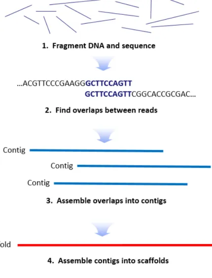
lactis LT 19-2 was sent to Macrogen coporation (Korea) for sequencing using an Illumina Hiseq 4000 platform (Illumina, USA). De novo assembly was performed by SOAPdenovo algorithm (Figure 3).
Then, sequence date analysis was conducted using Prokka, which is an integrated genomic pipeline including protein coding gene, rRNA, and tRNA. The protein coding genes were identified using Prodigal;
the rRNAs were predicted with RNAmmer; the tRNAs were predicted with Aragorn. Subsequently, the gene functions were annotated into COG (Clusters of Orthologous Groups) databases (http://www.ncbi.nlm.nih.gov/COG) and carbohydrate-active enzyme analysis was performed by dbCAN (DataBase for automated Carbohydrate-active enzyme ANotation) (Tatusov, Galperin et al. 2000)(Yin, Mao et al. 2012). Also, complete genome map was drawn by CGview based on CDSs (coding sequences) with COG, rRNA, tRNA, and G + C content (Stothard and Wishart 2005). For genomic comparison with other B. animalis subsp. lactis strains, BRIC (Blast
18
Ring Image Generator) was used (Alikhan, Petty et al. 2011).
Comparative whole genome was studied by average nucleotide identity based BLAST (ANIb). ANIb value was calculated by ANI calculator from the Kostas lab (http://enve-omics.ce.gatech.edu/ani).
19
Figure 3. Procedure steps for De novo asssmbly.
20
3.3. Relative quantification of gene expression
3.3.1. Primer design
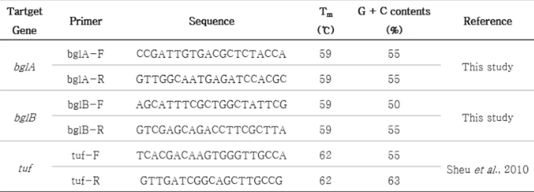
For evaluation of β-glucosidases encoding gene expression level of B. animalis subsp. lactis LT19-2, two step RT-qPCR (Reverse transcription-quantitative PCR) was performed. Elongation factor Tu (tuf) was used as housekeeping gene for the purpose of expression calibration of target genes (Sheu, Hwang et al. 2010). B. animalis subsp. lactis DSM 10140, type strain of B. animalis subsp. lactis, used as control strain. PCR primers for the target β-glucosidase genes and housekeeping gene are summarized in Table 1.
21
Table 1. Summary of primers used in this study.
22
3.3.2. RNA extraction and cDNA synthesis
Total RNA of B. animalis subsp. lactis LT19-2 and B. animalis subsp.
lactis DSM 10140 strain was extracted using Accuzol™ reagent (Bioneer, Korea). Pellet cells were lysed by repetitive pipetting in Accuzol™ reagent. Then, for phase separation, 200 ㎕ of cold chloroform was added and shake vigorously for 15 sec. Following centrifugation at 12,000 rpm for 15 min, the mixture seperates into a lower organic phase (green color), and interphase, and a colorless upper aqueous phase. RNA remains exclusively in the aqueous phase.
For cDNA synthesis, target RNA was reverse transcription using ReverTra Ace qPCR RT Master Mix with gDNA remover (Toyobo, Japan). cDNA synthesis carried out in 10 ㎕ of total volume. In a first step, 4 X DN master mix with gDNA remover was added to 0.2 ㎍ of total RNA template and nuclease-free water. After incubating at 37 ℃ for 5 min, 2 ㎕ of 5 X RT master mix II was added. cDNA synthesis was performed at 37 ℃ for 15 min followed by 50 ℃ for 5 min and 98 ℃ for 5 min.
23
3.3.3. qRT-PCR
qRT-PCR was performed using TOPreal™ qPCR Premix (Enzynomics, Korea). The assay contained 2 X TOPreal qPCR Premix (SYBR Green with high ROX), 10 pmol of forward and reverse primer and cDNA template. The qPCR was performed using thermal cycler (Bio-rad, USA) according to the following protocol: Initial denaturation of 95 ℃ for 10 min; 40 cycles of 95 ℃ for 10 min, 57 ℃ for 15 sec, and 72 ℃ for 15 sec. After the PCR, melting curve was drawn in the range of 50 ℃ to 95 ℃. Relative quantification of target genes was calculated using 2-△△Ct method (Livak and Schmittgen 2001).
24
3.4. Enzyme purification
3.4.1. Purification
B. animalis subsp. lactis LT 19-2 was cultured at 37 ℃ for 24 H in 3 L of MRS medium supplemented 0.05 % L-cysteine HCl (Sigma, USA) and incubated at 37 ℃ for 24 H anaerobically. Cultures were centrifuged at 15,000 rpm for 7 min at 4 ℃, and the harvested cell pellet was washed twice with 50 mM sodium phosphate buffer (pH 7.0). The pellet was resuspended in the same buffer. Cells were disrupted in mini bead beater 16 (Biospec, USA) with 0.1 mm zirconium beads for 45 sec. Cell debris was removed by centrifugation (15,000 rpm for 5 min at 4 ℃) and the supernatant was used as crude enzyme extract. Purification procedures were conducted at 4 ℃. The protein in the crude extract was precipitated with 20-80 % ammonium sulfate saturation. The pellets were suspended in 50 mM sodium phosphate buffer (pH 7.0) and dialyzed using Slide-A-Dialysis Cassette (Thermo scientific, USA) three times for following step. The dialyzed sample was loaded on a DEAE sepharose fast flow column (1.5 cm ⅹ 10 cm)(GE Healthcare, USA) equilibrated with the 50 mM sodium phosphate buffer (pH 7.0). The enzyme fractions was eluted
25
with a linear gradient of NaCl from 0.0 M to 0.5 M. The active fraction from DEAE sepharose column was concentrated through ultrafiltration and loaded to sephadex G-100 column (1.5 cm ⅹ 40 cm) (GE Healthcare, USA) equilibrated with 50 mM sodium buffer (pH 7.0).
The enzyme fractions were eluted with a same buffer.
3.4.2. Protein assay
Protein assay was conducted by Bradford method, using a bovine serum albumin (BSA) as a standard (Bio-rad, USA).
3.4.3. SDS-PAGE and Native-PAGE
The molecular weight of the purified enzyme was determined by SDS-PAGE (sodium dodecyl sulfate polyacrylamide gel electrophoresis) (Laemmli 1970). SDS-PAGE was performed with a mini-protein system (Bio-rad, USA) using 10 % resolving gel and 5 % stacking gel. Precision plus protein dual color system (Bio-rad, USA) was used as molecular weight marker. After electrophoresis, the gel was stained with Coomassie Brilliant R-250 (Bio-rad, USA). To calculate of molecular weight, relative migration was plotted against the log of molecular weight marker.
26
For activity staining, purified enzyme were loaded to native polyacrylamide gel (10 % resolving gel and 5 % stacking gel). Native- PAGE was performed in a pH 8.3 Tris-glycine buffer at 4 ℃. Activity staining was conducted as follows. After electrophoresis, the gel was washed in 50 mM sodium phosphate buffer (pH 7.0) and incubated in the same buffer containing 0.1 % esculin (Sigma, USA) and 0.25 % ammonium iron (Ⅲ) citrate (Sigma, USA) at 37 ℃ for 40 min (Fang, Fang et al. 2010)(Kwon, Lee et al. 1994).
27
3.5. Enzyme reactive conditions
3.5.1. pH and temperature
The optimal pH of the purified β-glucosidases was determined by incubating the purified enzyme at a pH range of 5.0-7.5 with 50 mM sodium phosphate buffer (pH 5.0-7.5) at 37 ℃. The effect of temperature on enzyme activity was evaluated at 32 ℃ to 52 ℃ in 50 mM sodium phosphate buffer (pH 7.0) (Han, Youn et al. 2014).
3.5.2. Metal ions
The effect of metal ions on the β-glucosidases activity was determined as follows. The enzyme assays were performed in the presence of various metal ions (1 mM), including CaCl2, CuSO4, MgCl2, MnSO4, KCl, and NaCl in 37 ℃ in 50 mM sodium phosphate buffer (pH 7.0) (Chang, Jo et al. 2012).
3.5.3. Enzyme kinetic analysis
Enzyme kinetics was conducted at 37 ℃ in 50 mM sodium phosphate (pH 7.0). Michaelis constant (Km) and the maximum reaction rate (Vmax) of the enzyme was estimated at different
28
concentrations of pNPG (0.312 to 20 mM), but that of the enzyme was constant. Because Michaelis-Menten plot is no useful for estimating Km and Vmax, the hyperbolic plot of Michaelis-Menten equation was converted to a linear plot of Lineweaver-Burk equation.
29
Chapter 4. Results
4.1. Isolation and identification of β-glucosidase producing bifidobacteria
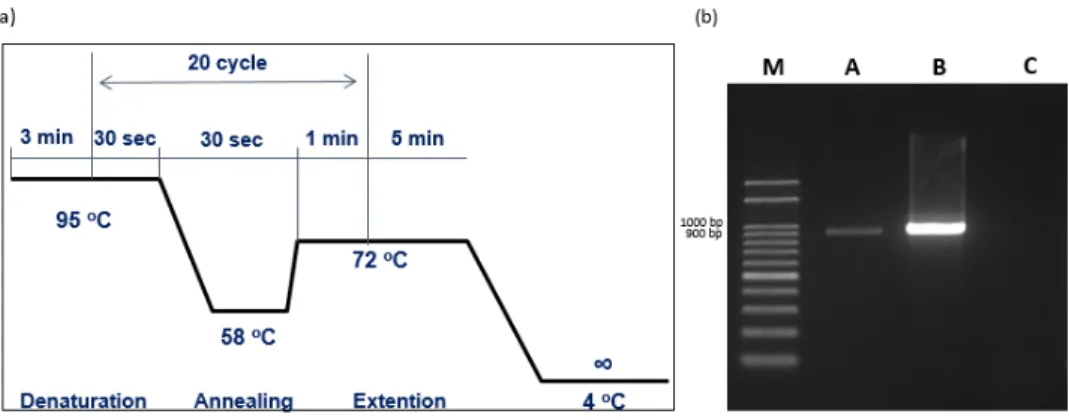
Bacteria were isolated from infant fecal samples using TOS- propionate medium. Subsequently, genus specific PCR targeting xfp gene was performed (Figure 4). After colorimetric assay, one isolate with the highest β-glucosidase activity was selected and identified as B. animalis subsp. lactis LT 19-2 by 16s rRNA sequencing and BLAST search.
30
Figure 4. PCR amplication for the detection of Bifidobacterium genus specific xfp gene (950 bp). (a) PCR conditions for amplication of xfp. (b) M : 100 bp DNA ladder line; A : B. animalis subsp. lactis LT19-2;
B : B. animalis subsp. lactis DSM 10140 (Positive control); C : Lactobacillus casei ATCC 393 (Nagative control).
31
4.2. Genomic analysis and characterization
Genome assembly results were summarized in Table 2. Scaffold N50 was 1,923,614 bp and this result showed that genome assembly was well progressed. General genomic features of B. animalis subsp.
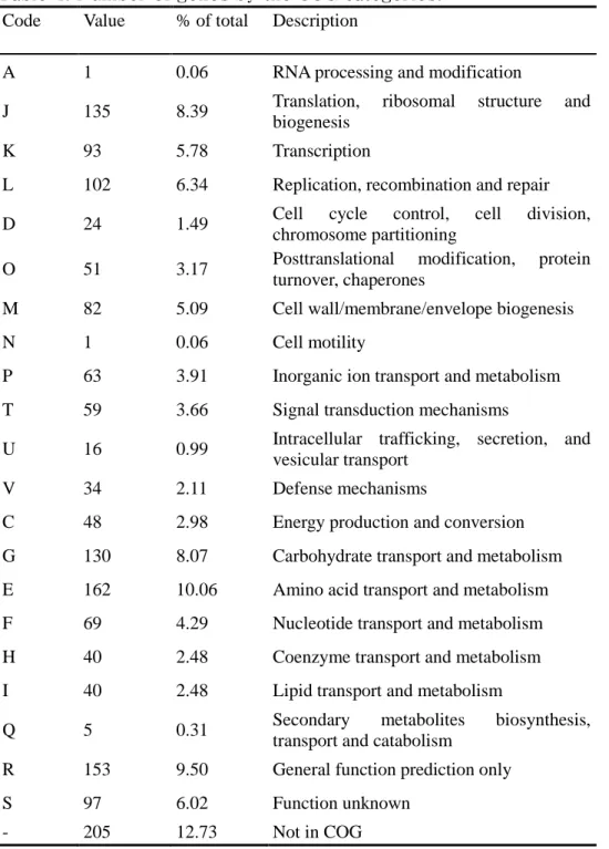
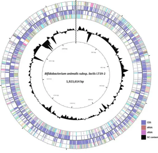
lactis LT19-2 and B. animalis subsp. lactis DSM 10140 were summarized in Table 3. B. animalis subsp. lactis LT19-2 had one chromosome with 1,923,614 bp and G + C content of 60.49 %. The chromosome contained total 1,610 of genes that were consisted of 1,551 of CDSs and 59 of RNA genes. RNA genes are 52 of tRNA, and 6 of rRNA. With COG analysis, CDSs were classified into functional categories (Table 4). ANIb values were calculated to 100 % with B.
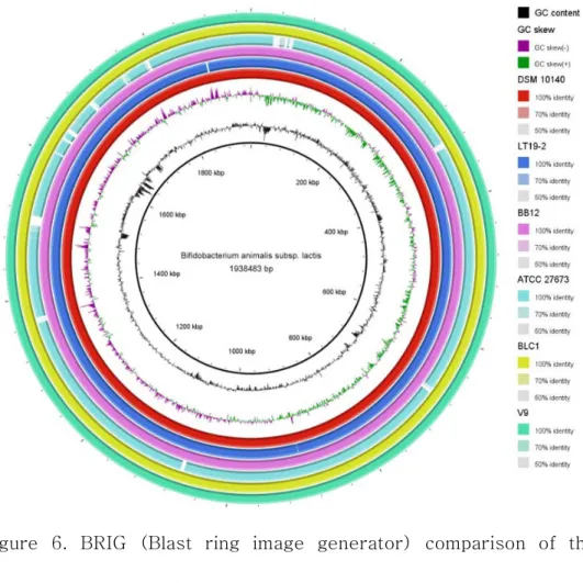
animalis subsp. lactis DSM 10140. Also, complete genomic map based COG categories was drawn in Figure 5. Genomic comparison with other B. animalis subsp. lactis strains (BB12, ATCC 27673, BLC1, and V9) was shown in Figure 6 (Garrigues, Johansen et al. 2010) (Loquasto, Barrangou et al. 2013) (Bottacini, Dal Bello et al. 2011) (Sun, Chen et al. 2010). The genomic comparison revealed that large regions of genome sequences among B. animalis subsp. lactis strains were highly conserved.
32
Also, whole genomic analysis of B. animalis subsp. lactis LT19-2 revealed two β-glucosidase (E.C 3.2.1.21) encoding genes, bglA and bglB. By dbCAN analysis, BglA and BglB were categorized to GH1 and GH3, respectively. The alignment of their amino acid sequences using Clustal W revealed that two β-glucosidase CDSs are highly conserved among B. animalis subsp. lactis strains (Figure 7 and Figure 8).
33
Table 2. Summary of genomic assembly of B. animalis subsp. lactis LT19-2.
Table 3. Comparison of genome annotation summary.
34
Table 4. Number of genes by the COG categories.
Code Value % of total Description
A 1 0.06 RNA processing and modification
J 135 8.39 Translation, ribosomal structure and
biogenesis
K 93 5.78 Transcription
L 102 6.34 Replication, recombination and repair
D 24 1.49 Cell cycle control, cell division,
chromosome partitioning
O 51 3.17 Posttranslational modification, protein turnover, chaperones
M 82 5.09 Cell wall/membrane/envelope biogenesis
N 1 0.06 Cell motility
P 63 3.91 Inorganic ion transport and metabolism T 59 3.66 Signal transduction mechanisms
U 16 0.99 Intracellular trafficking, secretion, and vesicular transport
V 34 2.11 Defense mechanisms
C 48 2.98 Energy production and conversion G 130 8.07 Carbohydrate transport and metabolism E 162 10.06 Amino acid transport and metabolism F 69 4.29 Nucleotide transport and metabolism H 40 2.48 Coenzyme transport and metabolism I 40 2.48 Lipid transport and metabolism
Q 5 0.31 Secondary metabolites biosynthesis,
transport and catabolism
R 153 9.50 General function prediction only
S 97 6.02 Function unknown
- 205 12.73 Not in COG
35
Figure 5. Complete genome map of B. animalis subsp. lactis LT19-2.
All CDSs were categorized to COG functional categories and colored differently.
36
Figure 6. BRIG (Blast ring image generator) comparison of the sequenced B. animalis subsp. lactis genomes. The six sequenced genomes are arranged as follows: DSM 10140 (reference genome), LT19-2, BB12, ATCC 27673, BLC1, and V9.
37
Figure 7. Alignment of amino acid sequences of BglA from B. animalis subsp. lactis LT19-2, DSM 10140, BB12, BLC1, V9, and ATCC 27673.
38
Figure 8. Alignment of amino acid sequences of BglB from B. animalis subsp. lactis LT19-2, DSM 10140, BB12, BLC1, V9, and ATCC 27673.
39
4.3. Relative quantification of β-glucosidase encoding genes expression
mRNA expression level of bglA and bglB β-glucosidase encoding gene were evaluated by two step RT-qPCR. mRNA expression level of bglA and bglB was higher than those of B. animalis subsp. lactis DSM 10140. The bglA and bglB were expressed approximately 4.9 fold and 10 fold more than that of DSM 10140, respectively.
40
Figure 9. Relative mRNA expression of (a) bglA and (b) bglB. Data are represented as mean ± SD and representative of three independent experiments. P values were analyzed by student’s t test analysis of variance (ANOVA)(*p < 0.05, **p < 0.01).
41
4.4. Enzyme Purification
4.4.1. Purification of β-glucosidase
The β-glucosidases from cell disruption of B. animalis subsp. lactis LT 19-2 was purified using ammonium sulfate precipitation, ion- exchange chromatography, and gel filtration chromatography. In ammonium sulfate precipitation, the crude enzyme was precipitated with 70 % saturated ammonium sulfate (Figure 10). Then, the precipitates was suspended in 50 mM sodium phosphate buffer and loaded to anion ion chromatography on DEAE sepharose Fast Flow column. The result was shown in figure 11. A fraction volume was 1 ml and one peak (fraction No. 26) with β-glucosidase activity was eluted after DEAE sepharose chromatography. This peak was eluted within fractions No. 45-49. The active fractions were concentrated and purified further using gel filtration chromatography on sephadex G-100 shown in figure 12. β-glucosidase peak was detected within fraction No. 10-28 (Figure 12). BglA was detected in fraction No.
10-13 and BglB was detected in fraction No. 20-24. The overall yield of the purification of BglA and BglB were 2.33 % with a purification fold of 10.60 and 2.17 % with a purification fold of 13.45, respectively.
42
The specific activity of the purified BglA and BglB was measured at 8.98 U/mg and 11.39 U/mg, respectively. Purification results of each step were summarized in Table 5.
43
Figure 10. β-glucosidase activity and protein concentration by ammonium sulfate precipitation.
Figure 11. β-glucosidase activity and protein concentration of fractions by DEAE sepharose Fast Flow column chromatography.
44
Figure 12. β-glucosidase activity and protein concentration of fractions by Sephadex G-100 column chromatography.
Table 5. Summary of purification of the β-glucosidases purified from B. animalis subsp. lactis LT19-2.
45
4.4.2. Molecular weight determination and activity staining
For the confirmation of enzyme purity and determination of molecular weight, SDS-PAGE was used. Molecular weight of the purified BglA and BglB was estimated to approximately 52 kDa and 84 kDa, respectively, based on relative migration of molecular weight marker (Figure 13). The purified BglA was detected after native PAGE using esculin as substrate (Figure 14), but purified BglB was not detected.
46
Figure 13. SDS-PAGE of BglA (52 kDa) and BglB (84 kDa). M : protein marker, A : crude extract, B : Ammonium sulfate precipitation, C : DEAE sepharose fast flow, D : Sephadex G-100 (BglA), E : Sephadex G-100 (BglB).
Figure 14. SDS-PAGE and Native-PAGE of purified (a) BglA and (b) BglB.
47
4.5. Optimization of enzyme reactive conditions
4.5.1. Effect of pH and temperature on the activity of β-glucosidases
The optimal pH of the purified BglA and BglB was estimated at the range of pH 5.0 - 7.5. Figure 15 shows the effect of pH on the activity of the enzyme. The optimal pH of purified BglA and BglB was pH 7.0 (Figure 15). The effect of temperature on the enzymes activity was evaluated from 32 ℃ to 52 ℃ at interval of five degree and optimal temperature of the enzymes was 37 ℃ (Figure 16).
48
Figure 15. Effect of pH on the (a) BglA and (b) BglB. Data are represented as mean ± SD and representative of three independent experiments.
.
Figure 16. Effect of temperature on the (a) BglA and (b) BglB. Data are represented as mean ± SD and representative of three independent experiments.
.
49
4.5.2. Effect of metal ions on the activity of β-glucosidases
The enzyme assays were performed in the presence of various metal ions (1 mM), including CaCl2, CuSO4, MgCl2, MnSO4, KCl, and NaCl in 37 ℃ in 50 mM sodium phosphate buffer (pH 7.0). BglA was significantly decreased by Cu2+, but BglB was not influenced by metal ions (Figure 17, 18).
50
Figure 17. Effect of metal ions on the BglA. All metal ions tested in this study was adjusted to 1 mM. Data are represented as mean ± SD and representative of three independent experiments. P values were analyzed by one-way analysis of variance (ANOVA)(***p < 0.001).
Figure 18. Effect of metal ions on the BglB. All metal ions tested in this study was adjusted to 1 mM. Data are represented as mean ± SD and representative of three independent experiments.
51
4.5.3. Enzyme kinetic analysis
Enzyme kinetic parameters (Vmax and Km) were calculated by linear plot of Lineweaver-Burk equation. Vmax and Km of BglA were 0.0092 μmol and 4.72 mM, respectively. Also, Vmax and Km of BglB were 0.0078 μmol and 1.67 mM, respectively.
52
Chapter 5. Discussion
Bifidobacteria were used as probiotics for their health promoting effects such as immune modulation. Also, they are one of the major human intestinal microbiota and play an important role in carbohydrate fermentation in GIT. β-glucosidase hydrolyzes β-glucosidic natural compounds and result in enhance of bioavailability and physiological effects of the compounds. Therefore, β-glucosidase producing bifidobacteria might lead to synergistic health benefits.
However, there was less research on screening of bifidobacteria with β-glucosidase. The purpose of this study was the screen of β- glucosidase producing bifidobacteria from infant feces and characterization of its β-glucosidase. By using screening procedures of selective media, genus specific PCR, and colorimetric assay, finally, B. animalis subsp. lactis LT19-2 was selected for its high level of β -glucosidase activity.
To analyze the genetic characteristics and to confirm the β- glucosidase encoding genes of B. animalis subsp. lactis LT19-2, whole genome sequencing was conducted. The genomic comparison with other B. animalis subsp. lactis strains revealed that large regions of genome sequences among B. animalis subsp. lactis strains were
53
highly conserved (Garrigues, Johansen et al. 2010) (Loquasto, Barrangou et al. 2013)(Bottacini, Dal Bello et al. 2011)(Sun, Chen et al. 2010). Also, Genome of B. animalis subsp. lactis LT19-2 was analyzed by COG categories and it had similar number of genes involved in carbohydrate metabolism to other bifidobacteria. Based on amino acid sequence similarities, β-glucosidases have been classified into several families. The family of most β-glucosidase belongs to either family 1 or family 3 (Henrissat 1998) (Henrissat, Callebaut et al. 1995). The two β-glucosidase encoding genes, bglA and bglB, were categorized to GH1 and GH3, respectively.
CDSs of β-glucosidase from B. animalis subsp. lactis LT19-2 were the same as type strain, B. animalis subsp. lactis DSM 10140 so we compared the mRNA expression level of bglA and bglB in B.
animalis subsp. lactis LT19-2 to B. animalis subsp. lactis DSM 10140 to confirm the difference. The mRNA of bglA and bglB of B. animalis subsp. lactis LT19-2 was expressed approximately 4.9 fold and 10 fold more than that of in B. animalis subsp. lactis DSM 10140, respectively. Through these results, B. animalis subsp. lactis LT19- 2 was estimated to metabolite the β-glucosidic compound actively than B. animalis subsp. lactis DSM 10140. These mRNA expression differences might be attribute to their differences of origin and
54
transcription regulation. B. animalis subsp. lactis DSM 10140 was isolated from fermented milk (Barrangou, Briczinski et al. 2009). In fermented milk, B. animalis subsp. lactis DSM 10140 might adapt to environment of lactose rich and they do not need to hydrolyze non- digestible carbohydrate such as human milk oligosaccharide that exists in human intestine. Also, several transcription regulation systems such as, transcription factors, promoter, or cis-acting element were likely to influence the mRNA expression.
Molecular weight of the purified BglA and BglB was estimated to approximately 52 kDa and 84 kDa, respectively. These results were different from other purified β-glucosidases. Molecular weight of purified β-glucosidase from L. brevis and Aspergillus niger was 330 kDa and 123 kDa, respectively (Michlmayr, Schümann et al. 2010) (Chang, Jo et al. 2012). Result of molecular weight from SDS-PAGE corresponded to protein sequence based whole genome sequence.
After native PAGE using esculin as substrate, the purified BglA was detected, but the BglB was not detected. Also, crude extract of B.
animalis subsp. lactis LT19-2 only showed BglA activity in native PAGE. Althought purified BglB showed activity in fraction eluted from sephadex G-100, several factors such as electrophoresis buffer
55
composition, cofactor, pH, and temperature might influence protein folding pattern and structure in native PAGE and resulted in the loss of intrinsic activity.
The highest activity of purified BglA and BglB was shown at 37 ℃ and pH 7.0 and decreased over the optimum conditions. Whereas, optimum temperature of other β-glucosidases derived from Weissella cibaria and L. brevis were 50 ℃ and 45 ℃, respectively.
(Hong, You et al. 2009) (Michlmayr, Schümann et al. 2010). Also, optimum pH of other β-glucosidases derived from Weissella cibaria and L. brevis were pH 7 and pH 5.5, respectively (Hong, You et al. 2009)(Michlmayr, Schümann et al. 2010). Throught these result, we estimated that B. animalis subsp. lactis LT19-2 was well adapted to environment of host. Additionally, the enzyme activity in the presence of various metal ions and enzyme kinetics were evaluated. BglA was significantly decreased by Cu2+, but BglB was not influenced by metal ions. Km of BglA and BglB was 4.72 mM and 1.67 mM, respectively.
Km for pNPG of BglB was lower than that of L. plantarum and L. casei (Sestelo, Poza et al. 2004) (Coulon, Chemardin et al. 1998). These result indicated that β-glucosidases of the B. animalis subsp. lactis LT19-2 had higher affinity to substrate.
56
In this study, B. animalis subsp. lactis LT19-2 with β-glucosidase activity was successfully screened. Additionally, to optimize the reactive condition of β-glucosidases, β-glucosidases from B.
animalis subsp. lactis LT19-2 were purified and investigated. The conversion of glucosides using probiotics such as Bifidobacterium might be valuable process in industry. Especially, β-glucosidase producing B. animalis subsp. lactis LT19-2 from infant feces has a possibility to increased bioavailability and physiological effects of natural compounds as well as indigenous probiotic effects of Bifidobacterium strain. Also, infant feces originated B. animalis subsp.
lactis LT19-2 might be adapted to host easily and it is an important factor for industrial application.
57
References
Alikhan, N.-F., N. K. Petty, N. L. B. Zakour and S. A. Beatson (2011).
"BLAST Ring Image Generator (BRIG): simple prokaryote genome comparisons." BMC genomics 12(1): 402.
Arunachalam, K., H. Gill and R. Chandra (2000). "Enhancement of natural immune function by dietary consumption of Bifidobacterium lactis (HNO19)." European Journal of Clinical Nutrition 54(3): 263.
Barrangou, R., E. P. Briczinski, L. L. Traeger, J. R. Loquasto, M.
Richards, P. Horvath, A. C. Coute-Monvoisin, G. Leyer, S. Rendulic, J.
L. Steele, J. R. Broadbent, T. Oberg, E. G. Dudley, S. Schuster, D. A.
Romero and R. F. Roberts (2009). "Comparison of the complete genome sequences of Bifidobacterium animalis subsp. lactis DSM 10140 and Bl-04." J Bacteriol 191(13): 4144-4151.
Bhatia, Y., S. Mishra and V. Bisaria (2002). "Microbial β- glucosidases: cloning, properties, and applications." Critical reviews in biotechnology 22(4): 375-407.
58
`
Bisaria, V. S. and T. K. Ghose (1981). "Biodegradation of cellulosic materials: substrates, microorganisms, enzymes and products."
Enzyme and Microbial Technology 3(2): 90-104.
Bottacini, F., F. Dal Bello, F. Turroni, C. Milani, S. Duranti, E. Foroni, A. Viappiani, F. Strati, D. Mora and D. van Sinderen (2011). "Complete genome sequence of Bifidobacterium animalis subsp. lactis BLC1."
Journal of bacteriology 193(22): 6387-6388.
Cairns, J. R. K. and A. Esen (2010). "β-Glucosidases." Cellular and Molecular Life Sciences 67(20): 3389-3405.
Cantarel, B. L., P. M. Coutinho, C. Rancurel, T. Bernard, V. Lombard and B. Henrissat (2009). "The Carbohydrate-Active EnZymes database (CAZy): an expert resource for glycogenomics." Nucleic acids research 37(suppl 1): D233-D238.
Chang, K. H., M. N. Jo, K.-T. Kim and H.-D. Paik (2012). "Purification and characterization of a ginsenoside Rb1-hydrolyzing β- glucosidase from Aspergillus niger KCCM 11239." International
59
journal of molecular sciences 13(9): 12140-12152.
Chichlowski, M., J. Guillaume De Lartigue, H. E. Raybould and D. A.
Mills (2012). "Bifidobacteria isolated from infants and cultured on human milk oligosaccharides affect intestinal epithelial function."
Journal of pediatric gastroenterology and nutrition 55(3): 321.
Coulon, S., P. Chemardin, Y. Gueguen, A. Arnaud and P. Galzy (1998).
"Purification and characterization of an intracellular β-glucosidase from Lactobacillus casei ATCC 393." Applied biochemistry and biotechnology 74(2): 105-114.
de Vrese, M. and J. Schrezenmeir (2008). Probiotics, prebiotics, and synbiotics. Food biotechnology, Springer: 1-66.
De Vries, W. and A. Stouthamer (1967). "Pathway of glucose fermentation in relation to the taxonomy of bifidobacteria." Journal of bacteriology 93(2): 574-576.
Eun-Ah, B., P. Sun-Young and K. Dong-Hyum (2000). "Constitutive β-gluccosidases hydrolyzing ginsenoside Rb1 and Rb2 from human intestinal bacteria." Biological and Pharmaceutical Bulletin 23(12):
60 1481-1485.
Fang, Z., W. Fang, J. Liu, Y. Hong, H. Peng, X. Zhang, B. Sun and Y.
Xiao (2010). "Cloning and characterization of a β-glucosidase from marine microbial metagenome with excellent glucose tolerance." J Microbiol Biotechnol 20(9): 1351-1358.
Furrie, E. (2006). "A molecular revolution in the study of intestinal microflora." Gut 55(2): 141-143.
Garrigues, C., E. Johansen and M. B. Pedersen (2010). "Complete genome sequence of Bifidobacterium animalis subsp. lactis BB-12, a widely consumed probiotic strain." J Bacteriol 192(9): 2467-2468.
Ghabbour, N., Z. Lamzira, P. Thonart, P. Cidalia, M. Markaoui and A.
Asehraou (2011). "Selection of oleuropein-degrading lactic acid bacteria strains isolated from fermenting Moroccan green olives."
grasas y aceites 62(1): 84-89.
Han, Y. R., S. Y. Youn, G. E. Ji and M. S. Park (2014). "Production of α-and β-galactosidases from Bifidobacterium longum subsp.
longum RD47." J Microbiol Biotechnol 24: 675-682.
61
Henrissat, B. (1991). "A classification of glycosyl hydrolases based on amino acid sequence similarities." Biochemical Journal 280(2):
309-316.
Henrissat, B. (1998). Glycosidase families, Portland Press Limited.
Henrissat, B., I. Callebaut, S. Fabrega, P. Lehn, J.-P. Mornon and G.
Davies (1995). "Conserved catalytic machinery and the prediction of a common fold for several families of glycosyl hydrolases."
Proceedings of the National Academy of Sciences 92(15): 7090-7094.
Henrissat, B. and G. Davies (1997). "Structural and sequence-based classification of glycoside hydrolases." Current opinion in structural biology 7(5): 637-644.
Hong, S.-W., L.-K. You, B.-M. Jung, W.-S. Kim and K.-S. Chung (2009). "Characterization of α-galactosidase and β-glucosidase by Weissella cibaria." Kor. J. Microbiol. Biotechnol 37: 204-212.
Jenkins, J., L. L. Leggio, G. Harris and R. Pickersgill (1995). "β- Glucosidase, β-galactosidase, family A cellulases, family F
62
xylanases and two barley glycanases form a superfamily of enzymes wit 8-fold β/α architecture and with two conserved glutamates near the carboxy-terminal ends of β-strands four and seven." FEBS letters 362(3): 281-285.
Kim, K.-A., I.-H. Jung, S.-H. Park, Y.-T. Ahn, C.-S. Huh and D.-H.
Kim (2013). "Comparative analysis of the gut microbiota in people with different levels of ginsenoside Rb1 degradation to compound K."
PLoS One 8(4): e62409.
Kim, S. H., W. Jeung, I.-D. Choi, J.-W. Jeong, D. E. Lee, C.-S. Huh, G.-B. Kim, S. S. Hong, J.-J. Shim and J. L. Lee (2016). "Lactic Acid Bacteria Improves Peyer's Patch Cell-Mediated Immunoglobulin A and Tight-Junction Expression in a Destructed Gut Microbial Environment." Journal of microbiology and biotechnology 26(6):
1035-1045.
Kwon, K.-S., J. Lee, H. G. Kang and Y. C. Hah (1994). "Detection of β-glucosidase activity in polyacrylamide gels with esculin as substrate." Applied and environmental microbiology 60(12): 4584- 4586.
63
Laemmli, U. K. (1970). "Cleavage of structural proteins during the assembly of the head of bacteriophage T4." nature 227: 680-685.
Livak, K. J. and T. D. Schmittgen (2001). "Analysis of relative gene expression data using real-time quantitative PCR and the 2(-Delta Delta C(T)) Method." Methods 25(4): 402-408.
Loquasto, J. R., R. Barrangou, E. G. Dudley, B. Stahl, C. Chen and R. F.
Roberts (2013). "Bifidobacterium animalis subsp. lactis ATCC 27673 is a genomically unique strain within its conserved subspecies."
Applied and environmental microbiology 79(22): 6903-6910.
Michlmayr, H., C. Schümann, N. Barreira Braz Da Silva, K. Kulbe and A. Del Hierro (2010). "Isolation and basic characterization of a β‐ glucosidase from a strain of Lactobacillus brevis isolated from a malolactic starter culture." Journal of applied microbiology 108(2):
550-559.
Park, J. K., L.-X. Wang, H. V. Patel and S. Roseman (2002).
"Molecular cloning and characterization of a unique β-glucosidase from Vibrio cholerae." Journal of Biological Chemistry 277(33):
29555-29560.
64
Quan, L.-H., J.-Y. Piao, J.-W. Min, D.-U. Yang, H. N. Lee and D. C.
Yang (2011). "Bioconversion of ginsenoside Rb1 into compound K by Leuconostoc citreum LH1 isolated from kimchi." Brazilian Journal of Microbiology 42(3): 1227-1237.
Ryu, D. D. and M. Mandels (1980). "Cellulases: biosynthesis and applications." Enzyme and Microbial Technology 2(2): 91-102.
Sela, D., J. Chapman, A. Adeuya, J. Kim, F. Chen, T. Whitehead, A.
Lapidus, D. Rokhsar, C. B. Lebrilla and J. B. German (2008). "The genome sequence of Bifidobacterium longum subsp. infantis reveals adaptations for milk utilization within the infant microbiome."
Proceedings of the National Academy of Sciences 105(48): 18964- 18969.
Sestelo, A., M. Poza and T. Villa (2004). "β-Glucosidase activity in a Lactobacillus plantarum wine strain." World Journal of Microbiology and Biotechnology 20(6): 633.
Setchell, K. D., N. M. Brown, L. Zimmer-Nechemias, W. T. Brashear,
65
B. E. Wolfe, A. S. Kirschner and J. E. Heubi (2002). "Evidence for lack of absorption of soy isoflavone glycosides in humans, supporting the crucial role of intestinal metabolism for bioavailability." The American journal of clinical nutrition 76(2): 447-453.
Sheu, S. J., W. Z. Hwang, Y. C. Chiang, W. H. Lin, H. C. Chen and H. Y.
Tsen (2010). "Use of Tuf Gene‐Based Primers for the PCR Detection of Probiotic Bifidobacterium Species and Enumeration of Bifidobacteria in Fermented Milk by Cultural and Quantitative Real‐ Time PCR Methods." Journal of food science 75(8).
Stothard, P. and D. S. Wishart (2005). "Circular genome visualization and exploration using CGView." Bioinformatics 21(4): 537-539.
Sun, Z., X. Chen, J. Wang, P. Gao, Z. Zhou, Y. Ren, T. Sun, L. Wang, H.
Meng and W. Chen (2010). "Complete genome sequence of probiotic Bifidobacterium animalis subsp. lactis strain V9." Journal of bacteriology 192(15): 4080-4081.
Tatusov, R. L., M. Y. Galperin, D. A. Natale and E. V. Koonin (2000).
"The COG database: a tool for genome-scale analysis of protein functions and evolution." Nucleic acids research 28(1): 33-36.
66
Tsangalis, D., J. Ashton, A. McGill and N. Shah (2002). "Enzymic Transformation of Isoflavone Phytoestrogens in Soymilk by β‐ Glucosidase‐Producing Bifidobacteria." Journal of Food Science 67(8):
3104-3113.
Varghese, J. N., M. Hrmova and G. B. Fincher (1999). "Three- dimensional structure of a barley β-D-glucan exohydrolase, a family 3 glycosyl hydrolase." Structure 7(2): 179-190.
Ventura, M., C. Canchaya, G. F. Fitzgerald, R. S. Gupta and D. van Sinderen (2007). "Genomics as a means to understand bacterial phylogeny and ecological adaptation: the case of bifidobacteria."
Antonie Van Leeuwenhoek 91(4): 351-372.
Ventura, M., S. O'Flaherty, M. J. Claesson, F. Turroni, T. R.
Klaenhammer, D. van Sinderen and P. W. O'Toole (2009). "Genome- scale analyses of health-promoting bacteria: probiogenomics." Nat Rev Microbiol 7(1): 61-71.
Ventura, M., F. Turroni and D. van Sinderen (2014). "Bifidobacteria of
67
the Human Gut: Our Special Friends." Diet-Microbe Interactions in the Gut: Effects on Human Health and Disease: 41.
Verdrengh, M., I. Jonsson, R. Holmdahl and A. Tarkowski (2003).
"Genistein as an anti-inflammatory agent." Inflammation Research 52(8): 341-346.
Yeo, N.-I., S.-K. Lee and G.-E. Ji (1993). "Characterization of alpha-galactosidase from Bifidobacterium sp. Int-57." Korean Journal of Food Science and Technology 25(6): 689-693.
Yin, Y., X. Mao, J. Yang, X. Chen, F. Mao and Y. Xu (2012). "dbCAN: a web resource for automated carbohydrate-active enzyme annotation."
Nucleic acids research 40(W1): W445-W451.
Youn, S. Y. and G. E. Ji (2012). "Identification of the β-glucosidase gene from Bifidobacterium animalis subsp. lactis and its expression in B. bifidum BGN4." Journal of microbiology and biotechnology 22(12):
1714-1723.
Zhao, J. H., Y. Arao, S. J. Sun, A. Kikuchi and F. Kayama (2006). "Oral
68
administration of soy-derived genistin suppresses lipopolysaccharide-induced acute liver inflammation but does not induce thymic atrophy in the rat." Life Sci 78(8): 812-819.
69
Abstract in Korean
β-glucosidase (E.C 3.2.1.21)는 β-glucosic 결합으로 이루어진 천 연물질을 가수분해하는 역할을 한다. 가수분해의 결과로 생성된 비배당체 는 생체이용률이 높으며, 항염증 및 항함효과 등의 잠재적인 생리효과가 크다. 인체 주요 장내미생물이자 프로바이오틱스인 비피도박테리아는 여 러가지 건강증진 효과가 있다. 또한 비피도박테리아는 당대사와 관련된 유전자를 다수 가지고 있어서, 장에서 당대사와 관련하여 중요한 역할을 한다. 이를 종합하여 볼 때, β-glucosidase를 생산하는 비피도박테리아 는 프로바이오틱스로서의 역할과 생리활성물질의 생체이용률을 높일 수 있다는 점에서 시너지효과를 기대할 수 있다. 그럼에도 불구하고, β- glucosidase를 생산하는 비피도박테리아의 대한 연구는 부족하다. 본 연 구는 β-glucosidase활성이 우수한 Bifidobacterium animalis subsp.
lactis LT19-2를 선발하고 특성을 규명하고 했다.
B. animalis subsp. lactis LT19-2의 genome 크기는 1,923,614 bp이 며 G + C 함량은 60.49 %이다. 1,610 개 유전자로 구성되어 있으며, coding 서열은 1,551 개, RNA 유전자수는 59 개로 분석되었다. B.
animalis subsp. lactis LT19-2의 유전체 분석결과 2개의 β- glucosidase gene (bglA, bglB)이 확인되었으며, BglA는 GH1, BglB는 GH3로 분류되었다.
B. animalis subsp. lactis LT19-2의 β-glucosidase 특성을 규명하기
70
위해 효소를 정제하였다. 정제 결과, BglA는 10.6 배, BglB는 13.25 배 정제되었다. 효소의 pH, 온도, 금속이온 등에 의한 최적 반응조건을 확인 하였으며, Lineweaver-Burk 식에 의해 Michaelis 상수를 계산하였다.
본 연구에서는 β-glucosidase 활성이 우수한 B. animalis subsp.
lactis LT19-2를 선발하였으며, β-glucosidase를 정제하여 특성을 확 인하였다. 추후 연구에서는 B. animalis subsp. lactis LT19-2를 직접적 으로 천연물과 같은 배당체에 적용함으로써 천연물 자체의 생리효과뿐만 아니라 비피도박테리아 자체의 프로바이오틱 효과도 기대해 볼 수 있다.
따라서 비피도박테리아와 같은 프로바이오틱스를 활용한 잠재적 생리활성 을 가진 배당체의 전환은 산업적 가치가 높기 때문에 지속적인 연구가 필 요하다.
주요어 : β-glucosidase, 비피도박테리움, 프로바이오틱스