Bacillus amyloliquefaciens HC188이 생산하는 혈전분해 효소의 정제 및 특성
신소희, 홍성욱, 정건섭*
연세대학교생명과학기술학부
Received : August 27, 2012 / Revised : September 24, 2012 / Accepted : September 25, 2012
서 론
청국장은우리나라전통대두발효식품중의하나로서콩 을삶고볏짚을덮어 40-45oC에서 2-3일간발효한다. 청국장 발효에관여하는주요미생물로서Bacillus subtilis [38, 39], B. licheniformis [15], B. amyloliquefaciens [2], B. pumilus
[29] 등이널리알려져있다. 청국장의영양성분으로는단백
질뿐만 아니라 isoflavone, phytic acid, trypsin inhibitor, 식이섬유, 올리고당등각종생리활성물질이함유되어있다 [16, 25, 41]. 이와같은생리활성물질로인한청국장의기 능성에는항돌연변이[49], 항고혈압[6], 콜레스테롤저하[21], 항산화[17], 면역증강[14], 혈전분해[26] 등에효과가있다고 보고되어있다.
최근현대인들은생활수준의향상과더불어건강에대한
관심도높아져가고있다. 남녀노소를불구하고성인병의발 병률이높아짐에따라성인병의예방과치료에대한관심도
높아지고있는실정이다. 우리나라통계청이발표한 2011년
사망원인통계결과에의하면한국인의 3대사망원인은암, 뇌혈관질환, 심장질환으로총사망자수의 47.4%라고보고 하였다. 이와같이고혈압, 동맥경화, 심장병, 뇌졸중과같은
심혈관계질환으로인한사망률이비교적높은편이다[27].
심혈관계질환은 fibrinogen이활성화된 thrombin에의해
서전환된불용성의중합체인혈전(fibrin)을형성하여발생
된다. 불용성의중합체인혈전은 plasminogen이활성화되어 plasmin에의하여분해되지만, 과도한혈전이생성되면혈전 용해(fibrinolysis)의불균형으로혈관내에혈전이축적되어혈 전증을유발하여심혈관계질환의원인이되는것으로알려져 있다[38]. 이러한 혈전증을 치료하기 위하여 plasminogen activator로써작용을하는 streptokinase[42], urokinase[28]
및 tissue plasminogen activator (t-PA)[8] 등의혈전용해제 가개발되어사용되고있다. 하지만이와같은제품은반감 기가짧을뿐만아니라전신출혈등의부작용을나타내며경 Purification and Characterization of a Fibrinolytic Enzyme Produced by Bacillus amyloliquefaciens HC188. Shin, So Hee, Sung Wook Hong, and Kun Sub Chung*. Division of Biological Science and Technology, Yonsei University, Wonju, Korea
A bacterium producing a fibrinolytic enzyme was isolated from Cheonggukjang. The bacterium was identified as a strain of Bacillus amyloliquefaciens by 16S rDNA analysis and designated as B. amyloliquefaciens HC188. The optimum culture medium appeared to be one containing 0.5% (w/v) maltose and 0.5% (w/v) soytone. Bacterial growth in the optimal medium at 37oC reached the stationary phase after 27 hours of incubation and the fibrinolytic enzyme showed optimum activity at 24 hours. The enzyme was purified by 20-80% ammonium sulfate precipitation, CM Sepharose fast flow ion exchange chromatog- raphy, and Sephacryl S-200HR column chromatography. Its specific activity was 38359.3 units/mg protein and the yield was 5.5% of the total activity of the crude extracts. The molecular weight was 24.7 kDa and the amino acids of the N-terminal sequence were AQSVPYGVSQIKAPA. The fibrinolytic enzyme activity had an optimum temperature of 40oC and an optimum pH of 8.0, and the enzyme was stable in the ranges 20-40oC and pH 6.0-8.0. Enzyme activity was increased by Ca2+ and Co2+
but inhibited by Cu2+, EDTA, and PMSF. It is suggested that the purified enzyme is a metallo-serine protease.
Keywords: Purification, fibrinolytic enzyme, Bacillus amyloliquefaciens, Cheonggukjang
*Corresponding author
Tel: +82-33-760-2252, Fax: +82-33-760-2183 E-mail: [email protected]
구용으로사용하기는부적합하며, 가격이매우높은단점을 가지고있다[31].
일본에서는전통식품인natto에서우수한혈전분해활성 을가지는 nattokinase를분리하여보고하였으며[12], 국내 에서도발효식품인청국장[22, 26], 메주[20], 된장[24], 김치 [36], 젖갈[18] 등에서 nattokinase와유사한특성의혈전분 해효소를생산하는미생물에관한연구결과가보고되어있 다. 또한, 혈전분해효소를생산하는미생물은주로Bacillus sp. 균주인것으로보고되어있다[9].
따라서식품섭취를통해혈전증을사전에예방하거나개선 할수있도록본연구에서는전통적방법으로제조한청국장 에서혈전분해효소활성이우수한Bacillus amyloliquefaciens 를선발및동정하였으며, 선발미생물이생산하는혈전분해 효소를분리및정제하고이화학적특성을조사하였다. Natto 나청국장에서분리한균주들과비교하였을때, 혈전분해효 소의분자량및 protease type에서상이한결과로확인하였 으므로이에관한연구결과를보고하고자한다.
재료 및 방법
미생물 분리 및 배지
본실험에서사용된청국장과볏짚시료들은한국전통식 방법으로제조하고있는풀목산농원(경기도여주군북내면), 미륵산농원(강원도원주시귀래면), 전통흥업토속된장(강원 도원주시흥업면), 원조김점례할머니(전라북도순창군순 창읍), 서일농원(경기도안성시일죽면)에서각각구입하여 실험에사용하였다. 미생물의분리를위하여볏짚과청국장 시료를각각취하여멸균생리식염수에 1:9 비율로혼합한 후, homogenizer (Stomacher 400, Seward, England)를사 용하여 10분동안균질화한후, 현탁액을제조하였다. 이현 탁액을멸균생리식염수로십진희석한후 tryptic soy agar (TSA; Difco, Detroit, MI, USA) plate에 100µl씩도말하여 37oC에서 14시간동안배양한후미생물집락의형태에따 라서로다른미생물을순수분리하였다.
효소활성 측정
분리미생물이생산하는단백질분해효소활성의측정은 isolated soy protein (ISP)을기질로하는 Anson[3] 개량법 을사용하여측정하였다. 분리미생물은액체배지(1% ISP, 1% glucose, 1% peptone, 0.5% yeast extract)에서 37oC, 24시간동안배양하였다. 배양액은원심분리(10,000 × g/10 min)한후, 상등액을조효소액으로사용하였다. 3% ISP 기 질용액(0.1 M phosphate buffer, pH 7.0) 150µl에조효소액 35µl을첨가하여 30oC에서 10분동안반응시킨후, 0.4 M trichloroacetic acid를가하여반응을중지시키고원심분리
(5,000 × g/15 min)하여상등액을취하였다. 상등액 100µl와 0.4 M Na2CO3 150µl 그리고 phenol reagent 50µl를혼합 하여 30oC에서 30분동안발색한후 650 nm에서흡광도를 측정하였다.
분리미생물이 생산하는 혈전분해 효소활성의 측정은 Astrup and Mullertz법[4]을 변형하여측정하였다. 0.1 M sodium phosphate buffer (pH 7.0) 5 ml에 fibrinogen (bovine;
MP biochemicals, France)을 0.6%가 되도록 용해시키고 thrombin (100 NIH/ml) (bovine; MP biochemicals, France) 100µl를 첨가하였다. 위와 동일한 완충용액에 녹인 1%
agarose 용액 5 ml을첨가하여충분히혼합한후, petri dish 에붓고실온에서 10 min 동안방치, 고형화시켜 fibrin plate 를 제조하였다. Fibrin plate에 paper disc (8 mm, Toyo Roshi, Japan)를올려놓고, TSB 배지에서 37oC, 24시간동 안배양하고원심분리 (10,000 × g/10 min)한배양상등액 10 µl를적가하여 37oC에서 12시간동안배양한후, 생성된투 명환의크기로혈전분해효소활성을측정하였다. 효소활성 unit은 plasmin (5 unit/ml) (bovine, Roche, Germany)을 2배씩희석하여 0.6% fibrin plate에 10µl씩적가하고 37oC 에서 12시간동안반응시킨후, 생성된투명대의크기로나 타내었다.
미생물의 동정
분리선발한미생물의동정은 Gram 염색법과현미경관
찰을통해 형태학적특성을관찰하여 Bergey's Manual of Systematic Bacteriology에따라분류하였다. 생화학적동정 을위하여 API 50 CHB kit (bioMerieux Co., France)으로
49개의탄소원에대한이용성을조사하고이결과를 API 50
CHB database V3.0 (http://apiweb.biomerieux.com)을 이용 하여잠정적으로동정하였다. 보다더정확한동정을위하여 16S ribosomal DNA gene sequencing을통하여동정하였 다. 선발미생물의 chromosomal DNA는 DNeasy tissue kit (Qiagen, Valecia, CA, USA)를사용하여 DNA를추출하였
다. 16S rDNA gene 증폭을 위하여 일반적으로 사용하는
27F (5'-AGAGTTTGATCATGGCTCAG-3')와 1492R (5'- GGATACCTTGTTACGACTT-3') primer를사용하였다. PCR 반응시 0.4 mM dNTP, 0.5 units Taq polymerase, 4 mM Mg2+이 함유된 Takara Perfect Premix (Takara, Japan) 10µl에 DNA template (20µg/ml) 1µl, forward primer (1.0µM)와 1µl reverse primer (1.0 µM)를각각 1µl씩넣 고나머지는증류수를첨가하여총부피가 20µl가되도록 제조하였다. PCR 증폭은 94oC에서 1분간 denaturation, 51oC에서 1분간 annealing, 72oC에서 1분 30초 동안 extension을 30 cycles 실시하였고, 72oC에서 5분간 최종 extension을 실시하는 조건에서 PCR (MinicyclerTM, MJ
research Inc., Waltham, MA, USA)로증폭하였다. 증폭된 약 1,400 bp의 fragment를 T vector (Invitrogen, Carlsbad, CA, USA)에결합시킨후형질전환하였다. T vector sequencing
primer를이용하여염기서열결정을수행하였으며그결과
는 BLAST (http://www.ncbi.nlm.nih.gov) program을이용하 여 GENBANK (NCBI, Bethesda, MD, USA)의 ribosomal DNA gene sequence와비교하여동정하였다.
분리된 미생물의 16S rDNA 염기서열을 CLUSTAL_X
program으로 multiple sequence alignment한후 phyloge- netic tree를작성하였다. 해당분석은 neighborhood-joining 알고리즘을바탕으로수행되었다[40, 44].
단백질 함량 측정
단백질함량은 Ohnishi 등[37]의정량법에따라실시하였 다. 시료용액(50µl)에 550µl의 biuret reagent (0.75 mM/L cupric sulfate, 94 mM/L sodium hydroxide)를넣어혼합한 후, 실온에 10분 동안 방치하였다. 그 후에 25µl의 folin- ciocalteu's phenol reagent (Sigma, catalog No. 690-2)를 넣고실온에 30분동안방치한후 725 nm에서흡광도를측 정하였다. 표준단백질로는 bovine serum albumin (Sigma, catalog No. 690-10)을사용하여 725 nm에서흡광도를측정 하였다.
효소의 분리 및 정제
미생물이생산하는혈전분해효소를정제하기위하여최적 배지 [0.5% maltose, 0.5% soytone, 0.7% K2HPO4, 0.15%
KH2PO4, 0.004% MnCl·4H2O, 0.05% MgSO4·7H2O, 0.03%
CaCl2·2H2O, 0.00025% FeSO4·7H2O, 0.0002% (NH4)2MoO4]에 1% 접종하여 37oC에서 24시간동안배양하였다. 배양액은 원심분리(10,000 × g/15 min)하여배양상등액을조효소액으 로사용하였다. 조효소액에황산암모늄을 20-80% 포화시킨 후, 침전분리한조효소액을소량의 10 mM sodium acetate buffer (pH 6.0)에용해한후, 동일한완충용액에 4oC에서 24 시간동안 dialysis (6-8 kDa cut-off)를수행하였다. 양이온 교환수지인 CM Sepharose Fast Flow (Pharmacia, Biotech.
AB Uppsala, Sweden) column (25 × 400 mm)을 10 mM sodium acetate buffer (pH 6.0)로평형화시킨 후, 투석한 농축조효소액을 loading하여 ion exchange chromatography 를수행하였다. 용출은 0-1 M NaCl를 linear gradient하게 1 ml/min 유속으로용출하여 5 ml씩분획하였다. 활성을나 타낸분획을모아동결건조하여농축시킨후, sephacryl S- 200HR (Pharmacia, AB Uppsala, Sweden) column (15 × 960 mm)을사용하여 gel filtration을실시하였다. 10 mM sodium acetate buffer (pH 6.0)로평형화시킨후, 용출속도
는 0.5 ml/min으로 3 ml씩분획하여활성을나타내는분획 을모아동결건조하였다.
SDS-PAGE에 의한 분자량 측정
정제한효소의분자량을측정하기위하여 sodium dodecyl sulfate-polyacrylamide gel electrophoresis (SDS-PAGE) 분석을 실시하였다[30]. Ion exchange chromatography와 gel chromatography에의해얻어진 시료는 sample buffer [12.5 mM Tris·HCl buffer (pH 6.8), 2% β-mercaptoethanol, 2% SDS, 20% glycerol, 0.02% bromophenol blue]을혼합 하여 90oC에서 5분동안열처리한후, 80 V에서 2시간동안 전기영동을하였다. 전기영동후 gel은 coomassie brilliant blue (Elpis Biotech Inc., Korea)로 1시간 동안 염색한 후 증류수로 탈색하였고, pre-stained protein marker (Elpis Biotech Inc., Daejeon, Korea)를이용하여분자량을측정하 였다.
Fibrin zymography 분석
0.12% (w/v) fibrinogen과 thrombin 100µl (10 NIH unit/ml) 을함유한 10% (w/v) polyacrylamide gel에정제한효소단 백질 20µg과 zymogram sample buffer [10% SDS, 20%
glycerol, 0.03% bromophenol blue, 0.5 M Tris·HCl (pH 6.8)]을혼합하여열처리하지않고 80 V에서 2시간전기 영동을수행하였다. 전기영동한후, 상온에서 2.5% Triton X-100이 함유된 50 mM Tris·HCl buffer (pH 7.4)에 30분 동안고정하였고, Triton X-100을제거하기위하여증류수 로 30분 동안 세척한 후, zymogram reaction buffer [30 mM NaN3, 30 mM Tris·HCl (pH 7.4)]에 넣어 37oC에서 12시간 동안반응시켰다. Polyacrylamide gel은 coomassie brilliant blue로 1시간동안염색한후, 증류수로탈색하여 확인하였다[22].
N-terminal 아미노산 서열분석
정제한효소단백질을 SDS-PAGE 전기영동한후, transfer buffer [10×transfer buffer (Tris 30.3 g, glycine 144.2 g;
1 L, pH 8.3) 100 ml, methanol 200 ml, 증류수 700 ml]에평형 시킨 polyvinylidene difluoride (PVDF) membrane (Bio- Rad Laboratories, Hercules, CA, USA)에 17 V, 40분동안 transfer 하였다. Transfer한 후, PVDF membrane은 commasie brilliant blue로염색하고 methanol로탈색하였
다. PVDF membrane을건조하여염색한효소단백질을확
인한후, Edman[11] 방법에따라 Protein/Peptide sequencer (Applied Biosystems Inc., CA, USA)를이용하여아미노산 서열을확인하였다.
최적반응 pH 및 pH 안정성
혈전분해효소의 pH에대한영향을조사하기위해 0.1 M
sodium acetate buffer (pH 4.0-6.0), 0.1 M sodium phosphate buffer (pH 6.0-8.0), 0.1 M Tris-HCl buffer (pH 8.0-10.0) 을사용하였다. 조효소액과정제한효소액각각 10µl와각 pH의 buffer (pH 4.0-10.0) 190µl씩 혼합한 후, 각 pH의 buffer로제조한 0.6% (w/v) fibrin plate에 10µl씩적가하고 37oC에서 12시간동안반응시킨후, 효소활성을최적반응 pH를조사하였다. pH 안정성은조효소액과정제한효소액 각각 10µl와각 pH의 buffer (pH 4.0-10.0) 190µl씩혼합하 여 37oC에서 5시간 동안 반응시킨 후, 0.6% (w/v) fibrin plate에 10µl를적가하고 37oC에서 12시간동안반응시킨 후, 잔존효소활성을조사하였다.
최적반응 온도 및 온도 안정성
혈전분해효소의온도에 대한 영향은 20부터 60oC까지 10oC 간격으로측정하였다. 최적반응온도는각온도에서조
효소액과정제한효소액을 12시간동안반응시킨후, 0.6%
(w/v) fibrin plate를사용하여효소활성을측정하였고, 온도 안정성은조효소액과정제한효소액을각온도에서 1시간동 안반응시킨후, 잔존효소활성을조사하였다.
금속이온과 저해제의 영향
정제한효소의 금속이온에 대한 영향을 알아보기위해 0.1 M Tris-HCl buffer (pH 8.0)에 Ca2+, Co2+, Cu2+, Fe2+, Mg2+, Mn2+, Zn2+금속이온들을최종농도가 2 mM과 5 mM 이되도록첨가한후, 잔존효소활성을조사하였다.
정제한효소의저해제에대한영향을알아보기위해일반 적인단백질분해효소 inhibitor로알려진 ethylenediamine- tetraacetic acid (EDTA), phenylmethanesulfonyl fluoride (PMSF), antipain, pepstatin A을 최종농도가 2 mM과 5 mM이되도록첨가한후, 잔존효소활성을조사하였다.
결과 및 고찰
혈전분해 효소 생산미생물의 분리 및 선발
전통방식으로제조한청국장과청국장발효에사용하는 볏짚을멸균생리식염수로각각십진희석하고 tryptic soy agar (TSA) 배지에도말하여배양한후, colony 형태가서로 다른미생물총 1,592주를순수분리하였다. 혈전분해효소 는단백질분해효소의일종으로알려져있어단백질분해효 소활성이높은미생물의탐색이선행되었다. 분리미생물은
1% ISP배지에서배양한후, 배양상등액을조효소액으로사
용하여단백질분해효소활성을측정한결과, 단백질분해효 소활성이우수한 72주를 1차선발하였다(data not shown). 단
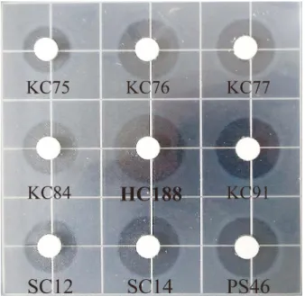
백질분해활성이우수한 72주를대상으로혈전분해효소활 성을 측정하였다. 0.6% (w/v) fibrin plate의 paper disc 주 위로생성된투명환의직경을비교하여혈전분해효소활성 이우수한분리미생물 9주를 2차선발하였다. 2차선발한미 생물은김점례할머니청국장에서분리한 5주(KC75, KC76, KC77, KC84, KC91), 흥업토속된장청국장에서분리한 1주 (HC188), 서일농원청국장에서분리한 2주(SC12, SC14), 풀 목산농원청국장에서분리한 1주(PS46)이었다. 동일한방법 으로 0.8% (w/v) fibrin plate를제조하여 2차선발한미생물 의혈전분해효소활성을측정한결과(Fig. 1), 혈전분해효소 활성이가장우수한 isolate HC188을최종선발하였다. 선발미생물의 동정
혈전분해효소활성이가장높은 isolate HC188를현미경
으로관찰한결과, Gram 양성의간균이었으며, 선발미생물
의당이용성검토를위하여 API 50 CHB kit test를실시한 결과, Bacillus subtilis/amyloliquefaciens와 84.7%의상동성 을나타내었다. 보다더정확한동정을위하여 16S rDNA 분 석을실시한결과, Bacillus amyloliquefaciens와 99%의상동 성을나타내었다. 따라서선발미생물을B. amyloliquefaciens HC188이라명명하였다(Fig. 2).
Fig. 1. Fibrinolytic activity of isolates producing protease.
Fibrinogen (0.6%) and thrombin solution (100 NIH/ml) in sodium phosphate buffer (pH 7.0) was poured into squre dish and 1%
agarose were slowly mixed in squre dish. The mixture was let to solidify for 10 min at room temperature. 10 µl of supernatant was dropped onto the paper disc and the fibrin plate was incu- bated for 12 hours at 37oC. Diameter of hydrolyzed clear zone was measured.
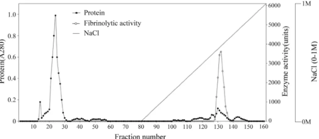
Fig. 3. Ion exchange chromatogram of the crude fibrinolytic enzyme on a CM-sepharose column.
The eluant was 10 mM sodium acetate buffer (pH 6.0) with a linear salt gradient to 1 M NaCl at a flow rate 1 ml/min. The volume of each fraction was 5 ml.
Fig. 2. Phylogenetic relationship of isolate HC188 through 16S rDNA gene sequence homology.
The number on each branch indicates the percentage of 1,000 replicates that included the branch. Sequences of isolate HC188 deter- mined in this study are shown in bold, and GenBank accession numbers are shown in parentheses for all reference sequences. The scale bar represents a 1% nucleotide substitution rate according to the Jukes-Cantor evolutionary distance.
Fig. 4. Sephacryl S-200HR column chromatogram of CM-sepharose the fibrinolytic enzyme fractions.
The enzyme was eluted with 10 mM sodium acetate buffer (pH 6.0) at a flow rate 0.5 ml/min. The volume of each fraction was 3 ml.
혈전분해 효소의 정제
B. amyloliquefaciens HC188가생산하는혈전분해효소 를정제하기위해최적배지에접종하여 37oC에서 24시간동 안배양하였다. 배양액을원심분리하여얻은배양상등액에 황산암모늄을첨가하여 20-80%로포화시킨후, 투석하였다. 조효소액은 ion-exchange chromatography인 CM-sepharose Fast Flow column에 loading하고 0-1 M NaCl을 linear gradient하게 1 fraction당 5 ml씩용출하였다(Fig. 3). 활성 분획이 있는 fraction no. 128-137를모아동결건조한후, 10 mM sodium acetate buffer (pH 6.0)에현탁하였다. 현 탁액을 sephacryl S-200HR gel filtration을수행한결과는
Fig. 4에 나타내었다. 혈전분해 효소활성을 나타내는
fraction no. 46-50을모아동결건조하였다. 전체적인효소 의정제과정은 Table 1에나타내었다. 최종정제한혈전분 해효소는배양상등액에비해약 50.7배효소활성이증가되 었으며, 이때효소단백질의회수율은 5.5%를나타내었다.
효소단백질의 분자량 측정과 fibrin zymography 정제한혈전분해효소의단일성을확인하고분자량을측 정하기위하여 SDS-polyacrylamide gel과 zymography 전 기영동을수행한결과는 Fig. 5에나타내었다. SDS-PAGE
와 zymography를 비교하여혈전분해효소활성이 나타난
band를확인한결과, 정제한혈전분해효소단백질의분자량 은약 22.3 kDa으로추정되었다. Kim 등[22]이정제한혈전 분해효소의분자량은 27 kDa으로확인하였고, Jo 등[20]은 메주로부터분리한B. amyloliquefaciens MJ5-41이생산하 는혈전분해효소의분자량은 27 kDa이었다. Lee 등[32]은 된장에서분리한Bacillus sp. KDO-13이생산한혈전분해 효소는 44 kDa이었고, Wang 등[45]은 Douchi에서분리정제 한 혈전분해 효소는 30 kDa이었으며, Jaouadi 등[19]은 Bacillus pumilus CBS가 생산하는 혈전분해 효소는 약
34 kDa으로보고하였다. 이는본연구에서청국장에서분리
한B. amyloliquefaciens HC188가생산하는혈전분해효소 는기존에연구보고된혈전분해효소들에비해서분자량이 적은것으로확인하였다.
N-terminal 아미노산 서열분석
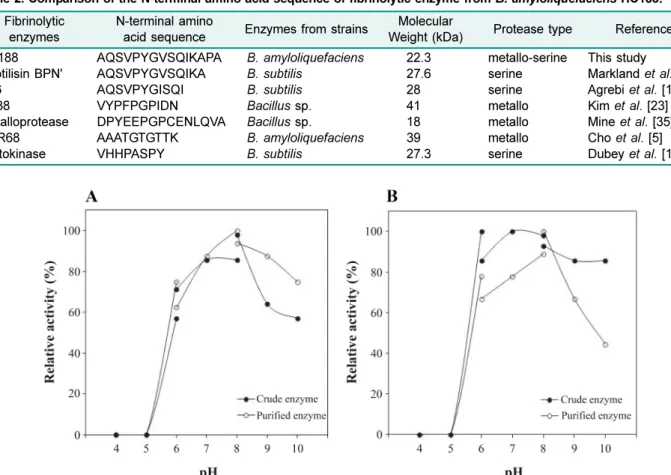
정제한혈전분해효소단백질의 N-terminal 아미노산서열 은 Ala-Gln-Ser-Val-Pro-Tyr-Gly-Val-Ser-Gln-Ile-Lys-Ala-Pro- Ala로분석되었다. Bacillus sp.가생산하는혈전분해효소 의 N-terminal 아미노산서열과비교한결과(Table 2), 혈전 분해효소인 KA38[23]과B. amyloliquefaciens가생산하는 혈전분해효소인 NPR68[5]의 N-terminal 아미노산서열과 는일치하지않은것을확인하였으며, B. subtilis가생산하 는혈전분해효소인 nattokinase의아미노산서열과도일치 하지않은것을확인하였다[11]. B. subtilis가생산하는혈 전분해효소인 subtilisin BPN[34]과 subtilisin A26[1]의아 미노산서열을비교하였을때각각 13개아미노산과 10개 아미노산이동일하여높은유사성(sequence homology)을가 지고있는 것으로나타났으나 subtilisin BPN와 subtilisin A26은B. subtilis가생산한혈전분해효소인반면에본연 구에서분리·정제한효소는B. amyloliquefaciens가생산 하였으며, 분자량및 protease type 등과비교해볼때상이 한결과로다른혈전분해효소인것으로사료되었다. Fig. 5. Determination of molecular weight and fibrin
zymography of the purified fibrinolytic enzyme from B. amy- loliquefaciens HC188.
A, SDS-PAGE; B, fibrin zymography. Lane M, marker proteins;
Lane 1, after CM sepharose FF; Lane 2, after gel filtration chro- matography. Zymography of purified fibrinolytic enzyme for activity staining was performed with 10% (w/v) polyacrylamide gel containing 0.12% (w/v) fibrinogen and thrombin (10 NIH unit/
ml) at 80 V for 2 hours. The gel was stained with coomassie bril- liant blue and destained with distilled water at room temperature for 1 h.
Table 1. Purification summary of the fibrinolytic enzyme from B. amyloliquefaciens HC188.
Total activity (units)
Total protein (mg)
Specific activity (unit/mg)
Purification (fold)
Yield (%)
Culture supernatant 378,920.5 501.3 755.9 1.0 100
Ammonium sulfate precipitation 128,250.0 28.9 3,250.8 4.3 33.8
CM-Sepharose FF column chromatography 32,645.5 1.2 26,220.2 34.7 8.6
Sephacryl-200HR column chromatography 20,986.4 0.5 38,359.3 50.7 5.5
최적반응 pH 및 pH 안정성
조효소액과정제한효소액에대한 pH의영향을알아보기 위해 pH 4.0-6.0은 0.1 M sodium acetate buffer, pH 6.0- 8.0은 0.1 M sodium phosphate buffer, pH 8.0-10.0은 0.1 M Tris-HCl buffer를사용하였다. 조효소액과정제한효소액 모두 pH 8.0에서최대활성을나타내었으며(Fig. 6A), pH 6.0- 8.0 사이에서안정함을나타내었다. 정제한효소액은 pH 9.0 에서는활성이 65%로급격히감소하였다(Fig. 6B). 조효소 액과정제한효소액의혈전분해활성은산성조건에서보다 알칼리조건에서비교적높게나타내었다. Choi 등[7]이분 리한Bacillus sp. DJ-2가생산한혈전분해효소는 pH 9.0에 서최대활성을나타낸다고보고하였으며, Lee 등[32]은된 장으로부터분리한Bacillus sp.가생산한혈전분해효소는 pH 6.0부터효소활성이 60% 이상증가하여 pH 8.0에서최 대활성을나타냈으며 pH 10.0에서는 80% 이상의효소활성 이유지되었다고보고하였다. Yang 등[46]은 pH가증가할 수록혈전분해효소활성도증가하여 pH 8.0에서최대활성을
나타내었으며 pH 10.0에서는 90% 이상효소활성이유지된 다고보고하였고, Kim 등[26]이분리한Bacillus sp. CK 11-
4가생산한혈전분해효소는 pH 6.0 이하에서는효소활성이
낮으나 pH 8.0에서최대활성을나타낸다고보고하였다. 발
효한청국장의 pH가 6.0-8.0 사이를나타내기때문에청국 장으로부터분리한미생물이생산하는혈전분해효소도 pH
6.0-8.0에서효소의최대활성과안정성을나타내는것으로
사료된다.
최적반응 온도 및 온도 안정성
조효소액과정제한효소액에대한온도의영향은 20oC부 터효소활성이점차증가하여 40oC에서최대활성을나타내 었다(Fig. 7A). 20oC에서 40oC 사이에서온도에대한안정성 을나타내었으나 50oC 이상부터활성이급격히감소하였고 60oC에서는 활성이 소실되었다(Fig. 7B). Bacillus subtilis KCK-7이생산한혈전분해효소는 60oC에서최대활성을나 타내었고 80oC에서도 20%의효소활성을유지하였다고보고 Fig. 6. Effect of pH on the activity and stability of the fibrinolytic enzyme from B. amyloliquefaciens HC188.
A, optimum pH; B, pH stability. For the pH stability, crude and purified enzyme were preincubated at various pH buffer for 5 h and the remaining activity was measured with 0.6% fibrin plate. The 0.1 M buffers were used as follows: sodium acetate (pH 4-6), sodium phosphate (pH 6-8), and Tris-HCl (pH 8-10).
Table 2. Comparison of the N-terminal amino acid sequence of fibrinolytic enzyme from B. amyloliquefaciens HC188.
Fibrinolytic enzymes
N-terminal amino
acid sequence Enzymes from strains Molecular
Weight (kDa) Protease type Reference HC188
Subtilisin BPN' A26
KA38
Metalloprotease NPR68 Nattokinase
AQSVPYGVSQIKAPA AQSVPYGVSQIKA AQSVPYGISQI VYPFPGPIDN DPYEEPGPCENLQVA AAATGTGTTK VHHPASPY
B. amyloliquefaciens B. subtilis
B. subtilis Bacillus sp.
Bacillus sp.
B. amyloliquefaciens B. subtilis
22.3 27.6 28 41 18 39 27.3
metallo-serine serine serine metallo metallo metallo serine
This study
Markland et al. [34]
Agrebi et al. [1]
Kim et al. [23]
Mine et al. [35]
Cho et al. [5]
Dubey et al. [10]
하였다[38]. B. licheniformis KJ-31이생산한혈전분해효소 는 40oC에서최대활성을나타내었고 60oC 이상에서는급격 히감소하는경향을나타내었다[18]. Kim 등[22]이분리한 B. amyloliquefaciens CH51이생산한혈전분해효소는 35- 45oC에서최대활성을나타내었다고보고하였는데, 이는본 연구결과와도유사하였다. Jo 등[20]은메주로부터분리한 B. amyloliquefaciens MJ5-41이 생산한 혈전분해 효소는 45oC에서최대활성을나타내었으나 50oC 이상에서는급격 히활성이감소되었다고보고하였다. Yeo 등[47]이분리한 Bacillus subtilis A1이생산한혈전분해효소는 20-50oC에 서효소의안정성을나타내었으나 60oC 이상에서는급격히 감소하였는데이는본연구결과와유사한경향을나타내었 다. 위와같은연구결과를통해청국장에서분리한미생물이 생산하는혈전분해효소활성의특성은 40-45oC에서발효하 는청국장의발효온도조건과유사한것으로사료된다. 금속이온 및 저해제에 대한 영향
정제한효소의금속이온과저해제에대한영향을알아보 기위해각각의금속이온과저해제를최종농도가 2 mM과 5 mM이되도록사용하여효소활성을측정하였다(Table 3).
2 mM과 5 mM 농도의 CoCl2와 CaCl2의금속이온과 5 mM 농도의 MgCl2와 MnCl2의금속이온이존재할때는효소활 성이증가하였으며, CuCl2의금속이온이 2 mM과 5 mM의 농도로존재할때는효소활성이감소하는결과를나타내었 다. 깊은바닷물로부터분리한B. subtilis ICTF-1이생산하 는혈전분해효소는 Ca+2, Mg+2, Mn+2등의금속이온이존 재할때효소활성이증가하였다고 Mahajan 등[33]은보고
하였으며, Hassanein 등[13]은B. subtilis K42가생산하는 혈전분해효소에 Co+2 금속이온이 존재할때, 효소활성이
137% 증가하였다고보고하였는데이는본연구의결과와유
사함을나타내었다. Yin 등[48]이분리한B. subtilis YJ1이 생산하는혈전분해효소는 Ba+2금속이온이존재할때, 효 Fig. 7. Effect of temperature on the activity and stability of the fibrinolytic enzyme from B. amyloliquefaciens HC188.
A, optimum temperature; B, temperature stability. For the temperature stability, crude and purified enzyme were preincubated at var- ious temperatures for 1 h and the remaining activity was measured.
Table 3. Effects of metal ions and inhibitors on the fibrin- olytic enzyme purified from B. amyloliquefaciens HC188.
Metal ions/
Inhibitors
Relative activity (%)
2 mM 5 mM
None 100.0 100.0
CaCl2 127.3 118.2
CoCl2 109.1 127.3
CuCl2 90.9 54.5
FeCl2 100.0 81.8
MgCl2 100.0 127.3
MnCl2 100.0 127.3
ZnCl2 72.7 109.1
EDTA 0.0 0.0
PMSF 0.0 0.0
Antipain 0.0 0.0
Pepstatin A 85.7 71.4
Purified enzyme was preincubated with 2 mM or 5 mM metal ions and inhibitors in 0.1 M sodium phosphate buffer (pH 8.0), respectively. The solutions were incubated at 37oC for 30 min and then dropped onto the paper disc. 0.6% fibrin plate was incubated at 37oC for 12 h and the remaining activity was measured.
소활성이증가하였으며, Cu+2, Zn+2, Fe+2의금속이온이존 재할때는효소활성이감소하는결과를나타내었다.
정제한효소의 inhibitor에대한영향을알아보기위하여
metalloprotease를억제하는 ethylenediaminetetraacetic acid (EDTA), serine protease를억제하는 phenylmethanesulfonyl fluoride (PMSF), serine/cystein protease를억제하는 antipain, 그리고 aspartyl protease를억제하는 pepstatin A을이용하 여효소활성을 측정하였다. 2 mM과 5 mM 농도의 EDTA, PMSF, Antipain의저해제가존재할때, 효소활성이감소하 는결과를나타내어 B. amyloliquefaciens HC188이생산하 는혈전분해효소는 metallo-serine protease로사료되었다. Wang 등[45]은중국의전통발효식품인 Douchi에서분리한 B. subtilis DC33이생산하는혈전분해효소가 PMSF에의
해저해를받았지만 EDTA에의해서는저해를받지 않아
serine protease라고 보고하였으며, Kim 등[23]이분리한 Bacillus sp. KA38와 Lee 등[32]이 분리한 Bacillus sp.
KOD-13이생산한혈전분해효소는 EDTA에의해효소활성
의저해를받아 metalloprotease라고보고하였다. Yang 등 [46]은 PMSF와 EDTA에의해저해를받아 metallo-serine
protease라고보고하였는데이는본연구의결과와도유사
하였다.
요 약
식품으로서안전하게섭취하여혈전증을사전에예방하거 나개선할수있도록하기위하여, 전통적방법으로제조한청 국장으로부터미생물을분리하여혈전분해효소활성이우수 한미생물을선발·동정한결과, Bacillus amyloliquefaciens
HC188이라명명하였다. 선발미생물이생산하는혈전분해
효소를분리및정제한결과, 50.7배의정제도와 5.5%의수 율을나타내었고, 혈전분해효소단백질의분자량은 22.3 kDa 이었으며, N-terminal 아미노산 서열은 Ala-Gln-Ser-Val- Pro-Tyr-Gly-Val-Ser-Gln-Ile-Lys-Ala-Pro-Ala로분석되었다. 효소의최적반응 pH와온도는 pH 8.0과 40oC로나타났으며, pH 6.0-8.0과 20-40oC 사이에서효소가비교적안정하였다. 금속이온에 대한 영향은 2 mM과 5 mM 농도의 CoCl2와 CaCl2의금속이온이존재할때효소활성이증가하였으며, inhibitor로서 EDTA와 PMSF에의해효소활성이저해되므 로청국장에서분리한B. amyloliquefaciens HC188가생성 한혈전분해효소는 metallo-serine protease로사료되었다.
Acknowledgments
This study was supported by the 2011 Technology Development Program for Agriculture and Forestry (TDPAF), Ministry for Food, Agriculture, Forestry and Fisheries, Republic of Korea.
References
1. Agrebi, R., A. Haddar, N. Hmidet, K. Jellouli, L. Manni, and M.
Nasri. 2009. BSF1 fibrinolytic enzyme from a marine bacte- rium Bacillus subtilis A26: Purification, biochemical and mole- cular characterization. Process Biochem. 44: 1252-1259.
2. Ahn, Y. S., Y. S. Kim, and D. H. Shin. 2006. Isolation, identifi- cation, and fermentation characteristics of Bacillus sp. with high protease activity from traditional Cheonggukjang. Korean. J.
Food. Sci. Technol. 38: 82-87.
3. Anson, M. L. 1939. The Estimation of pepsin, trypsin, papain, and cathepsin with hemoglobin. J. Gen. Physiol. 22: 78-89.
4. Astrup, T. and S. Müllertz. 1952. The fibrin plate method for esti- mating fibrinolytic activity. Arch. Biochem. Biophys. 40: 346- 347.
5. Cho, S. J., S. H. Oh., R. D. Pridmore, M. A. Juillerat, and C. H.
Lee. 2003. Purification and characterization of proteases from Bacillus amyloliquefaciens isolated from traditional soybean fermentation starter. J. Agric. Food Chem. 51: 7664-7670.
6. Cho, Y. J., W. S. Cha, S. K. Bok, M. U. Kim, S. S. Cheon, and U. K. Choi. 2000. Production and separation of anti-hypertensive peptide during Chunggugjang fermentation with Bacillus subtilis CH-1023. J. Korean Soc. Agric. Chem. Biotechnol. 43: 247- 252.
7. Choi, N. S., K. H. Yoo, J. H. Hahm, K. S. Yoon, K. T. Chang, B. H.
Hyun, P. J. Maeng, and S. H. Kim. 2005. Purification and char- acterization of a new peptidase, bacillopeptidase DJ-2, having fibrinolytic activity produced by Bacillus sp. DJ-2 from Doen- Jang. J. Microbiol. Biotechnol. 15: 72-79.
8. Collen, D. and H. R. Lijnen. 2009. The tissue-type plasmino- gen activator story. Arterioscler. Thromb. Vasc. Biol. 29: 1151- 1155.
9. Craik, C. S., S. Roczniak, C. Largman, and W. J. Rutter. 1987.
The catalytic role of the active site aspartic acid in serine pro- tease. Science. 237: 909-913.
10. Dubey, R., J. Kumar, D. Agrawala, T. Char, and P. Pusp. 2011.
Isolation, production, purification, assay and characterization of fibrinolytic enzyme (nattokinase, streptokinase and uroki- nase) from bacterial sources. Afr. J. Biotechnol. 10: 1408-1420.
11. Edman, P. and G. Begg. 1967. A protein sequenator. Euro- pean J. Biochem. 1: 80-91.
12. Fujita, M., K. Nomura, K. Hong, Y. Ito, A. Asada, and S. Nish- imuro. 1993. Purification and characterization of a strong fibrin- olytic enzyme (nattokinase) in the vegetable cheese Natto, a popular soybean fermented food in Japan. Biochem. Biophys.
Res. Comm. 197: 1340-1347.
13. Hassanein, W. A., E. Kotb, N. M. Awny, and Y. A. El-Zawahry.
2011. Fibrinolysis and anticoagulant potential of a metallo pro- tease produced by Bacillus subtilis K42. J. Biosci. 36: 773- 779.
14. Hong, S. W., J. Y. Kim, B. K. Lee, and K. S. Chung. 2006. The bacterial biological response modifier enriched Chungkook- jang fermentation. Korean J. Food Sci. Technol. 38: 548-553.