저작자표시-비영리-변경금지 2.0 대한민국 이용자는 아래의 조건을 따르는 경우에 한하여 자유롭게
l 이 저작물을 복제, 배포, 전송, 전시, 공연 및 방송할 수 있습니다. 다음과 같은 조건을 따라야 합니다:
l 귀하는, 이 저작물의 재이용이나 배포의 경우, 이 저작물에 적용된 이용허락조건 을 명확하게 나타내어야 합니다.
l 저작권자로부터 별도의 허가를 받으면 이러한 조건들은 적용되지 않습니다.
저작권법에 따른 이용자의 권리는 위의 내용에 의하여 영향을 받지 않습니다. 이것은 이용허락규약(Legal Code)을 이해하기 쉽게 요약한 것입니다.
Disclaimer
저작자표시. 귀하는 원저작자를 표시하여야 합니다.
비영리. 귀하는 이 저작물을 영리 목적으로 이용할 수 없습니다.
변경금지. 귀하는 이 저작물을 개작, 변형 또는 가공할 수 없습니다.
이학박사 학위논문
Molecular structure and immunological function of lipoteichoic acid purified
from Enterococcus faecalis
Enterococcus faecalis lipoteichoic acid의 분자구조와 면역학적 기능 연구
2015년 8월
서울대학교 대학원
치의학과 면역 및 분자미생물 전공
백정은
Molecular structure and immunological function of lipoteichoic acid purified
from Enterococcus faecalis
by
Jung Eun Baik
Under the supervision of Professor Seung Hyun Han, Ph. D.
A Thesis Submitted in Partial Fulfillment of the Requirements for the Degree of
Doctor of Philosophy
August 2015
School of Dentistry
The Graduate School
Seoul National University
ABSTRACT
Molecular structure and immunological function of lipoteichoic acid purified from Enterococcus faecalis
Jung Eun Baik Immunology and Molecular Microbiology Major Department of Dental Science The Graduate School Seoul National University
Enterococcus faecalis is a Gram-positive commensal bacterium that is frequently found in mucosal tissues of the oral cavity, gastrointestinal tract, and genital tract in humans. E. faecalis is one of the major opportunistic pathogens causing inflammatory diseases such as bacteremia and refractory apical periodontitis. Unlike Gram-negative bacteria that express lipopolysaccharide (LPS), a major etiologic component that causes inflammatory responses, Gram-positive bacteria express lipoteichoic acid (LTA) that is considered as the counterpart of LPS. Despite the importance of LTA, its molecular structure and immunological functions have been poorly characterized because the earlier LTA preparations were often contaminated and/or structurally damaged due to improper purification processes. Recently, a novel method has been introduced for purification of LTA using the sequential application of mild organic solvent extraction, hydrophobic-interaction column chromatography and ion-exchange column chromatography. The aims of the present study were (1) to purify highly pure and structurally intact LTA from E. faecalis, (2) to investigate its structural
and functional relationship in the modulation of innate immune responses, and (3) to elucidate the action mechanism of endodontic medicament against E.
faecalis and its LTA.
LTAs from E. faecalis, Staphylococcus aureus, Bacillus anthracis, and Lactobacillus plantarum were purified by butanol extraction followed by chromatography with the hydrophobic-interaction and ion-exchange columns using octyl-sepharose and diethlyaminoethyl (DEAE)-sepharose, respectively.
Endotoxin level in the LTA preparations was determined by Limulus amebocyte lysate (LAL) assay. The contamination of LTA preparations with nucleic acid was examined by using spectrophotometry. The contamination of LTA preparations with proteins was determined via coomassie blue and silver stainings. A murine macrophage cell-line (RAW 264.7) and bone marrow-derived macrophage (BMM) from wild-type or Toll-like receptor (TLR) 2-deficient C57/BL6 mice were used to determine the abilities of the purified LTAs to induce the production of tumor necrosis factor-alpha (TNF-α), interleukin (IL)-6, interferon gamma-inducible protein 10 (IP-10), macrophage inflammatory protein-1 alpha (MIP-1α), and monocyte chemoattractant protein-1 (MCP-1). To determine the production of proinflammatory mediators in macrophages, RAW 264.7 cells and BMMs were stimulated with purified LTAs in the presence or absence of recombinant mouse interferon-gamma (IFN-γ) and the production of TNF-α, IL-6, IP-10, MIP-1α, and MCP-1 in the culture supernatant was measured by enzyme-linked immunosorbent assay (ELISA). The nitric oxide (NO) production in LTA-stimulated macrophages was determined by reacting with an equal volume of Griess reagent. The ability to activate TLR2 or TLR4 by purified LTAs was determined by flow cytometry with nuclear factor kappa-light-chain-enhancer of activated B cells (NF-κB) reporter cell lines, CHO/CD14/TLR2 and CHO/CD14/TLR4, respectively. DNA-binding activity of NF-κB was determined by electrophoretic mobility shift assay (EMSA).
Backbone structure of LTAs was determined by Western blot analysis. The production of TNF-α in macrophages upon stimulation with heat-killed or
calcium hydroxide-killed E. faecalis was determined by ELISA. The immunostimulating abilities of intact E. faecalis LTA or calcium hydroxide-treated E. faecalis LTA to induce the production of NO, IP-10, and MIP-1α were determined by ELISA. TLR2-stimulating activity of intact E.
faecalis LTA or calcium hydroxide-treated E. faecalis LTA was determined by flow cytometry using CHO/CD14/TLR2. The structure of glycolipid in E.
faecalis LTA or in calcium hydroxide-treated E. faecalis LTA was analyzed by matrix-assisted laser desorption/ionization-time of flight (MALDI-TOF) mass spectrometry and thin layer chromatography (TLC).
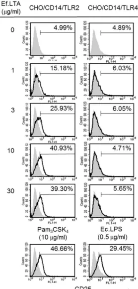
Upon exposure to E. faecalis LTA, RAW 264.7 cells significantly produced TNF-α in a concentration-dependent manner. Although E. faecalis LTA weakly induced the production of NO in RAW 264.7 cells, its ability to induce NO was remarkably increased in the presence of IFN-γ under the same condition. E. faecalis LTA preferentially activated TLR2 cells rather than TLR4. Concomitantly, E. faecalis LTA enhanced the DNA-binding activity of NF-κB, which plays an important role in the transcriptional activation of genes encoding inflammatory mediators. E. faecalis LTA was not able to induce TNF-α in macrophages lacking TLR2 while significantly induced TNF-α in wild-type macrophages. Compared with LTAs purified from other Gram-positive bacteria such as S. aureus, L. plantarum, and B. anthracis, E.
faecalis LTA showed a moderate potential in the activation of TLR2 and the induction of inflammatory mediators. E. faecalis LTA weakly interacted with anti-polyglycerophosphate antibody compared with other polyglycerophosphate-type LTAs such as S. aureus LTA, L. plantarum LTA, and B. anthracis LTA, implying that E. faecalis LTA might have a subtle structural difference in its polyglycerophosphate backbone compared with other polyglycerophosphate-type LTAs. The structural analysis using MALDI-TOF demonstrated that the glycolipids from E. faecalis LTA consist of dihexosyl diacylglycerol harboring fatty acids ranging from C16 to C22. Interestingly, unlike most LTAs which likely possess saturated and mono-unsaturated acyl chains, E. faecalis LTA contained mono- and
di-unsaturated fatty acids. Heat-killed E. faecalis potently induced the production of TNF-α in RAW 264.7 cells while such induction was significantly dampened by calcium hydroxide-killed E. faecalis under the same condition. Treatment of E. faecalis LTA with calcium hydroxide remarkably abrogated its ability to induce the production of proinflammatory mediators such as NO, TNF-α, IP-10, and MIP-1α in RAW 264.7 cells.
Furthermore, calcium hydroxide-treated E. faecalis LTA was not able to activate TLR2 signaling pathway. Structural analysis using MALDI-TOF and TLC showed that calcium hydroxide deacylated the glycolipid moiety of E.
faecalis LTA, implying that the lipid moiety of E. faecalis LTA is crucial for its inflammatory potential.
A novel purification method using mild organic solvent extraction, hydrophobic-interaction and ion-exchange chromatography provided a highly-pure and structurally-intact E. faecalis LTA preparation. E. faecalis LTA is one of the major immunostimulating components in the Gram-positive bacterial cell wall capable of activating the TLR2 signaling pathway. LTAs of E. faecalis have moderate inflammatory potential compared to LTAs purified from different bacterial species. E. faecalis LTA is a polyglycerophosphate-type LTA with subtle structural differences of substituent in repeating units and fatty acid composition. The lipid moiety of E. faecalis LTA plays a crucial role in determining its ability to induce the production of TNF-α, IL-6, IP-10, MIP-1α, and MCP-1 in macrophages by affecting TLR2 activation. Treatment with calcium hydroxide abrogates the inflammatory potential of E. faecalis LTA by inducing deacylation of the lipid moiety of LTA. Thus, development of medicament targeting either the lipid moiety of LTA or TLR2 signaling would be a good therapeutic candidate.
Collectively, E. faecalis LTA is an important virulence factor of E. faecalis for inducing inflammatory responses through TLR2 signaling pathway and its
lipid moiety is essential for its inflammatory activity.
Keywords: Lipoteichoic acid, Enterococcus faecalis, Gram-positive bacteria, Innate immune response, Toll-like receptor 2, Endodontic infection
Student number: 2007-22098
CONTENTS
Abstract Contents List of figures List of tables Abbreviations
Chapter I. Introduction
11.1. Enterococcus faecalis 1
1.1.1 General characteristics of E. faecalis 1
1.1.2. Virulence factors of E. faecalis 2
1.1.2.1. Secreted proteins 2
1.1.2.2. Surface proteins 3
1.1.2.3. Bacterial cell wall polysaccharide 4
1.2. LTA 6
1.2.1. Role of LTA in bacterial physiology 6
1.2.2. Structure and biosynthesis of LTA 9
1.2.3. Recognition of LTA in host innate immunity 13
1.2.4. Limitation of earlier studies for LTA 20
1.3. Endodontic infection and endodontic treatment 22
1.3.1. Primary root canal infection 22
1.3.2. Secondary/persistent root canal infection 23 1.3.3. Endodontic treatment and calcium hydroxide 23
1.4. Aim of the present study 25
Chapter II. Materials and Methods
262.1. Preparation of highly-pure and structurally-intact LTA 26
2.1.1. Reagents and chemicals 26
2.1.2. Bacterial strains and culture condition 27
2.1.3. Purification of LTA from Gram-positive bacteria 27
2.1.4. Phosphate assay 28
2.1.5. Limulus amebocyte lysate assay 29
2.1.6. Visualization of LTAs on SDS-PAGE gel using coomassie
blue staining, silver staining, and Western blotting 29
2.2. Structural analysis of E. faecalis LTA 30
2.2.1. Preparation of glycolipid anchor from E. faecalis LTA 30 2.2.2. Structural analysis of E. faecalis LTA using MALDI-TOF
and TLC 31
2.3. Functional analysis of LTA 32
2.3.1. Culture of RAW 264.7 32
2.3.2. Determination of nitric oxide 32
2.3.3. Enzyme-linked immunosorbent assay (ELISA) 32
2.3.4. Flow cytometric analysis 33
2.3.5. Electrophoretic mobility shift assay (EMSA) 33 2.3.6. Stimulation of bone marrow–derived macrophages 35 2.3.7. Preparation of killed whole cells by heat or calcium
hydroxide 35
2.3.8. Treatment of E. faecalis LTA with calcium hydroxide 36
2.3.9. Statistical Analysis 36
Chapter III. Results
373.1. Purification of highly-pure LTA from E. faecalis 37 3.2. Determination of immunological function of E. faecalis LTA 41 3.2.1. LTA from E. faecalis has inflammatory potential 41 3.2.2. TLR2 is crucial for activation of macrophage by E. faecalis
LTA 45
3.2.3. TLR2 is important for the inflammatory potential of E.
faecalis LTA in macrophage 47
3.2.4. E. faecalis LTA induces the activation of transcription
factor NF-κB 49
3.3. Structural and functional relationship of E. faecalis LTA 51
3.3.1. Highly-pure LTA was prepared from various Gram-positive
bacteria 51
3.3.2. E. faecalis LTA exhibits a modest ability to induce the production of NO, IL-6, MCP-1 in macrophages
comparable to those of LTA purified from other bacterial species
57
3.3.3. E. faecalis LTA has modest TLR2-stimulating activity comparable to other bacterial
LTAs
59 3.3.4. E. faecalis LTA more weakly interacts with
anti-polyglycerophosphate antibody compared to other bacterial LTA
61 3.3.5. Differential bacterial LTA exhibit subtle difference in their
backbone structure 64
3.4. Molecular mechanism of endodontic medicament 68 3.4.1. Calcium hydroxide inhibits the ability of E. faecalis to
induce TNF-α production in RAW 264.7 cells 68 3.4.2. Calcium hydroxide inhibits the ability of E. faecalis LTA to
induce the production of inflammatory mediators in murine macrophages
70 3.4.3. Attenuated induction of inflammatory mediators by
calcium hydroxide-treated E. faecalis LTA is due to the loss of TLR2-stimulating activity required for NF-κB activation
74 3.4.4. Calcium hydroxide deacylates the glycolipid moiety of
LTA from E. faecalis 76
Chapter IV. Discussion
81Chapter V. References
94List of publications
123국문초록
List of Figures
Figure 1. Structural classification of LTA 11
Figure 2. Biosynthesis of LTA 12
Figure 3. Bacterial cell wall structure 18 Figure 4. Elution profile of E. faecalis LTA from an
octyl-sepharose hydrophobic-interaction column followed by elution on a DEAE-sepharose anion-exchange column
39
Figure 5. Visualization of E. faecalis LTA using silver staining
40
Figure 6. Induction of TNF-α and NO productions by RAW 264.7 cells stimulated with E. faecalis LTA
43
Figure 7. Induction of TNF-α and NO productions by RAW 264.7 cells stimulated with E. faecalis LTA in the presence of polymyxin B
44
Figure 8. Activation of TLR2 by E. faecalis LTA 46 Figure 9. Activation of TLR2 on macrophage by E. faecalis
LTA
48
Figure 10. Enhanced DNA-binding activity of NF-κB by E.
faecalis LTA
50
Figure 11. Elution profiles of LTAs from octyl-sepharose hydrophobic-interaction column
53
Figure 12. Elution profiles of LTAs from DEAE-sepharose anion-exchange column
54 Figure 13. Visualization of LTA preparations using
coomassie blue staining and silver staining
55
Figure 14. Comparison of immunostimulating abilities of LTAs purified from various Gram-positive bacterial species.
58
Figure 15. Comparison of TLR2-activating ability among LTAs purified from various Gram-positive bacterial species
60
Figure 16. Characterization of backbone structure of LTAs 63 Figure 17. Experimental scheme for isolation of glycolipid
from E. faecalis LTA
65
Figure 18. Mass spectra obtained from the glycolipid anchor of E. faecalis LTA
66
Figure 19. Calcium hydroxide attenuates the ability of E.
faecalis to induce TNF-α production in RAW 264.7 cells
69
Figure 20. Calcium hydroxide inactivates the ability of LTA from E. faecalis to induce TNF-α production in RAW 264.7 cells
72
Figure 21. Calcium hydroxide abolishes the ability of E.
faecalis LTA to stimulate the expression of inflammatory mediators
73
Figure 22. Calcium hydroxide eliminates the ability of E.
faecalis LTA to stimulate TLR2
75
Figure 23. Experimental scheme for isolation of glycolipid from calcium hydroxide-treated E. faecalis LTA
78
Figure 24. Calcium hydroxide delipidates LTA of E. faecalis 79 Figure 25. Identifications of free fatty acids generated from
the glycolipids after calcium hydroxide treatment 80
Figure 26. Proposed structure and immunological functions of E. faecalis LTA
93
List of tables
Table 1. Known virulence factors of E. faecalis 5
Table 2. Known functions of LTA 8
Table 3. TLRs in human and mice 17
Table 4. Comparison of immunological properties between LTA and LPS
19
Table 5. Determination of impurities in LTA preparations
56
Table 6. Predictive structure of the glycolipid moiety of E. faecalis LTA
67
Table 7. Structural characterization of LTAs 88
Abbreviations
Ace adhesin of collagen from E. faecalis AS aggregation substance protein
BM bone marrow cell
BMM bone marrow-derived macrophage
Ca(OH)2 calcium hydroxide
CD14 cluster of differentiation 14 CD25 cluster of differentiation 25 CD36 cluster of differentiation 36
CFU colony forming uint
CpG cytosine-phosphate-guanine
DAG diacylglycerol
ELISA enzyme-linked immunosorbent assay
ElrA enterococcal leucine-rich-repeat-containing protein A EMSA electrophoretic mobility shift assay
Epa enterococcal polysaccharide antigen
FA fatty acid
NAG α-D-N-acetylglucosamine
GU guanine-uracil
IFN-γ interferon-gamma IL-6 interleukin-6
IP-10 interferon gamma-induced protein 10
LBP lipopolysaccharide binding protein
LPS lipopolysaccharide
LTA lipoteichoic acid
NF-κB
nuclear factor kappa-light-chain-enhancer of activated B cells
NO nitric oxide
MALDI-TOF
matrix assisted laser desorption/ionization time-of-flight mass spectrometry
MBL mannose-binding lectin
M-CSF macrophage colony-stimulating factor MCP-1 monocyte chemoattractant protein-1 MD-2 differentiation factor 2
MIP-1α macrophage inflammatory protein-1 alpha
MW molecular weight
MyD88 myeloid differentiation primary response gene 88 PAFR platelet activating factor receptor
PGN peptidoglycan
PMB polymyxin B
SrA scavenger receptor class A
TAG triacylglycerol
TCL thin layer chromatography TIR Toll/interleukin-1 receptor
TLR toll-like receptor
TNF-α tumor necrosis factor
TRIF TIR-domain-containing adapter-inducing interferon-β
WT wild-type
1
Chapter I. Introduction
1.1. Enterococcus faecalis
1.1.1. General characteristics of E. faecalis
Enterococcus faecalis is a Gram-positive, nonmotile, and facultative anaerobe that is a commensal in the mucus area of the oral cavity, the gastrointestinal tract, and the genital tract in humans [1]. Enterococci are one of the major opportunistic pathogens causing a variety of diseases including bacteremia, endocarditis, and refractory apical periodontitis. In recent years, these bacterial species have been ranked in the top three nosocomial bacterial pathogens frequently isolated from patients with bacteremia [2] and they also have been known as the most prevalent species isolated from failed endodontic root canals [3]. Moreover, enterococcal infections usually pose therapeutic difficulties because of their ability to persist in harsh environmental conditions and their resistance to multiple antibiotics.
According to previous reports, enterococci are able to grow in the range of 10°C and 45°C, in 6.5% NaCl broth, at pH 9.6, and they can survive at 60°C for 30 min [4]. In addition, they have resistance to lethal levels of bile salts, sodium dodecyl sulfate, detergents, hyperosmolarity, ethanol, azide, hydrogen peroxide, UV, irradiation heat, sodium hypochlorite, acidity and alkalinity [5-10]. Over the past two decades, enterococci have gained great attention due to the emergence of multi-drug resistant strain such as vancomycin-resistant enterococci (VRE) [11].
2
Moreover, enterococci have substantial attention because they have also served as donors of vancomycin resistance genes to other pathogens such as methicillin-resistant Staphylococcus aureus (MRSA) [12]. Currently, the molecular mechanism of enterococcal antibiotic resistance has been revealed that enterococci intrinsically have resistance to a number of antibiotics and also readily acquire additional resistance by uptaking exogenous genes that confer another antibiotic resistance [13]. To date, 12 enterococci species have been identified, and approximately 90% of the Enterococcus clinical isolates are Enterococcus faecalis.
1.1.2. Virulence factors of E. faecalis
Various etiologic virulence factors have been identified in E. faecalis. These virulence factors are either released from bacteria or expressed on the surface and they are related to bacterial adhesion, colonization, resistance to host defense, tissue destruction, and induction of inflammation (Table 1).
1.1.2.1. Secreted proteins
E. faecalis secrete several proteins into the extracellular space to destruct host tissues or to lyse target cells or molecules. Gelatinase is an extracellular protease involved in tissue destruction by hydrolyzing components of the extracellular
3
matrix such as gelatin, collagen, and fibrinogen [14]. Cytolysin is a secreted toxin produced by ~ 30% of E. faecalis strains, which can lyse not only host cells including red blood cells [15], polymorphonuclear leukocytes (PMNs), and macrophages [16], but also a broad range of Gram-positive, but not Gram-negative bacteria [17, 18]. Sex pheromones, small secreted hydrophobic peptides comprised of 7 - 8 amino acids, function as signaling peptides capable of regulating the expression of virulence factors such as cytolysin [19]. Some of the sex pheromones released from E. faecalis have been reported to induce abnormal production of superoxides and lysosomal enzymes in neutrophils resulting in tissue destruction [20, 21].
1.1.2.2. Surface proteins
E. faecalis expresses various virulence factors on its surface, which are likely to be involved in the transfer of plasmid DNA and bacterial adherence. Aggregation substance protein (AS) is a bacterial clumping factor that mediates the interaction between donor and recipient bacteria, facilitating plasmid exchange [22].
Enterococcal microbial surface component recognizing adhesive matrix molecule (MSCRAMM) is a family of bacterial adhesin that is involved in bacterial binding to host extracellular matrix [23]. Adhesin of collagen from E. faecalis (Ace) is a cell wall-anchored adhesin interacting with collagen and laminin [23, 24]. Another
4
enterococcal protein, enterococcal leucine-rich-repeat-containing protein (ElrA) has been reported to affect the adhesion of E. faecalis to macrophages [25].
1.1.2.3. Bacterial cell wall polysaccharides
Polysaccharides of bacterial cell walls are crucial for bacterial pathogenesis, mediating evasion of phagocytosis and inducing inflammatory responses.
Enterococcal polysaccharide antigen (Epa) is a rhamnose-containing polysaccharide facilitating biofilm formation and decreasing susceptibility to PMN-mediated killing [26-28]. Lipoteichoic acid (LTA), a Gram-positive bacterial membrane bound polysaccharide, seems to be a major etiologic factor that has a broad spectrum of pathogenic potentials: (i) LTA from Gram-positive bacteria triggers inflammatory responses and results in tissue damage [29], (ii) E. faecalis LTA plays an important role in biofilm formation, providing bacterial resistance to antimicrobial peptides, antibiotics, or disinfectants [30], and (iii) LTA acts as a representative target for generating opsonic antibodies during an infection with E.
faecalis [31].
5 Table 1. Known virulence factors of E. faecalis
Function Factor Reference
Bacterial adhesion and colonization
AS [32, 33]
Adhesins (Ace and ElrA) [23, 25]
Epa [27, 28]
LTA [34]
Resistance to host defense AS [35, 36]
Tissue destruction
LTA [37, 38]
Gelatinase [39, 40]
Cytolysin [41]
Induction of inflammation LTA [42, 43]
Sex pheromones [20, 21]
AS denotes aggregation substance protein
ElrA denotes enterococcal leucine-rich-repeat-containing protein A Epa denotes enterococcal polysaccharide antigen
Ace denotes adhesin of collagen from E. faecalis
6
1.2. LTA
1.2.1. Role of LTA in bacterial physiology
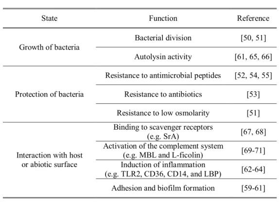
Although the exact role of LTA in bacterial physiology is still unclear, it is assumed that LTA is crucial for the (i) control of bacterial division and growth, (ii) protection of bacteria against environmental stress, (iii) interaction with host receptors or biomaterials (Table 2). According to several studies using LTA-deficient strains of Gram-positive bacteria including S. aureus, Bacillus subtilis, Bacillus anthracis, and Listeria monocytogenes, the strains lacking LTA result in growth defects such as aberrant cell division and septum formation, decreased autolysis, and reduced levels of peptidoglycan (PGN) hydrolases [44-46]
suggesting that LTA may be required for proper positioning of proteins related with the cell-division machinery. Indeed, LTA of S. aureus has a high affinity to autolysins, enzymes required for cleaving PGN during cell division [47].
LTA-deficiency in B. subtilis causes FtsZ, a key protein in bacterial division, to become incapable of assembling at the division site [48]. In addition to bacterial division, LTA is also responsible for protecting of bacteria from environmental stress such as osmotic pressure since S. aureus lacking LTA can survive under high-osmolality medium such as high concentration of NaCl or sucrose [49-51].
Moreover, LTA is important for the modulation of bacterial susceptibility to cationic antimicrobial substances such as antimicrobial peptides or enzymes including defensins and lysozyme as well as cationic antibiotics such as vancomycin by modifying D-alanine contents which results in decrease of negative net charge in the
7
cell envelops [52-56]. More recently, several regulatory systems such as sensor kinase ApsS have been shown to recognize defensins or other antimicrobial peptides and to modulate alanylation in the presence of antimicrobial challenge [57, 58]. On the other hand, LTA is also involved in bacterial interaction with host cells or biomaterials. For example, mutants of S. aureus, E. faecalis, L. monocytogenes, and Streptococcus pyrogenes deficient in dlt operon have shown decreased bacterial adherence and biofilm formation [59-61]. Indeed, passive immunization with monoclonal antibodies targeting the polyglycerophosphate moiety of LTA exhibited significant increases of mortality in a murine S. aureus peritonitis model. In addition, numerous studies have reported that LTA can activate host innate immune systems via Toll-like receptors (TLR) 2 [62-64]. However, the proinflammatory potency of LTA is still controversial because previously commercially available LTA have been reported to be contaminated with other bacterial cell wall components such as lipopolysaccharide (LPS), bacterial lipoprotein, and PGN.
8
Table 2. Known functions of LTA
State Function Reference
Growth of bacteria
Bacterial division [50, 51]
Autolysin activity [61, 65, 66]
Protection of bacteria
Resistance to antimicrobial peptides [52, 54, 55]
Resistance to antibiotics [53]
Resistance to low osmolarity [51]
Interaction with host or abiotic surface
Binding to scavenger receptors
(e.g. SrA) [67, 68]
Activation of the complement system
(e.g. MBL and L-ficolin) [69-71]
Induction of inflammation
(e.g. TLR2, CD36, CD14, and LBP) [62-64]
Adhesion and biofilm formation [59-61]
SrA denotes scavenger receptor class A MBL denotes mannose-binding lectin TLR2 denotes Toll-like receptor 2
CD36 denotes cluster of differentiation 36 CD14 denotes cluster of differentiation 14 LBP denotes lipopolysaccharide binding protein
9
1.2.2. Structure and biosynthesis of LTA
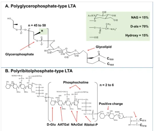
LTA is an alditolphosphate-containing polymer attached to cytoplasmic membranes of Gram-positive bacteria via glycolipid anchors. LTA is classified into two groups based on its chemical structures of the repetitive backbone (i.e., type I and II). Type I LTA, expressed in most Gram-positive bacteria including S. aureus, B. subtilis, E. faecalis, and Lactobacillus plantarum, is usually comprised of disaccharide-containing glycolipids linked to repeating polyglycerophosphate units, whose sn-2 position is esterified with hydroxyl, D-alanyl, or glycosyl substituent [64] (Figure 1A). Type II LTA, which is expressed by a small number of bacteria such as Streptococcus pneumoniae and Streptococcus oralis, is comprised of polyribitolphosphate repeating units linked to a positively charged glycolipid [72]
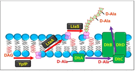
(Figure 1B). In addition to differences of backbone structure, type II LTA uniquely contains phosphocholine residue in its repeating units which plays an important roles in bacterial growth and virulence by mediating the interaction with choline-binding proteins (CBP) [73]. Recently, most enzymes involved in LTA synthesis have been identified including YpfP, LtaA, and LtaS [74] (Figure 2). The YpfP enzyme produces the glycolipid anchor in the bacterial cytoplasm by adding two glucose residues from UDP-glucose to diacylglycerol. Subsequently, the flippase, LtaA, translocates the glycolipid from the inner to the outer leaflet of the cytoplasmic membrane followed by extension of the glycerophosphate polymer on the glycolipid by LTA synthase, LtaS. In addition, further studies using mutagenesis
10
have also revealed that dltABCD gene cluster is responsible for modification of D-alanine after LTA or TA biosynthesis. The D-alanylation of LTA have reported to be crucial roles in the regulation of autolysis [75, 76], divalent cationic ions homeostasis through the interaction with Mg2+ [77, 78], biofilm formation [30], and bacterial resistance to cationic antimicrobial peptides by affecting their net charge [79].
11
Figure 1. Structural classification of LTA (A) Polyglycerophosphate-type LTA as found in S. aureus and (B) polyribitolphosphate-type LTA from S. pneumoniae.
Chemical structures of S. aureus LTA and S. pneumoniae LTA proposed by Morath., et al [80] and Gisch., et al [81], respectively. The repeating unit of S. pneumococcal
LTA consists of the
pseudopentasaccharide-2-acetamido-4-amino-2,4,6-trideoxygalactose (AATGal), glucose (D-Glu), and ribitol-phosphate (Ribitol-P) followed by two N-acetylgalactosamine (NAcGal) moieties, both substituted with phosphocholine in position O-6. R denotes backbone substitutions. n denotes number of repeating units,
12 NAG denotes N-acetylglocosamine.
Figure 2. Biosynthesis of LTA. Polyglycerophosphate-type LTA synthesis machinery of S. aureus. The glycolipid anchor dihexose-DAG is produced by YpfP and moved to the outside of the membrane by LtaA. The LtaS is an enzyme required for the producing the polyglycerophosphate chain. Coincidently, DltA ligates D-alanines onto DltC. The D-alanines are subsequently transported across the membrane by DltB. Then, DltD aids in the transfer of the D-alanines to glycerophosphate (GroP) of LTA. DAG denotes diacylglycerol. D-Ala denotes D-alanine.
13
1.2.3. Recognition of LTA in host innate immunity
The human body is constantly challenged by diverse bacteria, which not only have co-evolved with the host by affecting various physiological processes including nutrient production/absorption, gut homeostasis, tissue development, and host defense [82, 83],but also cause infectious diseases that are leading causes of death worldwide including sepsis and pneumonia. The Gram-positive bacteria lactobacilli, commonly found in the gastrointestinal tract, are representative commensal bacteria beneficial to the host as they enhance intestinal barrier function and produce antimicrobial substances [84, 85]. On the other hand, S. aureus is a versatile Gram-positive pathogen that causes a range of illnesses from minor skin infections to life-threatening infections such as sepsis, pneumonia, and endocarditis [86]. For maintaining homeostasis with bacteria, the host immune system has developed under the dual pressure of defending against pathogen and coexisting with commensal bacteria. Upon the exposure to bacteria, the host triggers both innate and adaptive immune systems. While adaptive immunity provides highly effective protection against particular pathogens by recognizing specific antigens during the late phase of infection, innate immunity is responsible for the protection of host at the early phase of infection by using germ line-encoded receptors that recognize a wide range of highly conserved microbial moieties, termed microbial-associated molecular patterns (MAMPs) from both commensal and pathogenic bacteria [87].
Among these germ line-encoded receptors, TLRs, members of interleukine-1 (IL-1)
14
superfamily of transmembrane receptors, are one of the key MAMP receptors capable of inducing innate immune responses against a broad range of microorganism including bacteria, yeast, and viruses [88]. To date, a total of 10 and 13 TLRs have been identified in human and mice, respectively. Each TLR forms a homo- or heterodimer complex and recognizes various bacterial MAMPs such as LTA, lipoprotein, LPS, flagellin, and bacterial DNA (Table 3).
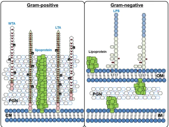
Bacteria can be classified into Gram-negative and Gram-positive according to their cell wall structures (Figure 3). Gram-negative bacteria are comprised of thin PGNs with two bilayer membranes including inner- and outer membranes which are primarily composed of phospholipids and LPS, respectively. In contrast, Gram-positive bacteria are surrounded by thick PGNs with a single cytoplasmic membrane and express LTA instead of LPS. Among various bacterial components, LPS, an amphiphilic molecule composed of hydrophobic lipid A and two distinct carbohydrate including highly conserved core polysaccharide and heterogeneous O-polysaccharide, is considered to be a major etiologic component of Gram-negative bacteria because it can stimulate host cells leading to cell death and the production of inflammatory mediators including cytokines, chemokines, and lipid metabolites [89]. Moreover, LPS alone is sufficient to cause septic shock when administered to mice [90]. Upon entering the blood stream, LPS forms a complex with LPS-binding protein (LBP) and soluble cluster of differentiation (CD) 14 [91,
15
92]. Then, the LPS in the complex is transferred to the myeloid differentiation factor 2 (MD-2) followed by interaction with TLR4 on target cells such as macrophages [93]. These interactions subsequently activate innate immune responses by triggering both myeloid differentiation primary response gene 88 (MyD88)-dependent and MyD88-independent signaling pathways which lead to the activation of nuclear factor-kappa B (NF-κB) transcription factor and the production of interferon (IFN)-β, respectively [94-96].
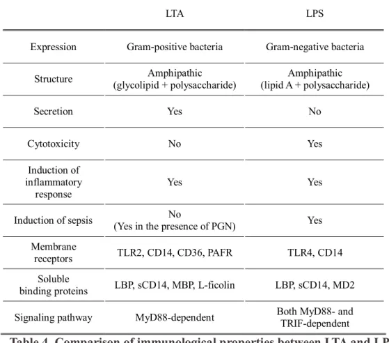
LTA of Gram-positive bacteria is considered to be a counterpart of the LPS of Gram-negative bacteria. LTA is an amphiphile consisted of hydrophobic glycolipids and hydrophilic polysaccharides that can induce various proinflammatory cytokines and chemokines [29, 62-64, 97]. Although both LTA and LPS share structural and immunological characteristics, they have distinctive properties (Table 4). For example, unlike LPS, LTA is actively released from the bacterial cell wall during bacterial division or bacteriolysis induced by external factors such as lysozymes, cationic peptides, and beta-lactam antibiotics [29, 98, 99]. In addition, the released LTA in the host bloodstream is recognized by not only LBP and CD14 but also mannose-binding protein (MBP), L-ficolin, CD36, and soluble TLR2 which are able to either facilitate LTA-mediated immune responses or neutralize the LTA as a scavenger [70, 71, 100-102]. Moreover, LTA is recognized by TLR2 and triggers a cell signaling cascade through the MyD88-dependent but not the
16
MyD88-independent pathway [103]. Furthermore, LPS by itself is a powerful agent that can provoke inflammatory responses, whereas LTA exhibits relatively weak induction of inflammatory responses that can be amplified in the presence of other bacterial components such as PGN [104]. Given these distinctions, host immune responses to LPS may not be directly applicable to those of LTA and Gram-positive bacteria.
17 Table 3. TLRs in human and mice
TLR Localization Species Ligands Recognized
pathogen TLR1 Extracellular Human and mice Triacyl lipopeptide Bacteria
TLR2 Extracellular Human and mice
Lipoproteins, PGN, LTA, zymosan, and
mannan
Bacteria
TLR3 Endolysosomal Human and mice dsRNA dsRNA
TLR4 Extracellular and
Endolysomal Human and mice LPS Gram-negative bacteria TLR5 Extracellular Human and mice Flagellin Bacteria TLR6 Extracellular Human and mice Diacylipopeptide,
LTA, and zymosan Bacteria TLR7 Endolysosomal Human and mice GU-rich ssRNA
and short dsRNA Viruses and bacteria TLR8 Endolysosomal Human and mice GU-rich ssRNA,
short dsRNA Viruses and bacteria TLR9 Endolysosomal Human and mice CpG DNA
Bacteria, viruses and
protozoan parasites
TLR10 Extracellular Human N.D. N.D.
TLR11 Endolysosomal Mice Profilin and flagellin
Apicomplexan parasites and
bacteria
TLR12 Endolysosomal Mice Profilin Apicomplexan
parasites TLR13 Endolysosomal Mice
Bacterial 23S rRNA with CGGAAAGACC
motif
Gram-negative and Gram-positive
bacteria GU denotes guanine-uracil
CpG denotes cytosine-phosphate-guanine N.D. denotes not determined
Referenced from Broz et al., [88]
18
Figure 3. Bacterial cell wall structure. (A) The Gram-positive bacterial cell wall is composed of thick PGN with a single cytoplasmic membrane. Teichoic acids (TA) are expressed as major polysaccharides of Gram-positive bacteria. These polymers are covalently anchored to PGN (wall teichoic acids; WTAs) or the cytoplasmic membrane (LTAs). Both WTAs and LTAs are often substituted with D-alanyl esters.
Gram-positive bacteria usually have diacylated lipoproteins in their cell wall surface.
(B) The Gram-negative bacterial cell wall is composed of a thin PGN with an inner and an outer membrane. They express LPS in the outer membrane as representative polysaccharide. Gram-negative bacterial usually have triacylated lipoproteins.
19
Table 4. Comparison of immunological properties between LTA and LPS PAFR denotes platelet activating factor receptor
TRIF denotes TIR-domain-containing adapter-inducing interferon-β
LTA LPS
Expression Gram-positive bacteria Gram-negative bacteria
Structure Amphipathic
(glycolipid + polysaccharide)
Amphipathic (lipid A + polysaccharide)
Secretion Yes No
Cytotoxicity No Yes
Induction of inflammatory
response Yes Yes
Induction of sepsis No
(Yes in the presence of PGN) Yes Membrane
receptors TLR2, CD14, CD36, PAFR TLR4, CD14 Soluble
binding proteins LBP, sCD14, MBP, L-ficolin LBP, sCD14, MD2 Signaling pathway MyD88-dependent Both MyD88- and TRIF-dependent
20
1.2.4. Limitation of earlier studies for LTA
Even though numerous studies have been reported to elucidate the role of LTA in host inflammatory responses during Gram-positive bacterial infection [29, 62-64, 97, 105], the biological properties of LTA and its inflammatory potency have been controversial because commercial LTA preparations used in early studies had been contaminated with endotoxins and/or they had been structurally damaged [106, 107].
Recent studies have reported that the non-hydrophobic phosphate containing fraction separated from commercially available S. aureus LTA using reverse-phase chromatography showed potent inflammatory activity [108, 109]. Moreover, re-purified LTA from commercially available S. aureus LTA had no inflammatory potential. More recent studies using nuclear magnetic resonance (NMR) have demonstrated that the commercial phenol-extracted S. aureus LTA showed dramatic loss of glycerophosphate repeats (approximately 48% of loss) and D-alanine residues (approximately 71% of loss), which are crucial for biological activity of LTA [80, 107]. In addition, the commercial S. aureus LTA was contaminated with considerable impurities including non-LTA (no phosphate, LPS), non-LPS (non-reactive to Limulus amebocyte lysate, bacterial DNA) [80]. This has led to contradictory immunological properties of LTA being reported due to the erroneous reflections of LTA-mediated immune responses. Recently, an upgraded purification method capable of extracting highly pure and structurally intact LTA was introduced [80, 107]. The method uses butanol, a mild organic solvent, to replace phenol and
21
serial applications of two different chromatographic columns such as a hydrophobic-interaction column and a subsequent ion-exchange column. Hence, the need for pure LTA has become a prerequisite to elucidate the role of LTA in bacterial pathogenesis and host immunity.
22
1.3. Endodontic infection and endodontic treatment
Endodontic infection is the infection of root canal and is a major etiological cause of apical periodontitis. Although certain chemical and physical factors are able to induce periradicular inflammation, microorganisms are essential for the endodontic infection and progression of apical periodontitis [110-112]. According to clinical conditions, endodontic infections can be divide into (1) primary root canal infection caused by initially infected microorganisms and (2) secondary/persistent root canal infection caused by very limited microorganisms that were members of the primary infection with resistance to intracanal antimicrobial procedure or with the ability to survive nutrient deprivation during endodontic treatment.
1.3.1. Primary root canal infection
Primary root canal infection is caused by microorganisms that initially colonize in the necrotic pulp tissue, which are generally polymicrobial infection with anaerobic bacteria and are composed of 10 to 30 species per canal [113]. Although several bacterial species belong to the genera Bacteroides, Porphyromonas, Prevotella, Fusobacterium, Treponema, Peptostreptococcus, Eubacterium, and Campylobacter usually isolated in the infected root canal, the bacterial profiles vary from individual to individual [114]. It indicates that primary root canal infection has heterogeneous etiology and multiple bacteria play a role in the progress of the disease.
23
1.3.2. Secondary/persistent root canal infection
Secondary/persistent root canal infection is caused by microorganisms that survived after endodontic treatment. It frequently leads to the progress of endodontic failure and apical periodontitis. The microbial profiles associated with secondary/persistent root canal infection are usually composed of a single species or relatively lower number of species compared with primary root canal infection [113, 115]. Bacterial species that were identified in secondary/persistent root canal belong to the genera Enterococcus, Streptococcus, Actinomyces, Propionibacterium, Staphylococcus, and Pseudomonas [114]. Among various microbial species identified in the secondary/persistent root canal infection, E. faecalis has gained considerable attention by its frequent recovery in root-filled teeth with apical periodontitis after endodontic treatment although they occupy only a small proportion of the flora in untreated canals [116-118]. E. faecalis can survive in harsh environmental conditions including intracanal irrigants and medications [119]
and can invade the dentinal tubules in variable depths [120] followed by colonization of the root canal [121, 122]. These abilities of E. faecalis may explain its ability to survive after a root canal treatment and to induce persistent apical periodontitis.
1.3.3. Endodontic treatment and calcium hydroxide
Since endodontic infection followed by apical periodontitis is an infectious
24
inflammatory disease, the major objectives of endodontic treatment are not only to eliminate viable bacteria in the root canal system but also to inactivate bacterial virulence factors that could potentially influence the progress of apical periodontitis.
Calcium hydroxide has been widely used as an endodontic medicament since the 1920s because of its highly effective bactericidal activity [123]. Calcium hydroxide releases hydroxide ions in aqueous solutions, giving rise to a high alkaline environment (approximately pH 12.5-12.8) in which most bacteria cannot survive [124]. In addition, accumulating reports suggest that calcium hydroxide can inactivate LPS [125], and that calcium hydroxide-treated LPS does not induce either the release of TNF-α from monocytes [126] or the stimulation of osteoclast formation, which is a major cell type responsible for alveolar bone loss [127]. Mass spectrometry demonstrated that calcium hydroxide detoxifies LPS by hydrolyzing fatty acids in the lipid A moiety [128]. Although the inactivation of LPS from Gram-negative bacteria by calcium hydroxide and other endodontic irrigants has been extensively investigated, the effects on LTA of Gram-positive bacteria have been poorly studied.
25
1.4. Aim of the present study
The aim of the present study is to investigate the structure and immunological functions of E. faecalis LTAs by (1) preparing highly-pure and structurally intact LTA from E. faecalis, (2) by studying its structural and functional relationship in innate immune responses, and (3) by elucidating the molecular mechanism of endodontic medicament against E. faecalis and its LTA.
26
Chapter II. Materials and Methods
2.1. Preparation of highly-pure and structurally-intact LTA
2.1.1. Reagents and chemicals
E. faecalis ATCC29212, S. aureus ATCC29213, and B. anthracis ATCC14185 were purchased from the American Type Culture Collection (Manassas, VA, USA).
L. plantarum KCTC 10887BP was obtained from Korean Collection for Type Culture (Daejon, Korea). Luria-bertani (LB), tryptic soy broth (TSB), brain heart infusion (BHI) and Mann, Rogosa, and Sharpe (MRS) media were purchased from BD Biosciences (Franklin, NJ, USA). octyl-sepharose CL-4B, DEAE-sepharose, Salmonella typhosa LPS, E. coli LPS, and polymyxin B were purchased from Sigma-Aldrich (St. Louis, MO, USA). Highly pure LPS from E. coli O111:B4, a synthetic lipopeptide stimulating TLR2, Pam2CSK4 and Pam3CSK4 were obtained from InVivoGen (San Diego, CA, USA). Spectra/Por 6 semi-permeable dialysis membrane was purchased from Spectrum® Laboratories Inc. (Ranch Dominquez, CA, USA). Endotoxin-free distilled water was obtained from Dai Han Pharm Co.
Ltd. (Seoul, Korea). Mouse recombinant interferon-gamma (mrIFN-γ) was obtained from R&D Systems (Minneapolis, MN, USA). All other reagents were purchased from Sigma-Aldrich unless otherwise indicated.
27
2.1.2. Bacterial strains and culture condition
E. faecalis ATCC29212 and L. plantarum KCTC 10887BP were grown in BHI medium and in MRS medium at 37oC without shaking overnight, respectively. S.
aureus ATCC29213 was cultured in TSB medium, and B. anthracis ATCC14185 in BHI medium supplemented with 0.7% bicarbonate at 37oC with shaking at 200 rpm overnight. After culturing these bacteria, they were centrifuged at 11,068 × g at 4oC for 10 min, washed with PBS three times and the pellets was kept until used.
2.1.3. Purification of LTA from Gram-positive bacteria
To purify LTAs from E. faecalis, S. aureus, L. plantarum, or B. anthracis, each bacterial pellet with approximately 80 to 100 g (wet weight) were resuspended in 0.1 M sodium citrate buffer (pH 4.7) and disrupted by ultrasonication (Labsonic P, B. Braun Biotech, Allentown, PA, USA) followed by mild organic solvent extraction with an equal volume of n-butanol for 30 min. After phase separation by using centrifugation, the aqueous phase was collected and subjected to dialysis in a semi-permeable dialysis membrane against endotoxin-free distilled water. The extract in 0.1M sodium acetate buffer (pH 4.7) containing 15% n-propanol was subjected to hydrophobic-interaction chromatography using octyl-sepharose CL-4B.
After removing the unbound materials with 15% n-propanol in 0.1 M sodium acetate buffer (pH 4.7), the bound materials were eluted with 35% n-propanol in 0.1
28
M of the sodium acetate buffer. After determining the column fractions containing LTA by using phosphate assay, the fractions were pooled and dialyzed against endotoxin-free distilled water. The LTA-containing fractions were further subjected to ion-exchange chromatography using DEAE-sepharose. After washing out the unbound materials with 30% n-propanol in 0.1 M sodium acetate buffer, the bound materials were eluted with a linear salt gradient (0-1 M NaCl in 0.1 M sodium acetate buffer containing 30% n-propanol). The fractions containing LTA were determined using the phosphate assay and further subjected to dialysis against endotoxin-free distilled water. After measuring the dry weight of the purified LTA, the LTA was dissolved in endotoxin-free water and kept at -80oC until use.
2.1.4. Phosphate assay
The phosphate contents of the eluted fractions or purified LTAs were determined by measuring inorganic phosphorus. Briefly, each sample was subjected to strong acidic digestion by boiling with a nitric acid-sulfuric acid mixture. The quantity of the phosphate in each sample was determined by measuring the optical density at 600 nm using a microtiter plate reader (VERSAmax; Molecular Devices, Sunnyvale, CA) after treatment of molybdate and stannous chloride. Sodium phosphate was used as a standard.
29
2.1.5. Limulus amebocyte lysate assay
The quantity of endotoxin was determined using a commercially available Limulus amebocyte lysate (LAL) test kit (QCL-1000; Cambrex Bio Science, Walkersville, MD, USA).
2.1.6. Visualization of LTAs on SDS-PAGE gel using coomassie blue staining, silver staining, and Western blotting
LTAs from E. faecalis, S. aureus, L. plantarum, or B. anthracis were mixed with 4
× sample buffer (8% SDS, 10% 2-mercaptoethanol, 30% glycerol, 0.02%
bromophenol blue in 0.25 M Tris-HCl, pH 6.8) and incubated at 100oC for 15 min.
The solution was then loaded into each well and run at 120 V for 1.5 h in 15%
SDS–PAGE gels. For coomassie blue staining, the SDS-PAGE gel was treated with commassie blue solution (0.1% coomassie blue R-250/10% acetic acid/45%
methanol) at room temperature for 1 h with gentle shaking. Then, the gel was incubated with de-staining solution (10% methanol/10% acetic acid) at room temperature with gentle agitation overnight. For silver staining, the SDS–PAGE gel was treated with fixation solution (50% methanol/12% acetic acid/0.0185% CH2O) for 1 h. Then, the gel was washed twice with 50% ethanol for 20 min each, after which the gel was treated with sensitizing solution (0.02% Na2S2O3·5H2O) for 1 min. After washing three times with distilled water for 20 sec each, the gel was
30
incubated with silver reaction solution (0.2% AgNO3/0.027% CH2O) for 1 h. After washing twice with non-pyrogenic water for 20 sec each, the gel was incubated with developing solution (6% Na2CO3/0.0004% Na2S2O3·5H2O/0.0185% CH2O) for 5 -10 min. After obtaining the desired band intensity, the gel was washed and incubated with stopping solution (50% methanol/12% acetic acid) to halt the silver reaction. For Western blot, the gel was electro-transferred to a PVDF membrane (Milipore, Bedford, MA, USA) and the membrane was blocked with 5% skim milk in TBS (50 mM Tris-HCl, 150 mM NaCl, pH 7.6) at room temperature for 1 h. After washing three times with TBST (50 mM Tris-HCl, 150 mM NaCl, 0.05% tween 20, pH 7.6) for 10 min at room temperature, the membrane was kept at 4°C overnight with mouse monoclonal anti-LTA antibody (clone: 55; Hycult biotech, Uden, Netherlands) in the blocking buffer. Subsequently, the membrane was washed three times with TBST for 10 min at room temperature, and incubated with horseradish peroxidase-conjugated goat anti-mouse IgG polyclonal antibody (West Grove, PA, USA) in the blocking buffer at room temperature for 1 h. The immunoreactive bands were detected with ECL Western blotting reagents (Neuronex Co.,Pohang, Korea).
2.2. Structural analysis of LTA
2.2.1. Preparation of glycolipid anchor from E. faecalis LTA
31
The lyophilized E. faecalis LTA (1 mg) were exposed to 98% acetic acid (1 ml) to cleave the phosphodiester bond on the LTA, then sonicated for 30 min, boiled at 100oC for 3 h, and lyophilized. Then, a solvent comprised of chloroform, methanol, and water (1:1:0.9, v/v/v) was added to the lyophilized products, and the chloroform phase containing the glycolipid anchors of E. faecalis LTA was obtained.
2.2.2. Structural analysis of E. faecalis LTA using matrix-assisted laser desorption/ionization-time of flight mass spectrometer (MALDI-TOF) and thin layer chromatography (TLC)
For MALDI-TOF analysis, the glycolipids of E. faecalis LTA were mixed with 30 mg/ml 2,5-dihydroxybenzoic acid (Sigma-Aldrich) in 70% acetonitrile, deposited onto a metallic sample holder and subsequently subjected to MALDI-TOF mass spectrometry using Biflex IV systems (Bruker Daltonics). For TLC, glycolipid from E. faecalis LTA (300 μg) treated with 25 mg/ml calcium hydroxide, 5.25% sodium hypochlorite or 0.2 N sodium hydroxide at 37oC for 60 min were deposited onto a silica gel TLC plate (Silica gel 60; Merck, Whitehouse Station, NJ) and developed with a chloroform/methanol/water (65:25:4, v/v/v). Then the glycolipids were visualized by spraying with 5% phosphomolybdic acid in ethanol followed by heating at 120°C.
32
2.3. Functional analysis of LTA
2.3.1. Culture of RAW 264.7 cells
A murine macrophage cell line, RAW 264.7 (TIB-71), was purchased from the American Type Culture Collection. The cells were cultured in Dulbecco modified Eagle’s medium (Invitrogen, Grand Island, NY) supplemented with 10% fetal bovine serum (FBS) (HyClone, Logan, UT), 100 U/ml of penicillin, and 100 μg/ml streptomycin at 37°C in a humidified incubator with 5% CO2.
2.3.2. Determination of nitric oxide
The production of nitric oxide (NO) in macrophages after stimulation with various stimuli was determined by reacting with equal volumes of the Griess reagent (1%
sulfanilamide, 0.1% naphthylethylenediamine dihydrochloride, and 2% phosphoric acid). After incubation for 5 min at room temperature, the absorbance was measured at 540 nm with the microtiter-plate reader. NaNO2 was used as a standard for nitrite concentrations.
2.3.3. Enzyme-linked immunosorbent assay (ELISA)
33
The amounts of tumor necrosis factor-alpha (TNF-α), interleukin (IL)-6, interferon gamma-induced protein 10 (IP-10), macrophage inflammatory protein-1 alpha (MIP-1α), and monocyte chemoattractant protein-1 (MCP-1) in culture supernatants of macrophages after stimulation with various stimuli for 24 h were determined by using a commercially available enzyme-linked immunosorbent assay (ELISA) kit (R&D Systems).
2.3.4. Flow cytometry
CHO/CD14/TLR2 and CHO/CD14/TLR4, two NF-κB reporter cell lines which are coexpressing CD14 and TLR2 or TLR4, respectively, were kindly provided by Dr.
Douglas Golenbock (Boston Medical Center, Boston medical center, Boston, MA, USA) and grown in Ham’s F-12 media containing 10% heat-inactivated FBS, 1 mg/ml G418, 400 μg/ml hygromycin, 100 U/ml penicillin, and 100 μg/ml streptomycin at 37ºC in a 5% CO2 humidified incubator. After stimulation of the cell lines with purified LTA or LPS, the ability to activate TLR2 or TLR4 was determined by measuring the expression of CD25 using a FACS Calibur with CellQuest software (BD Biosciences).
2.3.5. Electrophoretic mobility shift assay (EMSA)
34
RAW 264.7 cells cultured on a 60-mm culture dish were stimulated with 0.5 μg/ml of E. coli LPS or E. faecalis LTAs at 0, 3, 10, or 30 μg/ml for 90 min. To prepare nuclear extracts, the cells were lysed with hypotonic buffer [10 mM HEPES (pH 7.9), 1.5 mM MgCl2] on ice for 15 min and the nuclei were precipitated using centrifugation at 3000 × g for 5 min. After lysing the nuclear by treating with a hypertonic buffer (30 mM HEPES, 1.5 mM MgCl2, 450 mM KCl, 0.3 mM EDTA, 10% glycerol, 1 mM DTT, 1 mM PMSF, and 1 μg/ml each of aprotinin and leupeptin) on ice for 40 min, the nuclear extracts were collected by centrifugation and stored at -80oC until use. Double-stranded deoxyoligonucleotide probe containing the consensus recognition sites (in italics) of NF-κB was 5’-GATCTCAGAGGGGACTTTCCGAGAGA-3’ and synthesized oligonucleotides were end-labeled with [γ-32P]-dATP. Nuclear extracts were incubated with the
32P-labeled DNA probes in binding buffer (100 mM KCl, 30 mM HEPES, 1.5 mM MgCl2, 0.3 mM EDTA, 10% glycerol, 1 mM DTT, 1 mM PMSF, and 1 μg/ml each of aprotinin and leupeptin) containing 1 μg of poly(dI-dC) for 20 min at room temperature. Then, the mixtures were electrophoresed on a 4.8% polyacrylamide gel in 0.5 × TBE buffer (44.5 mM Tris, 44.5 mM boric acid, and 1 mM EDTA), after which the gel was dried and subjected to autoradiography. To confirm specific binding, one picomole of unlabeled probe was used as a “cold competitor.” The relative NF-κB-binding activity was quantitated by using a densitometer, BIO-PROFIL X-press Zoom 2000 (Vilber-Lourmat, Marnela-Vallee, France).
35
2.3.6. Stimulation of bone marrow-derived macrophages
TLR2-deficient mouse was kindly provided by Dr. Sizuo Akira (Osaka University, Osaka, Japan), and the wild-type C57BL/6 was purchased from Orient Bio Inc (Seoul, Korea). Bone marrow cells were isolated from the femur and the tibia of 6- to 8-week-old mice, adjusted to 1×10⁶ cells/ml, and differentiated to macrophages by incubation with 10% L929-cultured media in DMEM supplemented with 10%
FBS, 50 μM 2-mercaptoethanol, and antibiotics for 3 days. The cells were washed once with PBS, and the adherent cells were cultured in a fresh DMEM medium containing 10% FBS and antibiotics without L929-cultured media for 12 h. Then, the cells were stimulated with E. faecalis LTA (3 or 30 μg/ml), Pam3CSK4 (10 μg/ml), or S. typhosa LPS (0.5 μg/ml) for 16 h. At the end of the stimulation, the culture supernatants were used for the analysis of TNF-α production by the ELISA.
2.3.7. Preparation of killed whole cells by heat or calcium hydroxide
E. faecalis was cultured in BHI media at 37°C to mid-log phase (A600, 0.6).Bacterial cells were precipitated by centrifugation and washed three times with PBS.
Colony-forming units (CFU)/ml were re-determined by spectrophotometry based on a standard curve (A600 vs CFU). For the preparation of heat-killed E. faecalis, 1.0 ×
36
1010 CFU/ml was heated at 60°C for 60 min. For the preparation of calcium hydroxide-killed E. faecalis, 70 μl of 250 mg/ml of calcium hydroxide was added to 630 μl of 1.7 × 10¹⁰ CFU/ml of E. faecalis followed by incubation at 37°C for 24 h.
The mixture was neutralized to pH 7 by slowly adding 1 N HCl, and the final bacterial cell concentration was adjusted to 1.0×10¹⁰ CFU/ml. To ensure the bacteria were completely killed, the killed bacteria were plated on BHI media containing 1.5%
agar and cultured overnight at 37°C. No bacterial colonies were observed.
2.3.8. Treatment of E. faecalis LTA with calcium hydroxide
The E. faecalis LTA or endotoxin-free water, a control, was incubated with various concentrations of calcium hydroxide (0.25, 2.5, or 25 mg/ml) for 60 min or at 25 mg/ml for various time periods (short-term periods: 5, 10, 20, 40, or 60 min;
long-term periods: 1, 3, 6, 12, or 24 h). At the end of the incubation period, the mixture was neutralized to pH 7 by adding 1 N HCl. E. faecalis LTA was not lost during the treatment as confirmed by the measurement of phosphate contents in the E. faecalis LTA.
2.3.9. Statistical Analysis
All experiments including mass spectrometric analysis were performed at least two or three times. To analyze the statistics of in vitro experiments including the production of proinflammatory mediators in RAW 264.7 cells, comparisons
37
between experimental groups and control groups were compared using a Student’s t-test. Differences were considered significant when P < 0.05.
38
Chapter III. Results
3.1. Purification of highly-pure LTA from E. faecalis
LTA is one of the most important cell-wall virulence factors of Gram-positive bacteria, determining innate immunity. To examine the ability of LTA from E.
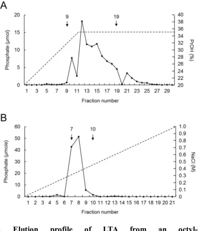
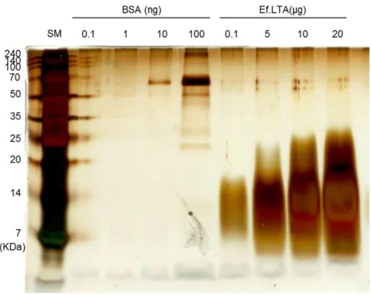
faecalis to induce inflammatory responses, LTA from E. faecalis was purified by using butanol extraction and octyl-sepharose chromatography, followed by DEAE-sepharose chromatography [80]. Column fractions were analyzed for phosphate contents, which have a linear correlation with LTA contents [107]. After purification by octyl-sepharose chromatography, fractions 9 to 19 were shown to contain abundant phosphate (Figure 4A) and were therefore pooled and further subjected to DEAE-sepharose column chromatography. As shown in Figure 4B, fractions 7 to 10, containing high amounts of phosphate, were pooled, lyophilized, quantified, and used for subsequent experiments. The purified LTAs were further examined for endotoxin contamination using the Limulus amebocyte lysate test. The level of endotoxin was less than 20.29 pg/mg of the purified LTA preparation, indicating that the amount of endotoxin in the LTA preparation was biologically irrelevant. To further determine protein contamination in LTA preparation, LTA purified from E. faecalis was subjected to silver staining. As shown in Figure 5, LTAs with heterogeneous size ranging from 7 to 25 kDa were detected in the purified LTA whereas no detectable protein band was observed. Furthermore, nucleic acid were not detected in one mg of the purified LTAs as determined by
39
spectrophotometry. These results indicate that E. faecalis LTAs purified by a novel purification method using mild organic solvent extraction, hydrophobic-interaction and ion-exchange chromatography were not contaminated with biologically active molecules such as nucleic acid, protein, and endotoxin.
40
Figure 4. Elution profile of LTA from an octyl-sepharose hydrophobic-interaction column followed by elution on a DEAE-sepharose anion-exchange column. LTA was prepared from E. faecalis by sequential application of butanol extraction, hydrophobic-interaction column chromatography, and ion-exchange column chromatography. (A) A phosphate assay was performed to measure the quantity of LTA after octyl-sepharose column chromatography.
Fractions 9 to 19 were pooled and subjected to DEAE-sepharose column chromatography. (B) A phosphate assay was performed to quantify of LTA aft