저작자표시-비영리-변경금지 2.0 대한민국 이용자는 아래의 조건을 따르는 경우에 한하여 자유롭게
l 이 저작물을 복제, 배포, 전송, 전시, 공연 및 방송할 수 있습니다. 다음과 같은 조건을 따라야 합니다:
l 귀하는, 이 저작물의 재이용이나 배포의 경우, 이 저작물에 적용된 이용허락조건 을 명확하게 나타내어야 합니다.
l 저작권자로부터 별도의 허가를 받으면 이러한 조건들은 적용되지 않습니다.
저작권법에 따른 이용자의 권리는 위의 내용에 의하여 영향을 받지 않습니다. 이것은 이용허락규약(Legal Code)을 이해하기 쉽게 요약한 것입니다.
Disclaimer
저작자표시. 귀하는 원저작자를 표시하여야 합니다.
비영리. 귀하는 이 저작물을 영리 목적으로 이용할 수 없습니다.
변경금지. 귀하는 이 저작물을 개작, 변형 또는 가공할 수 없습니다.
i
의학석사 학위논문
Outcomes of Repair of Coarctation of Aorta with Hypoplastic Arch Using Extended End-to-Side Anastomosis Technique
대동맥궁 형성저하가 동반된 대동맥 교악증 환아에서 확장 단측문합술의 효용성과 치료성적에 대한 연구
2017년 2월
서울대학교 대학원 의학과 임상의과학과
김 응 래
ii
ABSTRACT
Introduction: The optimal surgical repair technique for coarctation associated with aortic arch hypoplasia (CoA/AAH) in neonates and infants is controversial. This study evaluates our current strategy utilizing extended end-to-side anastomosis (EESA) under selective cerebral and myocardial perfusion (SCMP) in treating this group of patients.
Methods: Through a retrospective review, we analyzed the outcome of 87 infants who underwent surgical repair of CoA/AAH from January 2004 to December 2015.Patients with functional single ventricle were excluded.
Results: There were no early mortalities and 4 patients (4.6%) experienced
early complications. Eighty-five patients (97.7%) were followed-up during
a mean duration of 6.1
±3.53 years. There were 2 late mortalities (2.3%)
and 3 re-intervention (3.5%) of the aortic arch. Ten-year overall survival
and freedom from re-intervention for the entire cohort was 97.7% and
96.3%, respectively. At last follow-up, 4 patients (4.5%) showed a peak
velocity greater than 2.5m/s across the repair site.7patients (8.2%) were
hypertensive. Among 44 patients (50.6%) with post-operative CT data, 7
patients (15.9%) showed gothic shaped arches. In the present study, we
iii
could not correlate arch geometry with increased blood pressure.
Conclusions: Our strategy with EESA under SCMP is safe and effective for repairing CoA/AAH in neonates and infants. Concomitant repair of associated cardiac anomalies can be done without added risk. Surgical results are excellent with low rates of mortality, re-intervention, and late hypertension.
Keywords: Coarctation of Aorta, Hypoplastic Arch, Extended end-to-side anastomosis
Student number: 2011-21973
iv
CONTENTS
Abstract ...ii
Contents ... iv
List of Tables and Figures ... vi
List of Abbreviations ... vii
1. Introduction ... 1
2. Materials and Methods ... 5
2.1. Study subjects ... 5
2.2. Surgical technique ... 5
2.3. Data collection and follow-up methods ... 8
2.4. Blood pressure and arch geometry analysis ... 8
2.5. Statistical Analysis ... 11
3. Results ... 12
3.1. Patient demographics ... 12
3.2. Early post-operative results ... 14
3.3. Late post-operative results ... 14
v
3.4. Aortic Arch Geometry ... 18
Discussion ... 19
Conclusion ... 28
References ... 29
Abstract in Korean ... 35
vi
LIST OF TABLES AND FIGURES
Figure 1 European Congenital Heart Surgeons Association (ECHSA)
database search for all coarctation surgery in the year 2016. ... 4
Figure 2 Illerstration of extended end-to-side anastomosis technique 10
Figure 3 The geometric classification of the aortic arch ... 10
Figure 4 Height/width of the arch on the CT images ... 10
Table 1 Clinical characteristics of the whole cohort ... 13
Table 2 Early and late post-operative results ... 16
Figure 5 Kaplan-Meier plots showing (A) Overall survival and (B) freedom from re-intervention after surgical repair ... 17
Table 3 4 patients with high flow velocity in the latest echocardiogram ... 18
Table 4 Aortic arch geometry classification and H/W ratio ... 20
Table 5 Summary of various reports ... 26
vii
LIST OF ABBREVIATIONS
CoA, Coarctation of aorta
AAH, aortic arch hypoplasia
CoA/AAH, coarctation associated with aortic arch hypoplasia
EESA, extended end-to-side anastomosisSCMP, selective cerebral and myocardial perfusion
CT, computed tomography
BPP, blood pressure percentile
H/W ratio, height/width ratio
1
Introduction
Clarence Crafoord reported the first successful surgical correction of coarctation of aorta in 1945 [1]. During a surgery for patent ductus arteriosus, he was forced to clamp the aorta to control some bleeding and thereby cut off the blood flow to the descending thoracic aorta for some time. The patient recovered well from the surgery without any complication. Inspired by this experience, Clarence Crafoord performed the first surgical correction of coarctation while temporary clamping the proximal and distal aorta. Since then surgical management of this congenital anomaly has significantly improved.
Coarctation of aorta is unique compared to other congenital anomalies in that it is treated in a variety of surgical options. According to the European Congenital Heart Surgeons Association (ECHSA) database (Fig. 1), techniques such as patch angioplasty, subclavian flap aortoplasty, or graft interposition were used in 2016. The various surgical techniques can be classified in 4 perspectives. First, surgeon has to decide how to approach the targeted area. A left thoracotomy can provide a more clear view but it is difficult to apply cardiopulmonary bypass. A median approach via sternotomy, on the other hand, may be easy to apply can cardiopulmonary bypass but has limited visual access of the coarctation. Secondly, the manner of anastomosis can be a either end-to-end, or end-to-side. Also the various usages of flap or patch has resulted in numerous modifications. Lastly, surgeons have to decide whether they want to use selective cerebral perfusion or deep hypothermic circulatory arrest during the anastomosis.
2
Thanks to better understanding of the disease and improvement in surgical management, early results of surgical correction are excellent regardless of specific techniques. Neonatal and infantile coarctation associated with aortic arch hypoplasia (CoA/AAH), however, still presents significant challenges to the surgeon. These patients often require surgical correction at an early age, and their small size makes it technically difficult for the surgeon. And as a result, these patients are known to be more prone to recurrent stenosis after repair [2]. Although the general agreement is to achieve complete relief of arch obstruction to prevent recoarctation, there is still no consensus about the ideal surgical repair technique. Furthermore, aortic arch hypoplasia (AAH) is often combined with intracardiac anomalies [3]. The relative benefits of one- stage repair is still debatable and many centers prefer a staged approach for concurrent intracardiac anomalies [4].
Hypertension manifesting early in life is a well acknowledged long-term complication after CoA repair in infancy [5]. Although being a major risk factor for premature cardiovascular morbidity and mortality, the pathophysiology of late hypertension after coarctation repair is not fully understood. Ou and colleagues suggested that ‘gothic’ arch geometry may be strongly related to late hypertension after coarctoplasty [6].
Since 2004, our institute has adopted the extended end-to-side anastomosis (EESA) technique under selective cerebral and myocardial perfusion (SCMP) through a median sternotomy for all neonates and infants with CoA/AAH. In case of concurrent cardiac anomaly, one-stage repair
3
approach was taken. In this study, we review our overall experience with this surgical strategy to demonstrate the effectiveness and safety of our surgical strategy.
4
Figure 1 : European Congenital Heart Surgeons Association (ECHSA) database search for all coarctation surgery in the year 2016.
Abbreviations: n, number; CoA, coarctation of aorta; SCFA, subclavian flap aortoplasty; VSD, ventricular septal defect
5
Materials and methods
1. Study subjects
Through a retrospective review of the Seoul National University Children’s
Hospital surgical database, we identified infants less than 1 year of age who underwent surgical repair of CoA/AAH from January 1, 2004 to December 31, 2015. All patients were treated by a single surgeon using an EESA technique under SCMP. Along with the repair of CoA/AAH, concurrent repair of associated cardiac anomalies was done in one-stage. Perioperative and postoperative data including patient demographics, echocardiograms, operative notes, perioperative hemodynamics, and associated morbidity and mortality, were collected and analyzed. Patients with single ventricle physiology, however, were excluded from this cohort.
2. Surgical technique
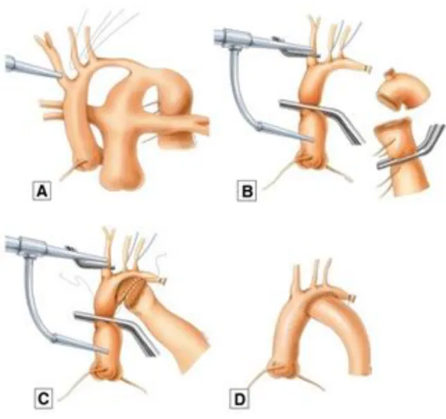
After median sternotomy, atrial cannula is inserted at the innominate artery (Fig. 2). Whenever possible, bicaval cannulation is utilized for effective venous drainage and rapid patient temperature control. Under cardiopulmonary bypass, the descending thoracic aorta and arch vessels are extensively dissected and fully mobilized. After cooling the patient to 28 °C, an additional small cannula is inserted at the aortic root for myocardial perfusion. The technique of SCMP has been described by us previously [7]. With the initiation of SCMP, the proximal part of the ascending aorta and innominate
6
artery is clamped, and the arch vessels are snared down. After dividing the isthmus and proximal descending aorta, all ductal tissue is excised including the ductus arteriosus. A longitudinal incision is made at the lesser curvature of the aortic arch. The proximal descending aorta is slightly beveled, and an end-to-side anastomosis is made as proximal as possible to bypass the isthmus and most of the hypoplastic arch. The whole procedure is done under a beating heart while maintaining coronary and brain perfusion. After anastomosis, all clamps and snares are removed, and cardiopulmonary bypass is increased to full support. If the patient has an associated intracardiac anomaly, the myocardial perfusion cannula is utilized to deliver cardioplegia and stop the heart.
7
Figure 2 : (A) An atrial cannula is inserted at the innominate artery to maintain cardiopulmonary bypass during extensive dissection. (B) An additional cannula is inserted at the aortic root and all ductal tissue is excised under selective cerebral and myocardial perfusion. (C) A longitudinal incision and anastomosis is made at the lesser curvature of the proximal aortic arch. (D) Completion of the end-to-side anastomosis technique for aortic arch repair.
3. Data collection and follow-up methods
8
Perioperative data including patient demographics, echocardiograms, operative notes, perioperative hemodynamics, and associated morbidity and mortality, were collected. Post-operative echocardiogram evaluations were performed just before discharge. After discharge, all patients were regularly studied by Doppler echocardiograms and blood pressure measurements. If possible, blood pressures from all 4 limbs were also measured to monitor for pressure gradients between the right arm and a lower extremity. Significant findings were defined as (1) 20 mmHg or greater blood pressure gradient between the right arm and a leg, or (2) 2.5m/s or higher estimated peak velocity across the anastomosis site was found on echocardiogram. If recurrent stenosis was suspected, computed tomography (CT) and/or cardiac catheterization was performed to evaluate the degree of stenosis and decide whether aortic re-intervention is required.
4. Blood pressure and arch geometry analysis
Blood pressures were adjusted into percentiles based on age and height percentile. Although there is a reference of blood pressure in South Korean pediatric population, its data only includes children with the age of 7 or older [8]. As our cohort includes many patients younger than 7, this data was not suitable for this research. Therefore, we referenced the data from the national institute of health of America and world health organization (WHO) for more elaborated blood pressure evaluation [9]. The WHO presented standards for growth based on height, weight and age. After standardizing our cohort population’s growth status, we adjusted their blood pressure according to their
9
age and height percentile [10]. Patients were considered hypertensive if either systolic or diastolic blood pressure percentile (BPP) was above the 95th percentile for age and height.
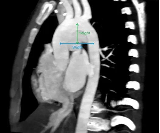
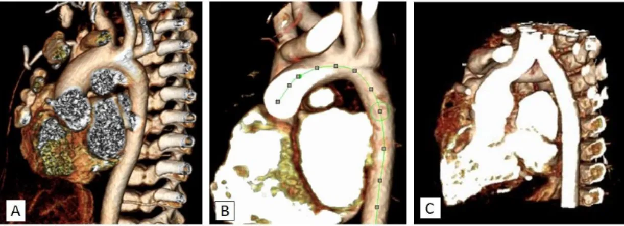
If post-operative CT was available, the repaired arches were classified as a roman, crenel or gothic shape based on the images (Fig. 2). The height and width of the arch was measured from the images and height/width (H/W) ratio was calculated to assess their geometry (Fig. 3). The width (W) was defined as the maximal horizontal distance between the midpoints of the ascending and descending aorta. The height (H) was defined as the maximal vertical distance between the W and the highest mid-point of the aortic arch. Both systolic and diastolic BPP was compared between the 3 arch shape groups
10
Figure 3 : The geometry of the aortic arch was classified as (A) roman, (B) crenel or (C) gothic based on its shape.
Figure 4 : Height/width (H/W) of the arch was measured based on the CT images.
5. Statistical Analysis
The data were analyzed using the Statistical Package for Social Science
11
software (SPSS release 14.0 for Windows; SPSS, Chicago, IL, USA). Survival and freedom from re-intervention rates were analyzed with Kaplan-Meier curves. Arch geometry and BPP were compared with a one-way analysis of variance and Tukey's multiple comparisons test. In all comparisons, variables with a P-value less than 0.05 were considered statistically significant.
12
Results
1. Patient demographics
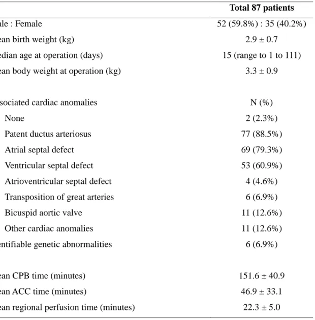
Between 2004 and 2015, 144 patients were diagnosed and treated for aortic coarctation. Among these, 87 patients (60.4%) had a hypoplastic aortic arch and underwent surgical repair at a median age of 15 days (range to 1 to 111 days; Table 1). Fifty-two out of 87 patients (59.8%) were male. Mean bodyweight was 2.9 ± 0.7 kg at birth and 3.3 ± 0.9 kg at the time of the operation. Only 2 patients (2.3%) had isolated CoA/AAH. Patent ductus arteriosus (n=77, 88.5%), atrial septal defect (n=69, 79.3%), and ventricular septal defect (n=53, 60.9%) were the most frequently associated cardiac anomalies. Eleven patients (12.6%) had a bicuspid aortic valve. Identifiable genetic abnormalities were found in 6 patients, which included Down (n=1), Turner’s (n=1), and Alagile syndrome (n=1) among others. Mean
cardiopulmonary bypass time and cross-clamp time was 151.6 ± 40.9 and
46.9 ± 33.1 minutes respectively. Mean regional perfusion time was 22.3 ± 5.0 minutes.
13
Table 1. Clinical characteristics of the whole cohort
Total 87 patients
Male : Female 52 (59.8%) : 35 (40.2%)
Mean birth weight (kg) 2.9 ± 0.7
Median age at operation (days) 15 (range to 1 to 111)
Mean body weight at operation (kg) 3.3 ± 0.9
Associated cardiac anomalies N (%)
None 2 (2.3%)
Patent ductus arteriosus 77 (88.5%)
Atrial septal defect 69 (79.3%)
Ventricular septal defect 53 (60.9%)
Atrioventricular septal defect 4 (4.6%)
Transposition of great arteries 6 (6.9%)
Bicuspid aortic valve 11 (12.6%)
Other cardiac anomalies 11 (12.6%)
Identifiable genetic abnormalities 6 (6.9%)
Mean CPB time (minutes) 151.6 ± 40.9
Mean ACC time (minutes) 46.9 ± 33.1
Mean regional perfusion time (minutes) 22.3 ± 5.0
14
2. Early post-operative results
There was no mortality within 30 days of surgery (Table 2). A total of 4 patients (4.6%) experienced acute complications. One patient had temporary recurrent laryngeal nerve paresis after the surgery. However, the patient showed no signs of aspiration and recovered from nerve paresis during follow-up. Another patient experienced chylothorax which improved after diet control. Left main bronchus compression was found in two patients resulting in prolonged mechanical ventilator support. After aortopecxy, both were able to wean off the ventilator.
There were no significant neurologic complications such as paraplegia or seizure. The median length of hospital stay was 16 days (range 8 to 102 days). At the time of discharge, no patients had residual coarctation by both physical examination and echocardiography.
3. Late post-operative results
Eighty-five out of 87 patients (97.7%) were followed up during a mean of 6.1 ± 3.5 years after surgery (Table 2). We lost follow-up in two patients, as
one foreign patient returned to his home country, and other patient emigrated abroad with his parents at 23 and 36 months after coarctation repair respectively. There were 2 cases (2.3%) of late mortalities during follow-up.
One in-hospital mortality occurred in a prematurely born baby with an extremely low birth weight (600g) and severe bronchopulmonary dysplasia.
Although the coarctation repair was successful, he died 2 months after surgery due to progression of bronchopulmonary dysplasia and respiratory
15
failure. Another patient died 3 month after coarctation repair due to an unidentified reason. Although there was no identifiable genetic disorder, the patient had multiple non-cardiac anomalies including macrocephaly, cleft palate, and foveal hypoplasia. She had no complications from the operation and was discharged from the hospital 18 days after. A follow-up echocardiogram study taken 1 month prior to her death showed no cardiac problems.
During follow-up, 7 patients showed signs of recoarctation on echocardiogram. Four patients (4.5%) with mild stenosis are regularly studied without intervention (Table 3). Patient A, C, and D were thought as mild coarctation and followed up without intervention. Cardiac catheterization Patient B showed that the actual pressure gradient was less than 10 mmHg across the repair site. Among the 4 patients, patient B and D were diagnosed with systemic hypertension. However, none of them showed significant blood pressure gradient between the right arm and a leg. Three patients (3.5%) underwent re-intervention for recurrent stenosis of the repair site. Two of them were born prematurely and weighed less than 1.5kg at the time of operation. Their small size caused some technical difficulties and both of them developed recoarctation within 4 month after surgery. Re-operation and balloon coarctoplasty were performed in these patients, respectively. The third patient had showed mild stenosis at the repair site 26 month after the surgery. During an operation for stenosis of bicuspid aortic valve, concurrent repair of the arch was performed. Ten-year overall survival and freedom from re-intervention for the entire cohort was 97.7% and 96.3%, respectively (Fig. 5).
16
Among the entire cohort, 7 patients (8.2%) were taking oral medication for hypertension at the last follow-up visit. Blood pressure measurements were recorded in 64 patients (75.3%) with a mean systolic and diastolic BPP of 65.88 ± 27 and 53.95 ± 26.38 respectively. Blood pressures from all 4 limbs were measured in 59 patients (69.4%) and 2 patients showed significant blood pressure gradient between the right arm and a leg.
17 Table 2. Early and late post-operative results
Early outcome
Early mortality 0
Median length of stay (days) 16 (range 8 to 102)
Complications 4 (4.6%)
Temporary RLN paresis 1
Chylothorax 1
Airway obstruction 2
Neurologic complication 0
Residual coarctation 0
Late outcome
Mean follow-up duration (years) 6.11 ±3.53
Late mortality 2 (2.3%)
Re-intervention 3 (3.5%)
Re-operation 2
Balloon coarctoplasty 1
Medication for systemic hypertension 7(8.2%)
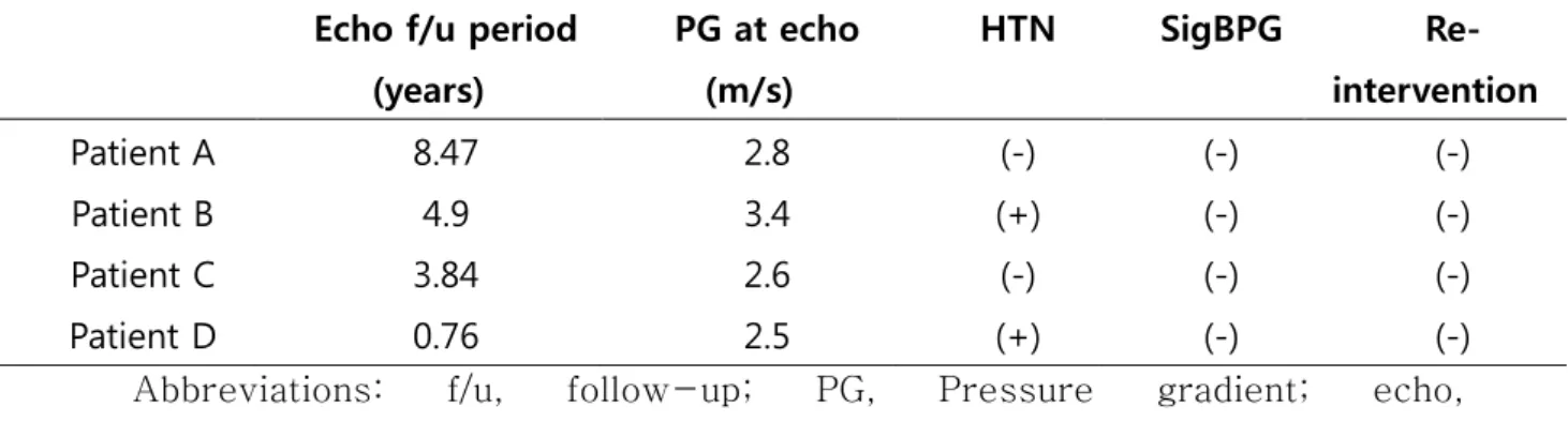
Significant peak velocity at last follow-up echocardiogram
4 (4.5%)
18
Figure 5 :(A) Overall survival and (B) freedom from re-intervention after surgical repair coarctation associated with aortic arch hypoplasia.
19
Table 3. 4 patients with high flow velocity in the latest echocardiogram Echo f/u period
(years)
PG at echo (m/s)
HTN SigBPG Re-
intervention
Patient A 8.47 2.8 (-) (-) (-)
Patient B 4.9 3.4 (+) (-) (-)
Patient C 3.84 2.6 (-) (-) (-)
Patient D 0.76 2.5 (+) (-) (-)
Abbreviations: f/u, follow-up; PG, Pressure gradient; echo, echocardiography; HTN, hypertension; SigBPG, Significant blood pressure gradient between the right arm and a leg
20
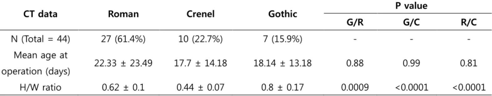
4. Aortic Arch Geometry
Post-operative CT data were available from 44 patients (50.6%) at a mean follow-up period of 2.8 ± 1.3 years (Table 4). The mean H/W ratio of the
entire cohort was 0.6 ± 0.2. Roman geometry was present in 27 patients (61.4%), crenel geometry in 10 (22.7%), and gothic geometry in 7 (15.9%) respectively. Mean age of repair showed no difference between three groups.
Aortic arches with gothic geometry showed a significantly higher H/W ratio when compared with roman or crenel morphologies (0.8±0.17 vs 0.62±0.1 vs
0.44±0.07, P<0.0001).
21
Table 4. Aortic arch geometry classification and H/W ratio
CT data Roman Crenel Gothic P value
G/R G/C R/C
N (Total = 44) 27 (61.4%) 10 (22.7%) 7 (15.9%) - - -
Mean age at
operation (days) 22.33 ± 23.49 17.7 ± 14.18 18.14 ± 13.18 0.88 0.99 0.81 H/W ratio 0.62 ± 0.1 0.44 ± 0.07 0.8 ± 0.17 0.0009 <0.0001 <0.0001
Abbreviations: CT, computed tomography; BP, Blood pressure; H/W ratio, Height/Width ratio; G/R, Gothic/Roman; G/C, Gothic/Crenel; R/C, Roman/Crenel
22
23
Discussion
The incidence of AAH in neonates and infants with CoA varies from 65% to 81%
[4, 5]. Thanks to advancements in perioperative care and surgical strategies, early result are excellent regardless of technique. The main concern nowadays is to reduce late complications including recoarctation and late hypertension. In a 40- year review on patients after coarctation repair, Dodge-Khatami and colleagues suggested simple end-to-end anastomosis and subclavian flap aortoplasty have a higher recurrence rate in neonates after coarctation repair [11]. Although it has been emphasized that extended end-to-end anastomosis relieves arch obstruction more effectively, the recoarctation rate is still in the range of 4 to 10% [4, 5, 11, 12].
In our opinion, there are three key factors that has to be met during the repair of neonatal or infantile CoA/AAH to ensure favorable long-term results: (1) extensive dissection and full mobilization of the entire thoracic aorta and arch vessels, (2) complete excision of ductal tissue, and (3) exclude the isthmus and most of the hypoplastic arch without using any kind of flap or patch. We believe that the EESA though a sternotomy approach is an ideal repair technique to satisfy all three points. One of the expected complications of this technique is bronchial compression by the aortic arch. Although prosthetic patch or subclavian flap aortoplasty can be an option to reduce this possibility, its long-term complications such as aneurysm, arm length discrepancy and claudication with exercise have been well documented [12, 13]. In our experience, full mobilization by extensive
24
dissection and careful positioning of the anastomosis site makes it possible to minimize bronchial complications without using patch or flap. Furthermore, aortopexy is a very effective method for relieving bronchial compression [14].
Considering the low prevalence of bronchial compression (2.3%), it is more important to prevent the long-term complications of patch or flap.
Extensive dissection and mobilization makes it also possible to resect all the ductal tissue. It has been well documented that remnant ductal tissue is an important factor for residual or early recurrent stenosis at the coarctation repair site [15, 16]. Usually the proximal part of descending thoracic aorta has to be resected with the patent ductus arteriosus due to the distal migration of ductal tissue. Insufficient dissection can lead to excessive tension and stretching of the surrounding vessels.
It remains highly debatable whether the hypoplastic arch grows after repair.
Histologic studies, however, have confirmed that a hypoplastic arch shows a significantly higher ratio of elastin lamellae and a decrease of α-actin–positive cells. This suggests diminished growth potential of the hypoplastic transverse arch resulting in un-favorable arch geometry even after proper surgical repair [17]. We believe that the initial repair should completely bypass the isthmus and hypoplastic distal arch to minimize the possibility of these complications. For this purpose, we adopted the end-to-side anastomosis over the end-to-end anastomosis. Several studies have shown good results with this technique in addressing hypoplastic arch [18-20] (Table 5). Our experience with EESA technique demonstrate similar or
25
better results with 10-year survival and freedom from re-intervention of 97.7%
and 96.3% respectively. Recoarctation (4.5%) and late hypertension (8.2%) rate is also low. This confirms the safety and efficacy of our strategy.
Due to the inevitable circulatory arrest during the repair of aortic arch, patients are exposed to the risk of various end-organ damages. The lack of blood flow to the descending thoracic aorta can increase the risk of kidney or spinal injuries [21, 22]. Despite myocardial protection and cardioplegia, stopping the heart puts stress to the myocardium. Furthermore, the possibility of increased neurologic damage after deep hypothermic circulatory arrest has been constantly suggested [23, 24].
To minimize the lack of perfusion, we utilized the SCMP method in various aortic arch repair operations since 2000 and demonstrated its feasibility and safety in multiple reports [7, 25-27]. Furthermore, our protocol of reginal cerebral perfusion has been proven to supply adequate and even perfusion flow to the whole brain [28, 29]. It is our belief that continuous cerebral perfusion with a nonworking beating heart using the dual-perfusion technique minimizes myocardial and neurological complications and morbidities.
Systemic hypertension is a well-acknowledged long-term complication after CoA repair in infancy with a prevalence ranging from 20% to 45% [30]. Although being a major risk factor for premature cardiovascular morbidity and mortality, the pathophysiology of late hypertension after coarctation repair is not fully understood. More recently, Ou and colleagues reported that ‘gothic’ arch geometry may be strongly related to late hypertension after coarctoplasty [6]. Based on
26
magnetic resonance imaging data, Olivieri and colleagues suggested that gothic shaped arches show an unsteady turbulent blood flow and thereby suffer from an unique wall shear stress point [31]. Although still on debate, the possible connection between gothic shapility Usually, H/W ratio was used as an index to underpin the different geometries between the 3 shape groups [32, 33]. In our study, the 3 groups also showed significant H/W ratio. Various studies have reported the ratio of gothic shaped arches from 27.3% to 60% [32-34]. On the other hand, only 15.6% of the repaired arches were gothic shaped.
The hemodynamic relation between intracardiac defect and AAH in CoA patients has been described by Rudolph and collegues [3]. They suggested that, by reducing blood flow through the ascending aorta and isthmus, intracardiac defects may induce the underdevelopment of the arch resulting in hypoplasia. In our review, 87.4% of the CoA/AAH patients had one or more concurrent intracardiac defect.
Nowadays, early mortalities after coarctoplasty are almost exclusively secondary to associated cardiac and non-cardiac anomalies. This provides the basis of a one- stage repair strategy. In a review of 307 neonates, Conte and associates suggested that one-stage repair of neonatal coarctation and associated complex heart defects through a median sternotomy produces better results than two-stage repair [4].
Several other studies have also supported the feasibility and superiority of concomitant repair of associated cardiac defects [35, 36]. We abandoned the thoracotomy approach as it has a significantly higher recurrence rate [2] and does not allow concurrent repair of other cardiac anomalies compared to median sternotomy. In our experience, both the coarctation and associated cardiac anomaly
27
could be sufficiently addressed through median sternotomy. Our operative and follow-up results showed that one-stage repair strategy through median sternotomy is safe and effective.
28
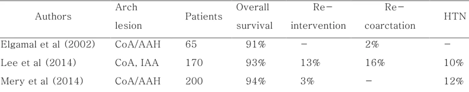
Table 5. Summary of various reports on surgical repair of coartation
Authors Arch
lesion Patients Overall survival
Re- intervention
Re-
coarctation HTN
Elgamal et al (2002) CoA/AAH 65 91% - 2% -
Lee et al (2014) CoA, IAA 170 93% 13% 16% 10%
Mery et al (2014) CoA/AAH 200 94% 3% - 12%
Abbreviations: CoA, coarctation of aorta; IAA, interrupted aortic arch; AAH, aortic arch hypoplasia; HTN, hypertension
29
Conclusion
In conclusion, the EESA technique combined with SCMP is a safe and effective method for repairing CoA/AAH in neonates and infants. By avoiding circulatory arrest, the possibility of myocardial and neurological damage can be minimized.
Concurrent repair of associated cardiac anomalies in a one-stage operation can be done without added risk. Our experience demonstrated excellent mid-term result with low rates of mortality, re-intervention, and late hypertension. Also we a greater lower ratio of gothic shaped arches. Based on these results, a one-stage repair with EESA and SCMP is the treatment of choice for neonatal and infantile CoA/HAA at our institution.
30
REFERENCES
1. Crafoord C, Nylin G. Congenital Coarctation of the Aorta and Its Surgical Treatment. J Thoracic Surg 1945;14:347–61.
2. Rakhra SS, Lee M, Iyengar AJ, Wheaton GR, Grigg L, Konstantinov IE et al.
Poor outcomes after surgery for coarctation repair with hypoplastic arch warrants more extensive initial surgery and close long-term follow-up.
Interactive cardiovascular and thoracic surgery 2013;16:31-6.
3. Rudolph AM, Heymann MA, Spitznas U. Hemodynamic considerations in the development of narrowing of the aorta. The American journal of cardiology 1972;30:514-25.
4. Conte S, Lacour-Gayet F, Serraf A, Sousa-Uva M, Bruniaux J, Touchot A et al. Surgical management of neonatal coarctation. The Journal of thoracic and cardiovascular surgery 1995;109:663-74; discussion 74-5.
5. Celermajer DS, Greaves K. Survivors of coarctation repair: fixed but not cured. Heart 2002;88:113-4.
6. Ou P, Bonnet D, Auriacombe L, Pedroni E, Balleux F, Sidi D et al. Late systemic hypertension and aortic arch geometry after successful repair of coarctation of the aorta. European heart journal 2004;25:1853-9.
7. Lim HG, Kim WH, Park CS, Chung ES, Lee CH, Lee JR et al. Usefulness of regional cerebral perfusion combined with coronary perfusion during one- stage total repair of aortic arch anomaly. Ann Thorac Surg 2010;90:50-7.
31
8. Lee CG, Moon JS, Choi J-M, Nam CM, Lee SY, Oh K et al. Normative blood pressure references for Korean children and adolescents. Korean Journal of Pediatrics 2008;51:33.
9. Onis M. WHO Child Growth Standards based on length/height, weight and age. Acta paediatrica 2006;95:76-85.
10. The Fourth Report on the Diagnosis, Evaluation, and Treatment of High Blood Pressure in Children and Adolescents. Pediatrics 2004;114:555-76.
11. Dodge‐Khatami A, Backer CL, Mavroudis C. Risk Factors for Recoarctation and Results of Reoperation: A 40‐Year Review. Journal of cardiac surgery 2000;15:369-77.
12. Vouhe PR, Trinquet F, Lecompte Y, Vernant F, Roux PM, Touati G et al.
Aortic coarctation with hypoplastic aortic arch. Results of extended end-to- end aortic arch anastomosis. The Journal of thoracic and cardiovascular surgery 1988;96:557-63.
13. Van Heurn L, Wong C, Spiegelhalter D, Sorensen K, De Leval M, Stark J et al. Surgical treatment of aortic coarctation in infants younger than three months: 1985 to 1990. The Journal of thoracic and cardiovascular surgery 1994;107:74-86.
14. Jang WS, Kim W-H, Choi K, Nam J, Kim J-T, Lee JR et al. Aortopexy with preoperative computed tomography and intraoperative bronchoscopy for patients with central airway obstruction after surgery for congenital heart disease: postoperative computed tomography results and clinical outcomes.
Pediatric cardiology 2014;35:914-21.
32
15. Wright GE, Nowak CA, Goldberg CS, Ohye RG, Bove EL, Rocchini AP.
Extended resection and end-to-end anastomosis for aortic coarctation in infants: results of a tailored surgical approach. Ann Thorac Surg 2005;80:1453-9.
16. Van Son JA, Falk V, Schneider P, Smedts F, Mohr FW. Repair of coarctation of the aorta in neonates and young infants. Journal of cardiac surgery 1997;12:139-46.
17. Machii M, Becker AE. Hypoplastic aortic arch morphology pertinent to growth after surgical correction of aortic coarctation. Ann Thorac Surg 1997;64:516-20.
18. Elgamal MA, McKenzie ED, Fraser CD, Jr. Aortic arch advancement: the optimal one-stage approach for surgical management of neonatal coarctation with arch hypoplasia. Ann Thorac Surg 2002;73:1267-72; discussion 72-3.
19. Lee MG, Brink J, Galati JC, Rakhra SS, Konstantinov IE, Cheung MM et al.
End-to-side repair for aortic arch lesions offers excellent chances to reach adulthood without reoperation. Ann Thorac Surg 2014;98:1405-11.
20. Mery CM, Guzman-Pruneda FA, Carberry KE, Watrin CH, McChesney GR, Chan JG et al. Aortic arch advancement for aortic coarctation and hypoplastic aortic arch in neonates and infants. Ann Thorac Surg 2014;98:625-33; discussion 33.
21. Brewer 3rd L, Fosburg R, Mulder G, Verska J. Spinal cord complications following surgery for coarctation of the aorta. A study of 66 cases. The Journal of thoracic and cardiovascular surgery 1972;64:368.
33
22. Jang WS, Kim W-H, Choi K, Nam J, Jung JC, Kwon BS et al. Incidence, risk factors and clinical outcomes for acute kidney injury after aortic arch repair in paediatric patients. European Journal of Cardio-Thoracic Surgery 2014:ezu132.
23. Greeley WJ, Kern F, Ungerleider R, Boyd 3rd J, Quill T, Smith L et al. The effect of hypothermic cardiopulmonary bypass and total circulatory arrest on cerebral metabolism in neonates, infants, and children. The Journal of thoracic and cardiovascular surgery 1991;101:783-94.
24. Gaynor JW, Nicolson SC, Jarvik GP, Wernovsky G, Montenegro LM, Burnham NB et al. Increasing duration of deep hypothermic circulatory arrest is associated with an increased incidence of postoperative electroencephalographic seizures. The Journal of thoracic and cardiovascular surgery 2005;130:1278-86.
25. Lim C, Kim W-H, Kim S-C, Rhyu J-W, Baek M-J, Oh S-S et al. Aortic arch reconstruction using regional perfusion without circulatory arrest.
European journal of cardio-thoracic surgery 2003;23:149-55.
26. Lim H-G, Kim W-H, Jang W-S, Lim C, Kwak JG, Lee C et al. One-stage total repair of aortic arch anomaly using regional perfusion. European journal of cardio-thoracic surgery 2007;31:242-8.
27. Lim H-G, Kim W-H, Park C-S, Chung E-S, Lee C-H, Lee JR et al.
Usefulness of regional cerebral perfusion combined with coronary perfusion during one-stage total repair of aortic arch anomaly. The Annals of thoracic surgery 2010;90:50-7.
34
28. Kwak JG, Kim W-H, Oh AY, Yoon TG, Kim H-S, Chae JH et al. Is unilateral brain regional perfusion neurologically safe during congenital aortic arch surgery? European Journal of Cardio-Thoracic Surgery 2007;32:751-5.
29. Kwak JG, Kim W-H, Kim JT, Kim I-O, Chae J-H. Changes of brain magnetic resonance imaging findings after congenital aortic arch anomaly repair using regional cerebral perfusion in neonates and young infants. The Annals of thoracic surgery 2010;90:1996-2000.
30. Sakurai T, Stickley J, Stümper O, Khan N, Jones TJ, Barron DJ et al. Repair
of isolated aortic coarctation over two decades: impact of surgical approach and associated arch hypoplasia. Interactive cardiovascular and thoracic surgery 2012:ivs265.
31. Olivieri LJ, de Zelicourt DA, Haggerty CM, Ratnayaka K, Cross RR, Yoganathan AP. Hemodynamic Modeling of Surgically Repaired Coarctation of the Aorta. Cardiovascular engineering and technology 2011;2:288-95.
32. Ou P, Mousseaux E, Celermajer DS, Pedroni E, Vouhe P, Sidi D et al. Aortic arch shape deformation after coarctation surgery: effect on blood pressure response. The Journal of thoracic and cardiovascular surgery 2006;132:1105-11.
33. Seo DM, Park J, Goo HW, Kim YH, Ko JK, Jhang WK. Surgical modification for preventing a gothic arch after aortic arch repair without the use of foreign material. Interactive cardiovascular and thoracic surgery
35 2015;20:504-9.
34. Liu JY, Jones B, Cheung MM, Galati JC, Koleff J, Konstantinov IE et al.
Favourable anatomy after end-to-side repair of interrupted aortic arch.
Heart, lung & circulation 2014;23:256-64.
35. Gaynor JW, Wernovsky G, Rychik J, Rome JJ, DeCampli WM, Spray TL.
Outcome following single-stage repair of coarctation with ventricular septal defect. European journal of cardio-thoracic surgery : official journal of the European Association for Cardio-thoracic Surgery 2000;18:62-7.
36. Planche C, Serraf A, Comas JV, Lacour-Gayet F, Bruniaux J, Touchot A.
Anatomic repair of transposition of great arteries with ventricular septal defect and aortic arch obstruction. One-stage versus two-stage procedure.
The Journal of thoracic and cardiovascular surgery 1993;105:925-33.
36
초 록
대동맥궁 형성저하가 동반된 대동맥 교악증 환아에서 확장 단측문합술의 효용성과 치료성적에 대한 연구
김 응 래 의학과, 임상의과학과 서울대학교 대학원
서론: 최근 대동맥 교악증(coarctation of aorta)의 치료성적은 매우 우수한 것으로 보고되고 있으나 대동맥궁 형성부전(aortic arch hypoplasia)이 동반된 신생아의 경우 수술적 치료의 난이도가 높고 장기적인 합병증에 취약한 것으로 알려져 있다. 본
연구는 상기 환자군을 대상으로 서울대학교 어린이병원 소아흉부외과에서 2004 년
부터 적용해온 확장 단측문합술(extended end-to-side anastomosis)의 치료 성적을 분 석하고 그 우수성을 검증하고자 한다.
방법: 2004년부터 2015년까지 모두 대동맥궁 형성부전이 동반된 대동맥 교악증으 로 1세 이전에 수술적 치료를 시행 받은 87명의 환아들에 대한 후향적 의무기록 분석을 통해 연구를 진행하였다.
결과
:수술 후 30 일 이내의 조기사망은 없었으며 4 명 (5.89%) 의 환아에서 합병증
이 발생하여 추가적인 치료를 요하였다 . 퇴원 후 85 명 (97.7%) 의 환아들의 평균 추
적관찰 기간은 6.1년이었으며 2명의 심장문제와 무관한 만기사망이 있었다. 이 기
간 동안 3명의 환아가 재협착으로 인해 재수술(n=2)을 받거나 대동맥 확장 시술
37
(n=1) 을 요하였다 . 최종 추적관찰 당시 모두 4 명 (4.5%) 의 환아에서 수술이나 시술
을 요하지 않는 경등도의 재협착이 보고되었으며 7명(8.2%)의 환아가 고혈압 진 단으로 약을 복용하고 있었다.
결론: 확장 단측문합술은 대동맥궁 형성부전이 동반된 대동맥 교악증으로 인해 신생아기에 수술적 치료를 요하는 환자군에서 매우 우수한 치료성적과 낮은 합병 증 비율을 보여주는 효과적인 수술법이다.
주요어: 대동맥 교악증, 대동맥궁 형성부전, 수술적 교정술, 고혈압, 재협착, 확장 단측 문합술
학 번 : 2011-21973