저작자표시-비영리-변경금지 2.0 대한민국 이용자는 아래의 조건을 따르는 경우에 한하여 자유롭게
l 이 저작물을 복제, 배포, 전송, 전시, 공연 및 방송할 수 있습니다. 다음과 같은 조건을 따라야 합니다:
l 귀하는, 이 저작물의 재이용이나 배포의 경우, 이 저작물에 적용된 이용허락조건 을 명확하게 나타내어야 합니다.
l 저작권자로부터 별도의 허가를 받으면 이러한 조건들은 적용되지 않습니다.
저작권법에 따른 이용자의 권리는 위의 내용에 의하여 영향을 받지 않습니다. 이것은 이용허락규약(Legal Code)을 이해하기 쉽게 요약한 것입니다.
Disclaimer
저작자표시. 귀하는 원저작자를 표시하여야 합니다.
비영리. 귀하는 이 저작물을 영리 목적으로 이용할 수 없습니다.
변경금지. 귀하는 이 저작물을 개작, 변형 또는 가공할 수 없습니다.
문학석사 학위논문
Sex Differences in the Voluntary Control of Pain Empathy:
An EEG Study.
고통 공감의 의지적 조절과 그 성차:
EEG 연구
2021년
서울대학교 대학원
협동과정 인지과학 전공
최 수 연
8월
Sex Differences in the Voluntary Control of Pain Empathy:
An EEG Study.
고통 공감의 의지적 조절과 그 성차:
EEG 연구
지도 교수 이성은
이 논문을 문학석사 학위논문으로 제출함
2021년 4월
서울대학교 대학원
협동과정 인지과학 전공
최 수 연
최수연의 문학석사 학위논문을 인준함
2021년 6월
위 원 장 홍화정
부위원장 이성은
위 원 권영성
Abstract
Contrary to the long-held belief that empathy is automatic and reflexive, recent evidence has begun to emphasize the role of top-down processes in empathic experience. That is, empathy is increasingly conceived as motivational in nature. The present study aims to investigate whether the effect of motivation for empathy is modulated by biological sex. 24 subjects (14 men, 10 women) viewed pictures of painful situations either passively or actively trying to up- or down-regulate empathy. As an index of empathic resonance with the target’s pain, I measured the EEG mu suppression. The results showed that men, as expected, exhibited the strongest mu suppression during up-regulation. For women, however, the strongest mu suppression occurred while trying to down-regulate pain empathy. One possible interpretation of such results for women is that their active inhibiting efforts “backfired”, paradoxically leading to the greatest level of vicarious pain. In conclusion, women appear to experience greater difficulty voluntarily modulating pain empathy. Empathy for men, on the other hand, appears to be more motivational and flexible in nature. These findings contribute a line of neural evidence to the Primary Caretaker Hypothesis, which posits that empathy may have evolved from offspring care, a role predominantly associated by female primates. Theoretical and practical implications are discussed.
Keyword : Empathy, Pain, Motivation, Sex Differences, EEG, Mu Rhythm Student Number : 2018-24349
Table of Contents
Abstract i
Table of Contents iii
List of Tables v
List of Figures vi
1.Introduction 1
2.Literature Review 7
2.1. The Nature of Empathy 7
2.1.1. Empathy and Its Neurobiological Basis 7 2.1.2. Pain Empathy and Its Neurobiological Basis 10
2.2. Theories of Empathy 12
2.2.1. Automaticity as a Theme in the Empathy Literature 12 2.2.2. Motivation as a Theme in the Empathy Literature 15 2.2.3. Sex-related Effects in Motivated Empathy 19 2.3. Research Objectives and Hypotheses 23
3.Methodology 24
3.1. Electroencephalogram (EEG) 24
3.2. Time-Frequency Analysis 25
3.3. Mu Rhythm 26
3.4. Alpha Rhythm 29
3.5. Overview of the Study 30
4.Expereiment 31
4.1. Participants 31
4.2. General Procedures 32
4.3. Picture Stimuli 35
4.4. EEG Data Acquisition 37
4.5. EEG Data Pre-processing and Time-Frequency Analysis 38
4.6. Statistical Analysis 40
5.Results 41
5.1. Behavioral Results 41
5.2. EEG Results 42 5.3. Correlation of Mu Suppressions and Self-report Measures of
Pain Empathy 46
6. Discussion 47
6.1. Summary and Interpretation of the Results 47 6.2. Theoretical Contributions and Practical Implications 54 6.3. Limitations and Future Directions 55
7. Overall Conclusions 56
References 58
Appendix 74
Abstract in Korean 78
List of Tables
Table 1. The Descriptive Statistics (Mean Values and Standard Deviations) of Pain Intensity for the Final Set of Pictures... ....36 Table 2. Behavioral Measures of Empathy (on a Scale of 1-5) and Pain Sensitivity (on a Scale of 1-10) in the Male and Female Subgroups. 42 Table 3. Suppression Indices in the Central Regions and Their Pairwise
Comparisons…… 45
Table 4. Suppression Indices in the Occipital Regions and Their Pairwise
Comparisons… 45
List of Figures
Figure 1. An Example of Facial Mimicry in Infants (A) and Juvenile Gelada
Baboons (B). 14
Figure 2. Simulation of Mu Suppression in the 8-13-Hz Frequency Band...29 Figure 3. Sequence of Events in One Trial in the Empathy Modulation
Paradigm 35
Figure 4. An Example of Painful Pictures ((A) Hand, (B) Foot) Used in the
Study. 37
Figure 5. Mu Sppression Indices for the Increase, Decrease, and Watch Conditions for Men (red) and Women (blue) in Central (left) and Occipital
(right) Regions 43
Figure 6. Raw Time-Frequency Spectra for the Increase, Decrease, Watch Conditions for Men and Women in the Central Brain Regions.. 46 Figure 7. Correlations Between the Mu Suppressions of Pain Empathy and the Subjective Ratings of the Pain Sensitivity Scale in Male (left) and
Female (right) Subgroups. 47
1. Introduction
Empathy refers to a cognitive phenomenon of sharing others’
affective states (Decety, & Lamm, 2006). We experience empathy when we tear up while watching a bereaved person’s cry on television, or when we wince and writhe while watching someone undergoing a painful medical procedure. In other words, through empathy we “penetrate the world of the other” using our own bodily resources (Gallese, 2003, p. 174).
Sharing others’ emotions is essential in social functioning. Empathy promotes prosocial behavior and strengthens relationships (Zaki, & Cikara, 2015). Failures in experiencing empathy have been associated with various deleterious consequences including cases of neglect, discrimination, and physical/psychological aggression (Cikara, 2015; Zaki, & Cikara, 2015).
Given its far-reaching social benefits, empathy has attracted much scholarly attention across various academic fields. Specifically, advances in knowledge on the nature of empathy have been made in disciplines such as developmental/social psychology, ethology, and neuroscience.
In many of these disciplines, empathy has been regarded as an automatic and reflexive phenomenon (e.g., Hatfield, Forbes, & Rapson, 2013; Lipps, 1903; Smith, 1790/2002). In Adam Smith’s early description of empathy, it was stated that “when we see a stroke aimed and just ready to fall on the leg or arm of another person, we naturally shrink back our own leg or our own arm” (Smith, 1790/2002, p. 4; emphasis added).
Automaticity, since then, has been a dominating theme in the empathy literature for more than two centuries (Zaki, 2014).
This focus on automaticity, however, has severely limited opportunities to investigate the role of top-down processes (e.g., an observer’s choice) in empathic experience. Despite the lack of attention in academia, there is ample evidence in reality, which suggests that empathy often comes under voluntary control. For instance, when confronted with socially distant others in pain, empathy is quickly suppressed in otherwise empathic individuals (Cheng, Chen, Lin, Chou, & Decety, 2010; Cikara, &
Van Bavel, 2014). In addition to social distance, empathy has been shown to be modulated by other contextual factors such as a priori attitudes towards the target in pain (Decety, Echols, & Correll, 2009). These observations question the view that empathy is a pure bottom-up reflex.
Recently, there have been long overdue attempts in academia to frame empathy as a phenomenon, modulated by motivation (e.g., Groat, &
Shane, 2019; Zaki, 2014). From this perspective, people have at least some degree of voluntary control to determine the degree to which their emotions resonate with those of the other. That is, motivated models of empathy posit that empathy, which appears to be automatic is, in fact, a choice. The present study aims to advance knowledge on the role of higher-level cognition in empathy.
Specifically, the present paper seeks to explore potential sex
differences in the role of motivation in experiencing empathy. This research interest originates from the assumption that empathy is rooted in women’s role of childrearing (Babchuk, Hames, & Thompson, 1985; Christov-Moore, Simpson,Coudé Grigaityte, Iacoboni, & Ferrari, 2014). Specifically, female primates who have shown considerate and sensitive attitudes toward others’
mental states could have been naturally selected due to their significant evolutionary advantages related to offspring care and reproduction. It can, thus, be claimed that women might have evolved to share others’ emotions more automatically and universally, regardless of the situation, than men (Christov-Moore et al., 2014). For men who were mainly involved in hunting, empathy might have been a less advantageous response.
A body of empirical research has attested to the female advantage in empathy. According to a series of behavioral studies, women, compared to men, showed greater mimicry while observing facial expressions (Dimberg,
& Lundquist, 1990; Lundqvist, 1995), were more accurate and faster in their attempt to recognize facial expressions (Babchuk et al., 1985; Hampson, Vananders, & Mullin, 2006; Thayer, & Johnson, 2000), and were more inequality-averse in resource-sharing tasks (Andreoni, & Vesterlund, 2001).
In addition, such findings have been replicated in studies, conducted with infants (Sagi, & Hoffman, 1976), animals (Langford, 2006) as well as adult volunteers, measuring their brain responses (Cheng, Lee, Yang, Lin, Hung,
& Decety, 2008; Yang,Decety, Lee,Chen, & Cheng, 2009).
Looking at the other side of the coin, this female advantage in empathy means that men have demonstrated less empathy than women across studies, conducted with a variety of populations and methodologies.
Importantly, though, male empathy increased in cases of interacting with certain social targets such as familiar or favorably-perceived others (e.g., Singer, Seymour, Odoherty, Stephan, Dolan, & Frith 2006). In other words, men appear to have evolved to share others’ emotions more selectively. This makes evolutionary sense, considering that empathy could have been more advantageous for male hunters when it depended on situational contexts, choice and motivation.
In sum, the existing literature implies that there is a greater likelihood of aspects of higher-level cognition, such as motivation, being involved in male empathy. Currently, however, no controlled experiments to investigate such a possibility have been conducted yet. In this sense, the existing literature is limited such that it provides only superficial descriptions of sex differences in empathic tendencies, while the nature of such differences remains yet to be understood. By employing an active empathy regulation paradigm, the present study aims to directly test the possibility that not only do men have more control over the external emotional expressions, but they may also have more control over the internal experience of empathy itself (Brehm et al., 1984; Christov-Moore et al., 2014).
This research will contribute to the literature in three important ways. First, I advance the literature on motivated empathy by contributing another line of empirical evidence. Recently, a burgeoning literature across various disciplines has demonstrated that motivation pervades information processing of nearly all stages. That is, information processing as early occurring as low-level perception (i.e., what people see or hear at the most fundamental level) can be shaped, biased, and distorted by aspects of higher-level cognition, such as motivation and goals (Hughes, & Zaki, 2015). In a similar vein, an increasing number of studies have focused on the importance of motivation in sharing another’s emotions, which often feels automatic. The current study will present yet another opportunity to expand empirical knowledge on motivated cognition in general, and motivated empathy in particular.
Second, I advance the literature on motivated empathy by examining potential sex differences for the first time. Christov-Moore and colleagues (2014) stated that revealing sex-related effects in empathy may be a process of identifying the nature of empathy, such as its origin and evolutionary history. Understanding this nature can ultimately provide clues for solving problems, associated with empathic failures, which are emerging as global issues in this era. The literature, as such, has been littered with calls for more research on sex differences in empathy (e.g., Christov-Moore et al., 2014). The current study aims to serve as one attempt to respond to
these calls. Its findings are expected to contribute important insights to inform and develop potential treatments for empathy disorders (e.g., psychopathy, autism), which are more prevalent among men than women (Baron-Cohen, 2002).
Third, I advance the literature on motivated empathy by approaching the phenomenon at a neurobiological level using electroencephalogram (EEG) for the first time. Empathy is a social virtue—members of society tend to practice it and have been observed to artificially increase it under experimental conditions. Such a tendency has been frequently observed in behavioral research where artificial manipulation of reactions is possible (Klein, & Hodges, 2001).
Measurements of sensitive constructs such as empathy, therefore, may be more accurate when the scale taps into implicit cognitions or brain responses (Koopman, Howe, Johnson, Tan, & Chang, 2013). The current study adopts a neurological approach to measure empathic reactions while minimizing social desirability responses of the study subject.
Thus far, only a handful of studies have explored the role of motivation in empathy by measuring brain reactions (e.g., Arbuckle, &
Shane, 2016; Jimenez, Abdelgabar,Angelis, Mckay, Keysers, & Gazzola., 2020; Meffert, Gazzola, Boer, Bartels, & Keysers 2013; Shane, & Groat, 2018). The extant literature is further limited in that i) all of the previous studies examined the phenomenon of interest through functional magnetic
resonance imaging (fMRI) only, and ii) potential sex-related effects were never explored. By conducting an EEG experiment, the present study aims to provide a line of converging evidence for the importance of motivation in empathy while, for the first time, shedding light on the sex differences in the phenomenon.
In the next chapter, I first review the general phenomenon of empathy with a focus on its neurobiological basis. Next, I examine the theories of empathy and how they have changed over time—early theories are discussed first, stressing its automatic nature, which will be followed by recent ones emphasizing motivation. I then discuss a possibility of sex- related effects in motivated empathy and provide the rationale for such a study. Lastly, I describe the methodology employed in the study as well as the overview of this project, which clarifies the research questions and hypotheses.
2. Literature Review
2.1. The Nature of Empathy
2.1.1. Empathy and Its Neurobiological Basis
Empathy refers to a cognitive phenomenon of sharing and vicariously experiencing others’ affective states (Decety, & Lamm, 2006).
With the rapid advances in neuroscientific technology in the 1990s, scholars’
interest to unveil the mystery of the neural basis of empathy has skyrocketed.
From the neurological perspective, empathy occurs when the brain regions
involved in a specific emotion are vicariously activated in an observer, who witnesses others’ emotional experiences (Decety, 2011). For instance, experiencing pain first-hand and sharing the noxious painful experience of another often activate the same regions in the bilateral anterior insula (AI), left anterior cingulate cortex (ACC) as well as the somatosensory, frontal and parietal areas (Legrain, Iannetti, Plaghki, & Mouraux, 2011). Likewise, feeling disgust oneself and sharing the unpleasant experience by observing disgusted faces often activate the same regions in the anterior cingulate cortex and the anterior insula (Wicker, Keysers, Plailly, Royet, Gallese, &
Rizzolatti, 2003). In short, through the neuroscientific lens, empathy is understood as neural simulation or resonance that occurs in the observer during an emotional encounter.
It has been reported that in some cases, the vicarious emotional experiences in the observer are triggered by vicarious activations of motor regions, specific to that given emotion (Carr, Iacoboni, Dubeau, Mazziotta,
& Lenzi, 2003; Jabbi, & Keysers, 2008). To further elaborate on this neural circuit, an understanding of the critical features of the central motor regions should be preceded.
The central motor regions are believed to be an integral part of the mirror neuron system ①(Rizzolatti, Fadiga, Gallese, & Fogassi, 1996). A mirror neuron is a nerve cell that fires not only when an animal performs an act him/herself but also when he/she observes the same act performed by another (Rizzolatti, & Craighero, 2004). Hence, the neuron mirrors the action of another, as if the observer were him/herself acting. As such, in the brain of a person witnessing another's emotional experience (e.g., sadness), behavior associated with that emotion (e.g., a sad facial expression) could be emulated first through the mirror neurons in the observer. This vicarious activation of emotional behavior (e.g., a sad facial expression) could then be mapped onto a specific emotion corresponding to that behavior (e.g., sadness) (Carr et al., 2003; Jabbi et al., 2008).
Indeed, it has been shown in fMRI research that the two brain regions, associated with behavioral and emotional simulation respectively are functionally and causally linked through the anterior insular (AI) (Carr
① A mirror neuron is a nerve cell that fires not only when an animal conducts an act, but also when he/she observes the same act conducted by others (Rizzolatti, & Craighero, 2004). Mirror neurons were first discovered in the macaque monkey through invasive single-cell recordings (Rizzolatti et al., 1996). There have been numerous attempts to identify the human homologues of mirror neurons using noninvasive brain imaging methods including fMRI. Meta-analyses of fMRI studies showed that the identified regions—the ventral premotor cortex, the inferior parietal lobe, and the inferior frontal gyrus—exhibit mirroring properties, entertaining the possibility of a mirror neuron system—as opposed to mirror neurons—in humans. Interestingly, the mirror system in humans is activated not only during an observation of bodily movements but also of movements involving mouths and faces (i.e., emotional facial expressions). Furthermore, the human mirror system becomes active through multiple sensory channels, including auditory and tactile domains (Christov-Moore et al., 2014).
Accordingly, a burgeoning number of studies on the human mirror system are focusing on its functional significance for complex social and communicative abilities. For example, it has been claimed that the involvement of a perception-action model extends beyond the motor domain onto higher-cognitive function, namely empathy (Carr et al., 2003). While it is still controversial whether the mirror system serves as the neural basis of empathy, it is generally accepted that empathy has its evolutionarily roots in experience sharing/behavioral mimicry demonstrated by non-human primates
et al., 2003; Jabbi et al., 2008). While this motor involvement/mediation may not be a prerequisite for giving rise to empathy (Decety, 2011), such a neural mechanism appears to make one reasonable and intuitive route, considering that important signals for the initial detection of others’
emotions are often behavioral (e.g., facial expression, bodily movement) (Jabbi et al., 2008).
2.1.2. Pain Empathy and Its Neurobiological Basis
Thus far, a considerable number of attempts to identify the neural basis of empathy have focused on the emotion of pain, given that there is extensive knowledge on the neural basis of first-hand experience of pain (compared to other emotions).
Neural networks implicated in pain (i.e., also known as the pain matrix) include insular and cingulate areas, as well as somatosensory, frontal and parietal areas (Legrain et al., 2011). These regions are anatomically connected, while subserving different aspects of pain (Decety, 2011). Specifically, the affective-motivational components of pain (i.e., the subjective unpleasantness or discomfort) are represented in the limbic system (e.g., anterior cingulate cortex (ACC) and anterior insula (AI)), while the sensory-discriminative dimensions of pain (i.e., the bodily location, intensity and duration of the aversive stimulus) are represented in the primary (S1) and secondary (S2) sensory cortices (Decety, 2011). It has been repeatedly documented that the pain matrix (in part or in its entirety)
can be vicariously instigated in the observer at a mere perception of pain (i.e., in the absence of first-hand experience) (Akitsuki, & Decety, 2009;
Botvinick, Jha, Bylsma, Fabian, Solomon, & Prkachin et al., 2005; Cheng et al., 2008; Decety, & Michalska, 2010; Decety, Michalska, & Akitsuki., 2008; Jackson, Rainville, & Decety, 2005). Accordingly, measurements of limbic as well as somatosensory activation in the observer have served as reliable indices of pain empathy in numerous neuroscience studies using fMRI (Lamm, Batson, & Decety, 2007), EEG (Bufalari, Aprile, Avenanti, Di Russo, & Aglioti, 2007; Hoenen, Lübke, & Pause., 2015; Yang et al., 2009), and magnetoencephalography (MEG) (Cheng, Yang, Lin, Lee, &
Decety, 2008).
In keeping with many prior studies in the literature, I adopt ‘pain’ in the current study as the focal emotion in examining empathy. Accordingly, a review of the relevant literature for the remainder of the study will be focused on, but not limited to pain. Specifically, I will discuss theories of empathy in generic terms, as the theories may be applied indiscriminately to a variety of emotions, regardless of the specific type. For a discussion of empirical evidence, however, I will primarily present research which focuses on pain. Such an organization is part of an effort to make the hypotheses of the current work firmly grounded in both theories and empirical evidence.
2.2. Theories of Empathy
2.2.1. Automaticity as a Theme in Empathy Literature
Empathy refers to the cognitive process that enables a person to vicariously feel the emotions of another. It has thus far been regarded largely as an automatic phenomenon, awareness in academia and popular belief notwithstanding (Zaki, 2014). In other words, it has long been considered that people cannot control their empathic experience, because this “so-called” temperament or ability (e.g., the empathy quotient (EQ), Baron-Cohen, & Wheelwright, 2004) is as natural as a simple reflex. The beginning of this line of thinking can be traced back to Adam Smith’s philosophical description of fellow feeling in the late 18th century. Smith stated that “when we see a stroke aimed and just ready to fall on the leg or arm of another person, we naturally retract our own leg or arm” (Smith, 1790/2002, p. 4; emphasis added). Over the course of the next two centuries, this idea has been consolidated by scholars such as Robert Vischer (1873) and Theodor Lipps (1903). They coined and developed the term Einfühlung, which was later translated to empathy by Titchener (1909). Einfühlung refers to the phenomenon in which a person is immersed in arts or others’
affective states. Vischer described Einfühlung as an “unconscious displacement of one’s bodily form—and thereby also of the soul—into the form” of art objects (Vischer, 1873, p. vii; emphasis added). Lipps, likewise, described Einfühlung as follows: “Observers share and comprehend targets’
affective states, immediately and simultaneously, with perception” (Lipps, 1903, p. 713; emphasis added)
Such a view is still strong and prominent in academia.
Contemporary researchers believe that the developmental precursors of empathy can be found in early infancy (Alexander, & Wilcox, 2012), while the evolutionary precursors can be found in other social animals (Preston, &
De Waal, 2002) both in the form of behavioral mimicry. Thus far, an impressive number of documents in various academic fields have provided evidence of automaticity of empathy and its developmental/evolutionary precursors. For example, developmental psychologists have emphasized the innateness of facial mimicry in infants, which refers to the newborn’s behavior of mimicking the mother’s facial expressions (Figure 1(A)) (e.g., Bernieri, Reznick, & Rosenthal, 1988). In the fields of evolutionary psychology and animal ecology, researchers have stressed empathy’s primitiveness through examples such as contagious yawning in apes (Figure 1(B)) (e.g., Palagi, Leone, Mancini, & Ferrari, 2009). Moreover, several brain and cognitive science studies have reported the existence of mirror neurons, which get reflexively activated in a person when he/she senses the motion of another (e.g., Iacoboni, Molnar-Szakacs, Gallese, Buccino, Mazziotta, & Rizzolatti, 2005; Rizzolatti, Fadiga, Matelli, Bettinardi, Paulesu, Perani, & Fazio, 1996). Taken together, empathy has been regarded as a process that is difficult to be adjusted through the intervention of
higher-level cognition; accordingly, its subconscious, automatic, and universal properties, regardless of the situation, have been highlighted. It has also been suggested that empathy has acquired its primitive properties because someone with empathy has an evolutionary advantage for managing his/her social life. With this perspective dominating academia, researchers have shown little interest in investigating the possibility of voluntary control of empathy (Zaki, 2014).
Figure 1. An example of facial mimicry in infants (A) (Meltzoff, & Moore, 1989) and juvenile gelada baboons (B) (Christov-Moore et al., 2014).
However, the importance of the observer’s choice and motivation should not be underestimated in empathic experience. That is, there exist times when someone otherwise completely empathetic does not automatically share others’ emotions. For example, athletes are unlikely to
share their injured opponents’ pain—in fact, they may feel joy in such situations as they would likely relate such injuries to a win or loss. Likewise, a doctor might not feel a severely injured patient’s pain, although his/her pain empathy is likely to be intact outside the surgical room. For both athletes and doctors, it is this temporary sense of disconnection on duty that helps them to perform their job more effectively.
The present study aims to advance knowledge on this context- dependency of empathy. Across many academic domains in recent years, research has increasingly converged on the idea that even a process traditionally regarded as the most fundamental (e.g., low-level perception for objective information gathering) can be shaped, biased and distorted by aspects of higher-level cognition, such as motivation and goals (Hughes, &
Zaki, 2015). In a similar vein, evidence is emerging, which acknowledges the role of top-down influences in sharing others’ emotion. In the next chapter, I specifically discuss this novel lens of motivation through which to examine empathy.
2.2.2. Motivation as a Theme in Empathy Literature
Recently, there have been long overdue attempts in academia to frame empathy as a phenomenon, modulated by motivation (e.g., Groat, &
Shane, 2019; Zaki, 2014). Zaki’s motivated account of empathy (2014), among others, has garnered a lion’s share of academic attention. According to this theory, empathy is often motivational and highly context-dependent.
People use various strategies to adjust empathy according to their situational needs, and such motivation affecting empathy ultimately varies depending on evolutionary goals such as survival and reproduction. For example, people are motivated to boost empathy for their children to the maximum possible extent, and to suppress it when it comes to their enemies because empathy for enemies can result in unwanted physical and psychological cost.
Zaki also presented specific strategies, frequently used in modulating empathy and classified them into three broad categories. For the convenience of explanation, the discussion here will be limited to a situation in which individuals are motivated to reduce empathy. The first strategy to reduce empathy is to physically avoid a situation that might induce empathy per se (i.e., physical avoidance). For example, upon detecting a beggar far ahead, a person may physically avoid the potentially empathy-evoking situation by choosing to take a different path and prevent empathy altogether. The second strategy is to psychologically divert attention from another's emotions (i.e., cognitive avoidance). For example, the U.S.
soldiers at the Abu Ghraib prison could propably avoid empathy in the face of the ruthless insults by denying the fact that the Iraqi prisoners could experience complex, uniquely human emotions such as humiliation and shame. The third strategy is to reappraise the emotions experienced by another (i.e., reappraisal). That is, by having a reappraisal that the degree of emotions expressed by the target is an exaggeration of what he/she is
actually experiencing on the inside, it is possible for people to reduce empathy.
Since the publication of Zaki’s work, several attempts have been made to find empirical support for the motivated model of empathy. When corroborating psychological theories in the empathy literature, it is important to do so at the neurological level because of the social-desirability bias. That is, responses may be driven by how empathic people would like to appear, as opposed to how empathic they really are (Klein, & Hodges, 2001). Such a bias is more commonly observed i) in behavioral studies where artificial manipulation of reactions is possible, and ii) among women given that empathy is largely associated with stereotypical feminine roles (Klein, & Hodges, 2001).
In keeping with the best practice recommendations, a few groups of neuroscientists have recently employed fMRI to directly examine the idea of voluntary control (i.e., motivated component) of empathy. For example, Jimenez and colleagues (2020) asked healthy subjects to watch movie clips in which various emotions were expressed while making active efforts to modulate empathy. The results showed that the direct instructions to up- regulate empathy boosted brain activity in the limbic (including putamen and cingulate) and sensorimotor regions (including premotor, primary/secondary somatosensory, and inferior parietal cortices).
Two other groups of neuroscientists (Arbuckle, & Shane, 2016;
Shane, & Groat, 2018) have tested antisocial offenders. Both groups of these researchers found that actively trying to empathize with the painful experience of another boosted vicarious activations in the offenders in the brain regions associated with pain processing including ACC, AI, as well as S1 and S2. These studies provided critical neural evidence that reduced empathy is at least partially attributable to a lack of motivation, as opposed to a lack of inherent ability. That is, even pathologically reduced empathy could be up-regulated, given sufficient motivation.
While experimental evidence in neuroscience has begun to support the motivated framework of empathy, the extant literature still has several limitations. First, the extant literature has, thus far, focused on the question of whether people could up-regulate empathy when making deliberate efforts. As such, it lacks in an understanding of down-regulation of empathy, although there exist various situations in everyday life where empathy must be effectively suppressed. To fill in this gap, the present study adopts a modified version of standard emotion-regulation paradigm ②(Ochsner, Silvers, & Buhle., 2012) where healthy subjects are tested for their capacity to both up- and down- regulate pain empathy.
Another limitation of the extant literature concerns the methodology.
② In the emotion regulation paradigm, subjects are asked to regulate (i.e., increase or decrease) their upcoming emotional response toward emotionally charged (i.e., positive or negative) pictures with the help of different emotion regulation strategies (e.g., distancing or reinterpreting).(Ochsner et al., 2012)
All of the aforementioned studies (i.e., Arbuckle, & Shane, 2016; Jimenez et al., 2020; Shane, & Groat, 2018) approached motivated empathy through fMRI only. In keeping with the principle of converging evidence in science, the present study examines the phenomenon through another neuroscientific approach, namely EEG. The present study is expected to contribute another line of neural evidence to the burgeoning literature on motivated empathy.
Finally, the extant literature is limited in that potential sex-related effects in the phenomenon have never been explored. Thus far, only male participants (for both general (Jimenez et al., 2020) and clinical (Arbuckle,
& Shane, 2016; Shane, & Groat, 2018) populations) have been examined.
Empathy, however, is sex-biased by nature (Babchuk et al., 1985). This necessitates research that examines how the role of motivation may vary in empathic experience for the two sexes. Drawing upon the Primary Caretaker Hypothesis, I specifically predict that the degree to which people have voluntary control over empathy may depend on biological sex.
2.2.3. Sex-Related Effects in Motivated Empathy
Primary Caretaker Hypothesis posits that empathy may have evolved from offspring care, a task which was predominantly undertaken by female primates in the old days (Babchuk et al., 1985). Empathy was likely a critical cognitive function for female primates, who have historically been responsible for child rearing. In this sense, empathic mothers might have had higher reproductive success compared to emotionally detached ones,
given their sensitivity to the needs of offspring. In contrast, empathy could have been rather detrimental at times for male primates, who were primarily involved in hunting.
To experience empathy to their benefit, men and women would have been evolutionarily equipped with different biological and psychological mechanisms. One experimental study supporting such a notion (Hall, Hutton, & Morgan, 2010) examined the face processing of men and women through eye-tracking experiments, where attention to the eyes was measured during a facial expression recognition task. The results showed that women, compared with men, looked more at the eyes of the target, and were more accurate and faster in their facial expression recognition. The frequent processing of the eye may be an evolutionarily- acquired mechanism to boost empathic experiences in women. On the other hand, the relative lack of processing of the eye in men may allow them to maintain an adequate distance from another’s emotions. Under circumstances in which empathetic motives become salient, however, men may use a number of empathic strategies to boost their empathy (e.g., by processing the eye area more frequently).
Another study (Brehm et al., 1984) supporting the notion that men and women may be equipped with different mechanisms associated with empathy tested early adolescents. The authors showed that male children (6- to 7-year-old) donated higher rates of resources to those in need when asked
to imagine themselves in the target’s situation, whereas female children were equally empathic whether instructed to do so or not. These results imply that women from early ages tend to “put themselves in the other person’s shoes'' and this projection of oneself into another may be one of their inherent tactics to boost empathy. In other words, women may have evolved to possess certain strategies/tactics for their pervasive and universal experience of empathy. Taken together, empathy in females may be more automatic, reflexive, and universal, regardless of their motivation, as an evolutionarily selected response. For men, on the other hand, empathy may function as a more controllable and context-dependent response (Brehm et al., 1984; Christov-Moore et al., 2014).
A body of research in neuroscience lands indirect support to this possibility. In one such study (Singer et al., 2006), male and female participants were engaged in an economic game, where two confederates played along either fairly or unfairly. Afterwards, brain activity was recorded with fMRI while these same participants viewed their partners (confederates) inflicted with pain. The results showed that women exhibited similar levels of empathy, regardless of their partners' cooperativeness, as indicated by hemodynamic activity in regions associated with pain processing. Empathy in men, in contrast, decreased significantly when observing non-cooperative confederates in pain compared to the cooperative counterparts.
In another study (Yang et al., 2009), male and female participants viewed pictures depicting painful and non-painful situations, while their brain activity was monitored with EEG. The results indicated that men, but not women, showed a significant decrease in somatosensory cortical activation while viewing non-painful pictures, compared to painful ones.
Similar patterns have been observed in animals (Langford, 2006).
Langford (2006) tested male and female rodents in a study where the rodents demonstrated increased writhing while observing familiar others inflicted with pain. However, when paired with an unfamiliar conspecific in pain, only males showed a significant decrease in their writhing, demonstrating greater context-dependency.
This empathic flexibility in males has been further supported by several lines of research conducted with clinical populations. For example, studies have documented that there is a negative association between empathic control and depression (Thoma, Zalewski, von Reventlow, Norra, Juckel, & Daum, 2011), a disorder more prevalent in women than men (Weissman, 1996). On the contrary, it has been shown that psychopathy and autism, disorders known to be associated with a lack of empathy (Gillespie, McCleery, & Oberman, 2014; Senju, Kikuchi, Akechi, Hasegawa, Tojo, &
Osanai,, 2009), are more prevalent in men than women (Baron-Cohen, Knickmeyer, & Belmonte, 2005; Cale, & Lilienfeld, 2002).
Taken together, the extant literature indirectly but consistently lands
support to the possibility that there is a greater likelihood of aspects of higher cognition such as motivation being involved in male empathy. In many societies, men are socialized to control their external expressions of emotion (Eisenberg, Schaller, Fabes, & Bustamante, 1988). Considering the extensive evidence across many academic fields reviewed thus far, I predict that men might also better control their internal experience of empathy itself.
To our knowledge, however, no controlled experiments to directly test such a hypothesis have been conducted yet. In fact, calls have been raised in the literature to fill in this missing piece of the empathy puzzle (e.g., Christov-Moore et al., 2014). In response to these calls, the present study engages male and female volunteers in an active empathy-modulation paradigm—a modified version of a standard emotion-regulation paradigm (Ochsner, Silvers, & Buhle., 2012)—while measuring their real-time brain activity associated with pain processing through EEG.
2.3. Research Objectives and Hypotheses
In the present study,
1. I aim to contribute to the literature on motivated empathy by
examining people’s capacity for both up- and down-regulation of empathy.
2. I aim to contribute to the literature on motivated empathy by examining the phenomenon with EEG for the first time.
3. I aim to contribute to the literature on motivated empathy by
examining sex-related effects in the phenomenon for the first time.
In line with theory and prior research, I hypothesize that men will demonstrate more empathic control than women, indicated by the brain activities measured through EEG. In other words, men will be more successful in their attempts to “turn up” or “turn down” their neural activity associated with pain, based on their motives to empathize with the target or to remain detached. For women, the pain-related neural activity is expected to be more consistent regardless of motivation. Therefore, the difference in the degree of female empathy between the situations in which they are motivated to enhance or reduce empathy will be smaller than that of men.
3. Methodology
3.1. Electroencephalogram (EEG)
Electroencephalogram (EEG) is a neurophysiological method, which measures real-time electrical activity in the brain (Luck, 2014). When information is processed in the human brain, an electrical current is generated through the workings of synapses between neurons, the fundamental units of the nervous system. An electrical current in a single synapse is very tiny. However, when synapses between a multitude of cells are at work, the electrical current generated in all the synapses is summated, increasing the voltage, and is sent up to the skull. The electrical current delivered to the skull flows to the scalp and can be measured by placing electrodes on the surface of the scalp. Based on the electrical waves
measured this way, brain functions can be identified on the basis of the level of neural activity. In short, EEG is used to investigate cognitive processes by measuring the electrical current generated through the workings of neuronal synapses while information is being processed in the brain (Luck, 2014).
EEG is non-invasive, highly accessible and inexpensive.
Additionally, the temporal resolution of the EEG is in milliseconds, and thus, it can assess the immediate responses in the brain that are constantly changing in the presence of a stimulus (Luck, 2014). This feature distinguishes EEG from fMRI, which has an excellent spatial resolution but a poor temporal resolution.
The present study is the first attempt to examine sex differences in the voluntary modulation of empathy using EEG. By utilizing the features of EEG, the study will provide the first-ever opportunity to present temporal trends in the voluntary modulation of empathy.
3.2. Time-Frequency Analysis
Time-Frequency Analysis (TF analysis) is an analytical method, used to interpret the brain waves collected through EEG. TF analysis is based on the following principle regarding the brain waves: Brain waves recorded through EEG can be expressed in the summation of sine waves of various frequencies, which reflect the activation of various neuronal regions (Cohen, 2014). Frequency ranges, commonly analyzed in the TF analysis
include the delta waves (<4 Hz), theta waves (4-8 Hz), alpha waves (8-13 Hz), beta waves (13-30 Hz), and gamma waves (>30 Hz) (Basar, Schürmann, Demiralp, Başar-Eroglu, & Ademoglu 2001). Different levels of amplitude/power in the brain waves in each frequency band over time are indicative of the degree of activation of a specific brain region (and a specific cognitive function associated with it) and real-time changes in it (Cohen, 2014). Given these features of TF analysis, neural activities in a specific frequency band corresponding to the purpose of the investigator can be extracted.
Specifically, TF analysis is performed in the following procedure.
First, the researcher presents a particular stimulus while simultaneously recording brainwaves in the study participants using EEG. After pre- processing such as filtering, the brainwave data are segmented into specific time intervals, based on the timing of stimulus onset. Subsequently, a technique such as the short-time Fourier transformation or the wavelet transformation is applied, through which decomposed signals are displayed in spectrograms to reveal how the amplitude in each frequency band changes over time. Then, the spectrograms are averaged across all the trials, and the power values for a frequency band of interest are extracted for statistical analyses.
3.3. Mu Rhythm
In the present attempt, I focus on the mu rhythm as the
methodological index. Mu rhythm refers to the cortical oscillations in the frequency range of 8-13 Hz and shares the same frequency band with the alpha rhythm (Niedermeyer, & Lopes, 2005). The mu rhythm is differentiated from the alpha rhythm with respect to its source of origin; it is generated in the sensorimotor cortex (Niedermeyer, & Lopes, 2005). A change in its amplitude can, therefore, be used as an index of the activity of the sensorimotor cortex. Specifically, its power increases during sensorimotor “idling” or physical rest (Pfurtscheller et al., 1996), while sensorimotor activity (i.e., movement or touch) desynchronizes the firing of the region, reducing the mu power (i.e., mu suppression) (Muthukumaraswamy, & Johnson 2004). The mu rhythm is also indicative of vicarious sensorimotor activity because it is suppressed in response to observed movements (Babiloni, Babiloni, Carducci, Cincotti, Cocozza, Percio, & Rossini, 2002; Muthukumaraswamy et al., 2004; Pineda, 2005) or observed touch (Yang et al.,2009; Hoenen et al., 2015) (Figure 2).
More recently, the mu rhythm has been utilized as an index of pain empathy. As described in Chapter 2.1.1, empathy, from the neurological perspective, refers to the vicarious neural activation (i.e., neural resonance) that occurs in response to a target’s emotional experiences. In the case of pain empathy, both the affective-motivational components (i.e., the subjective unpleasantness or discomfort) and the sensory-discriminative dimensions (i.e., the intensity, duration, and bodily location of the noxious
stimulus) can be emulated in the observer (Akitsuki, & Decety, 2009;
Botvinick et al., 2005; Cheng et al., 2008; Decety, & Michalska, 2010;
Decety et al., 2008; Jackson et al., 2005). Accordingly, the degree of pain empathy can be assessed by measuring the mu rhythm in the observer, which is indicative of the level of activation in the somatosensory regions.
Numerous studies have previously used the mu rhythm as a reliable index of empathy for pain (e.g., Cheng et al., 2010; Hoenen et al., 2015; Yang et al., 2009). The present study focuses on the mu rhythm to evaluate the extent to which study participants project themselves into a painful situation experienced by another. The mu rhythm can be observed from the Cz, C3 and C4 electrodes in the international 10–20 system (Hoenen et al., 2015).
Figure 2. Simulation of mu suppression in the 8-13-Hz frequency band.
During action observation as well as execution, there is a reduction in amplitude of the mu rhythm relative to the baseline. The action event is highlighted in green in the middle of the timeline (Fox, Bakermans- Kranenburg, Yoo, Bowman, Cannon, Vanderwert, Ferrari, & van IJzendoorn, 2016)
3.4. Alpha Rhythm
One thing of import regarding the mu rhythm is that it could easily be confounded with the alpha rhythm, characterized by the same 8-13 Hz range, but detected over the visual cortex in the posterior occipital lobe (Klimesch, 1999; Pfurtscheller, 1992). The alpha rhythm is indicative of the level of activation of the visual cortex. Specifically, its power decreases (i.e.,
alpha suppression) while visual information is actively being processed, whereas its power markedly increases if the eyes are closed and therefore visual information is blocked (Pfurtscheller, 1992). In addition to visual processing, the alpha rhythm is also suppressed when a person is engaged in solving a mathematical problem or performing a task that requires focused attention (Klimesch, 1999, Klimesch et al., 2012). Hence, alpha suppression has also been utilized as an index of attentional engagement (Fox et al., 2016). Because of the frequency overlap, alpha waves could become a confounding variable in the mu rhythm research. To preclude this possibility, studies using the mu rhythm typically measure and analyze the alpha rhythm as well (e.g., Hoenen et al., 2015). Furthermore, it has recently been suggested that mu suppression experiments should implement designs that tightly control for attentional engagement (Hobson, & Bishop, 2016). In keeping with these recommendations, the present study also measures the alpha rhythm, which can be recorded over the Oz, O1 and O2 electrodes in the international 10–20 system (Hoenen et al., 2015).
3.5. Overview of Studies
The present study, by employing EEG, aims to clarify whether biological sex predicts the degree to which people have regulatory control over pain empathy in an active empathy regulation paradigm. In line with theory and prior research, I predict that while both men and women will be capable of regulating pain empathy to some degree, men will demonstrate
more efficiency in empathic control. Specifically, I predict that men will be more successful at their attempts to “turn up” or “turn down” their vicarious somatosensory activation (indicated by mu suppression), based on their motives to empathize with the target or remain detached. In other words, for women, the difference in the level of mu suppression between the conditions in which they are motivated to up- or down-regulate empathy will be smaller than that of men.
4. Experiment 4.1. Participants
Originally, 25 undergraduate/graduate students from Seoul National University were recruited for the study via a participant-recruiting website.
One group of the original sample consisted of 10 females (M age 21.4, SD 3.3), while the other group, matched for age, educational level, and handedness, consisted of 15 males (M age 23.6, SD 2.4). All subjects confirmed that they were free of any neurological or psychiatric medications.
They also reported having no visual/hearing impairments or trypanophobia (i.e., needle phobia; as the painful stimuli used in the study contained images of needles and knives). Participants were monetarily compensated for their participation. Prior to data analysis, one man was excluded due to excessive artifacts, leading to an insufficient amount of clean EEG data. The final sample, therefore, was composed of 14 male and 10 female university students.
4.2. General Procedures
Prior to the experiment, participants were sent an online personality questionnaire with psychometrically validated measures to complete. They were a series of self-report measures of empathic tendency (i.e., the emotional contagion scale (ECS; Doherty, 1997), the empathizing quotient (EQ; Wakabayashi et al., 2006), and the interpersonal reactivity index (IRI;
Davis, 1980, only the subscales for perspective-taking and empathic concern were included)). Participants also completed the pain sensitivity questionnaire (PSQ; Sullivan, & Bishop, 1995) as a psychologically relevant control variable. That is, PSQ was completed to tear out the amount of variance in mu suppression that might be driven by pain sensitivity.
On the day participants signed up for, they attended an offline session on campus. They were informed about the nature of the study, assured of confidentiality, and secured their informed consent. Seated inside an acoustically and electromagnetically shielded room, participants performed an empathy-regulation task, while their brain activity was simultaneously recorded through EEG. In this empathy-regulation task, they were presented with images depicting people in various painful situations.
In three different blocks, they were either instructed i) to respond naturally, ii) to boost or iii) to reduce their empathic reaction for the target in the painful pictures. Specifically, in the "INCREASE" block, participants were instructed to maximize their empathic feelings towards the target’s pain. In
the "DECREASE" block, they were instructed to minimize their empathic feelings towards the target’s pain. In the "WATCH" block, these same participants were instructed to view the target in pain naturally as if they were watching their daily cartoons. The “WATCH” block was designed to measure the participants’ baseline empathy level for the target’s pain.
Importantly, the male and female participants were not provided with any specific strategies or tactics (as described in Chapter 2.2.2) as to how they should up- or down-regulate their empathic reaction. The present study was intended to serve as an initial foray to investigate whether men have more voluntary control over pain empathy than women per se (i.e., in any manner whatsoever). Depending on the results of the current study, subsequent work may further explore specific strategies and/or tactics employed by the two sexes for their empathic control.
Each block began with an instruction describing the task ahead.
Then a fixation cross appeared in the middle of the screen for 4000 ms. For the next 4000 ms, a picture (out of a total of 36 in each block) depicting a person’s hand or foot in a painful situation was presented. Following an intertrial interval (i.e., a black screen of 500 ms, 1000 ms, or 1500 ms), the next trial began (Figure 3). The order of the three blocks was randomized among the participants. The task, therefore, included three different blocks of trial types/conditions (i.e., WATCH, INCREASE, and DECREASE) for both male and female participants. After each block, the word “break”
appeared, allowing participants some time to move their eyes and take rest.
When the participants were ready to resume the experiment, they pressed the spacebar to proceed with the next block. Participants were asked to minimize blinking and bodily movement when performing the tasks except during breaks.
A confounding factor in the experiment (and therefore a factor that should be controlled for) was the degree to which participants paid attention to the task in each block. For example, in order to suppress empathy in the DECREASE block, a subject could simply divert attention from the painful pictures. If the degree of attentional engagement varies between conditions, mu suppression may be overshadowed by occipital alpha suppression (reflecting attentional engagement) and become difficult to interpret (Hobson, & Bishop, 2016). A question may arise with respect to the validity of interpretation of central mu suppressions.
To ensure that participants equally attended to the stimuli across blocks, I used a continuous performance task. Specifically, two photographs appeared after each block, and the participants were instructed to correctly identify whether they had seen the pictures or not in the preceding session.
By informing the participants ahead about this continuous performance task, it was ensured that they pay an equal amount of attention to the task regardless of the experimental condition.
Figure 3. Sequence of events in one trial in the empathy modulation paradigm
4.3. Picture stimuli
Originally, a total of 120 digital color pictures depicting a right hand or foot in painful situations were prepared. 48 of them were taken from a previous study (Jackson et al., 2005) while the remaining 72 were newly shot for the current study. The painful pictures depicted commonly- occurring noxious events including mechanical, thermal and pressure pain.
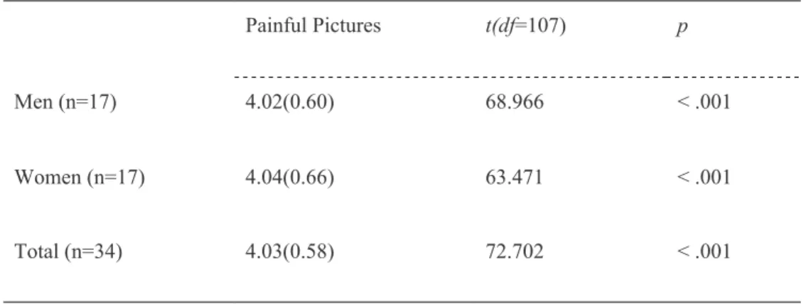
To validate the effectiveness of the painful pictures, pain intensity of the pictures was assessed by 34 undergraduate/graduate students (17 males)
from Seoul National University in a pilot study using a 7-point Likert scale (0 = no pain, 6 = unbearable pain). Pictures rated below 3 were eliminated, and a total of 108 pictures (Mean 4.03, SD 0.58) were selected as empathy- evoking stimuli. Examples of photographs used in the experiment are presented in Figure 5. The descriptive statistics of pain intensity for the final set of pictures are summarized in Table 1. There were no sex differences in the pain intensity ratings (p = .82).
The 108 pictures were randomly divided into three different sets.
The average pain intensities of the three sets of the pictures were assessed and confirmed to have no significant differences (p = .25). The order of the three picture sets was counterbalanced.
Table 1. The descriptive statistics (mean values and standard deviations) of pain intensity for the final set of pictures
Painful Pictures t(df=107) p
Men (n=17) 4.02(0.60) 68.966 < .001
Women (n=17) 4.04(0.66) 63.471 < .001
Total (n=34) 4.03(0.58) 72.702 < .001
Figure 4. An example of painful pictures ((A) hand, (B) foot) used in the study. It describes the physical pain commonly encountered in everyday life.
4.4. EEG Data acquisition
EEG data were recorded from the 61 electrodes embedded in a whole-head elastic cap (the international 10–20 method of electrode placement, Brain Products GmbH, Germany). For bipolar vertical and horizontal electro-oculograms (EOG), additional disc electrodes were attached to the areas below the left eye and around the external canthi, respectively. The Cz was used as the reference electrode. After the placements of the cap, each electrode site was injected with electrolytic gel.
Then, the skin surface was slightly abraded to minimize the electrode-skin impedance. Before and after testing, it was ensured that the impedances on all electrode sites were less than 10 kΩ. EEG was amplified via actiCHamp (Brain Products GmbH, Germany), and recorded using a BrainVision Recorder (Brain Products GmbH, Germany). The data were recorded at a sampling rate of 500 Hz.
4.5. EEG Data Preprocessing and Time-Frequency Analysis
Prior to analyzing the EEG data, the number of correct answers was counted on the continuous performance task for each participant. All participants correctly identified the pictures which they had/had not seen in each block more than 88.89% of the time (i.e., 8 out of the 9 pictures).
Furthermore, there were no significant differences in the number of correct answers across conditions (for men, p = .34, for women, p = .32) or sexes (p
= .68). Data from all participants, as such, were included in the analysis.
Data preprocessing and main analysis were all conducted using the Brain Vision Analyzer 2.2 (Brain Products). EEG records were initially re- referenced to the average of the two mastoids, and a band-pass filter of 0.5~40 Hz was applied. Through an ICA procedure, eye blinks and movements were corrected semi-automatically (Jung et al., 2000). Upon completing the EOG correction, further analysis was conducted only with the data without eye blink/movement artifacts. In each experimental block, the data were segmented into epochs of 8000 ms. Each segment started at
the onset of the fixation cross (i.e., baseline data) and ended at the termination of the picture (i.e., experimental data). The segmented data were baseline-corrected to the mean value of the signal in the time window of - 200-0 ms before the stimulus (picture) onset. Remaining artifacts exceeding +_100 μV in amplitude within a 200-second window were identified on the relevant sites (i.e., C3, C4, Cz, O1, O2, Oz). Epochs containing these artifacts were removed. The EEG records of one man were excluded, as they contained excessive movement artifacts.
TF (time-frequency) analysis was performed for each segment via continuous complex Morlet wavelet transformation in the frequency range of 5-30 Hz in logarithmic steps of 1 Hz. The Morlet parameter was set to 5.
The absolute power values were averaged across trials at each time- frequency point at each electrode site. Then the suppression indices were calculated for each data point of the time-frequency spectrum. Suppression indices in the present study were computed as the log-transformed ratios of the power during the experimental data relative to the power during the baseline (Cohen, 2014). Epochs starting 500 ms after the onset of the fixation cross (3500 ms) or the picture (3500 ms) were used for baseline and experimental data, respectively. The first 500 ms were removed to minimize the attention transients, caused by stimulus initiation and termination. I used the ratio, as opposed to simple subtraction, for the purpose of controlling potential variability in absolute EEG power between the subjects. This
individual variability is attributable to differences in electrode impedance and scalp thickness. For each of the three conditions for male and female participants, an average suppression value of the three electrodes was computed in the central region (i.e., Cz, C3, C4) to obtain a single mu suppression value (e.g., see Hoenen et al., 2015; Karakale et al., 2019 for similar procedures). The same procedure was done in the three occipital electrodes (i.e., Oz, O1, O2) to obtain a single alpha suppression value.
Consequently, six suppression indices for INCREASE, DECREASE and WATCH at the central and occipital areas for each subject were obtained.
Ratio data, however, have a non-normal distribution due to lower bounding. As such, I used a log transform for further statistical analysis. A log ratio of smaller than zero reflects a decrease of the power, while a log ratio greater than zero reflects an increase of the power.
4.6. Statistical Analysis
I used R studio (R Studio Team., 2016) for all the statistical analyses. In the analyses, data from 24 participants were used. For both sexes, two brain regions (i.e., central, occipital) and three conditions (i.e., INCREASE, DECREASE, WATCH) were examined. Prior to testing the hypotheses, one-sided t tests against zero for each condition were conducted to make sure that suppression occurred during the experimental compared to the baseline segment (all p-values < .05, Bonferroni-corrected). Upon validating suppression for all the brain regions and conditions, a 2 x 3 x 2
mixed design ANOVA was performed to compare the suppression indices in 8–13 Hz across sex (men, women), empathy modulation (increase, decrease, watch), and brain region (central, occipital). In correcting the degrees of freedom, the Greenhouse-Geisser epsilon values (G-GE) were used whenever necessary.
5. Results
5.1. Behavioral Results
The between-group analysis revealed no significant sex differences for the scores in all of the behavioral measures of empathy (i.e., the empathizing quotient (EQ), the emotional contagion scale (ECS), and the interpersonal reactivity index (IRI)). Furthermore, there were no outliers or significant differences in pain sensitivity between the male and female participants, indicating that this individual-difference variable was effectively controlled across the two sexes. These results are summarized in Table 2.
Table 2. Behavioral measures of empathy (on a scale of 1-5) and pain sensitivity (on a scale of 1-10) in the male and female subgroups.
5.2. EEG Results
The mix-design ANOVA results revealed that there was a significant three-way interaction effect between sex, condition, and brain region (F(2,44) = 3.458, p = .047, η2③ = 0.01). The three-way interaction effects are depicted in Figure 5. As indicated in the figure, mu and alpha rhythms over the central and occipital regions showed significant different suppression patterns, warranting further investigation at each brain region.
③ a generalized eta squared, as reported in R
Figure 5. Mu suppression indices for the increase, decrease, and watch conditions for men (red) and women (blue) in central (left) and occipital (right) regions.
The two-way interaction term between sex and condition at the central regions was further probed in a two-way mixed-design ANOVA.
Upon confirming its significance (F(2,44) = 20.12, p < .001, η2 = .10), pairwise t-tests were conducted to investigate the effect of condition on mu suppression in men and women, separately (i.e., sim