저작자표시-비영리-변경금지 2.0 대한민국 이용자는 아래의 조건을 따르는 경우에 한하여 자유롭게
l 이 저작물을 복제, 배포, 전송, 전시, 공연 및 방송할 수 있습니다. 다음과 같은 조건을 따라야 합니다:
l 귀하는, 이 저작물의 재이용이나 배포의 경우, 이 저작물에 적용된 이용허락조건 을 명확하게 나타내어야 합니다.
l 저작권자로부터 별도의 허가를 받으면 이러한 조건들은 적용되지 않습니다.
저작권법에 따른 이용자의 권리는 위의 내용에 의하여 영향을 받지 않습니다. 이것은 이용허락규약(Legal Code)을 이해하기 쉽게 요약한 것입니다.
Disclaimer
저작자표시. 귀하는 원저작자를 표시하여야 합니다.
비영리. 귀하는 이 저작물을 영리 목적으로 이용할 수 없습니다.
변경금지. 귀하는 이 저작물을 개작, 변형 또는 가공할 수 없습니다.
치의과학석사학위논문
Chondrgenic differentiation of human bone marrow-derived mesenchymal stem
cells by novel small-molecule inducers 새로운 저분자 화합물에 의한 인간 골수
유래 줄기세포의 선택적 연골 분화
2014년 8월
서울대학교 대학원
치의과학과 치의재생생명공학전공
조 태 준
Chondrgenic differentiation of human bone marrow-derived mesenchymal stem cells by
novel small-molecule inducers
새로운 저분자 화합물에 의한 인간 골수 유래 줄기세포의 선택적 연골 분화
지도교수 조 재 진
이 논문을 치의과학 석사학위논문으로 제출함
2014년 5월 서울대학교 대학원
치의과학과 치의재생생명공학전공 조 태 준
조태준의 석사학위논문을 인준함 2014년 6월
위 원 장 (인)
부위원장 (인)
위 원 (인)
Abstract
Chondrogenic differentiation of human bone marrow-derived mesenchymal stem cells by
novel small-molecule inducers
Cho Tae Jun
Dental Regenerative Biotechnology Major The Graduate School, Seoul National University
(Directed by Prof. Cho, Jaejin)
To treat cartilage defection, mesenchymal stem cells (MSCs) are considered as promising alternative cell source. Especially bone marrow derived MSCs have best characterized adult stem cells and have been used in various chondrogenic differentiation researches.
Inducing chondrogenesis from human bone marrow derived MSCs is formed in combination of defined media and TGF-β3. We also found small molecules 20B09, 20F08 which have the effect to cells by high throughput screening (HTS) method. The purpose of the experiment is to confirm whether these small molecules are able to replace TGF-β3. We measured the chondrogenic differentiation ability of 20B09, 20F08 after physical triggering by centrifugation.
After 7 and 14 days, three dimensional chondrogenic clusters were
measured by GAG assay, RT-PCR, specical staining such as Safranin-O and Von Kossa. Furthermore, we measured collagenⅠ,
Ⅱ by immunohistochemistry. Macroscopic results for 14 days were not different from TGF-β3 groups and small molecule groups. GAG assay showed that 20B09 groups were the faster enrichment of GAG contents than TGF-β3 at 7 days, but this trend was reversed at 14 days. Histological analysis results showed that TGF-β3 and 20B09 both allowed the efficient expression of proteoglycan by Safranin-O. Immunohistochemistry results showed that both collagen
Ⅰ and collagenⅡ observed the high expression in 20B09, 20F08 treated groups, compared to similar expression levels in TGF-β3 treated groups. RT-PCR assay results supported the trend too.
TGF-β3 groups showed a strong effect to chondrogenesis but transcripts for hypertrophy and osteogenesis were also increased. On the other hand, the selected small molecules 20B09, 20F08 did not.
Therefore, small molecules can selectively induce the chondrogenic differentiation of hBM-MSCs. A detailed mechanistic research and target identification are currently under way and will be reported in due course. This experiment using small molecules is considered as the best substance to solve signaling problems on chondrogenesis.
keywords : small molecule, TGF-β3, human bone marrow, mesenchymal stem cell, chondrogenic differentiation.
Student number: 2009-23606
TABLE OF CONTENTS
Introduction ...4
Materials and Methods ...6
Results ...13
Discussion ...19
References ...21
Figures and Tables ...25
Abstracts (Korean) ...33
Introduction
Osteoarthritis (OA), which is caused by cartilage defects, is one of the most common chronic diseases. Approximately 13.9% of adults aged 25 and older and 33.6% of those over 65 suffer from OA that seriously interferes with the performance of daily living activities (1).
Therefore, various biomedical strategies, such as subchondral drilling, microfractures and autologous chondrocyte implantations (ACI), have been studied and adapted for the treatment of damaged cartilage (2, 3). For example, surgeons have been performing microfractures on patients in order to induce human bone marrow-derived mesenchymal stem cells (hBM-MSCs) to migrate from the subchondral bone to the region of the cartilage defect or implantations of autologous cultured chondrocytes into the cartilage defect (3). However, the repair material that is the product of microfractures and ACIs is often fibrocartilage that has a weak capacity for resisting physical damage compared to natural hyaline cartilage due to a lack of sufficient hydroelastic characteristics (4). Thus, there is a huge medical demand for the treatment of patients with osteoarthritis that is of sufficient clinical consequence. MSCs are multipotent cells that can differentiate into many kinds of organ cell types, including cartilage, bone and adipose tissue (5). In particular, MSCs can be differentiated into hyaline cartilage that is abundant in glycosaminoglycan (GAG) and type II collagen as a result of chondrogenesis in the specific media containing chondrogenic inducers such as transforming growth factor-
βs (TGF-βs) superfamily, which includes TGF-β1–3 and bone morphogenetic protein (BMP) (6). For this reason, cell-based repair with the directed chondrogenesis of MSCs has been considered as a potentially promising therapy for improving the clinical outcome of patients with OA (7). However, a high dose of TGF-β can cause undesirable side effects, including synovial fibrosis and osteophyte formation, which culminate in the loss of joint function (8), cancer, metastasis (9) and kidney fibrosis. Furthermore, TGF-β is not specific for the chondrogenic differentiation of MSCs and cannot be used for OA treatment (10, 11). In fact, there have been several ground-breaking reports in the field of cell-based regenerative medicine of small molecules that can selectively regulate either the proliferation or differentiation of stem cells through the temporal perturbation of target cellular proteins instead of through non-specific differentiation methods (12, 13). Thus, the identification of potent and specific small molecule modulators that can be credible replacing TGF-β will provide the mechanistic insight into the developmental chondrogenesis of MSCs, which will lead to the discovery of novel therapeutic methods for patients with OA and related diseases.
Materials and Methods
Synthetic procedure and characterization of small-molecules library
Small-molecule inducers were obtained from department of chemistry, Seoul National University, which was synthesized in laboratory. The reaction steps for library construction were performed in parallel using the FlexChem Synthesis System from SciGene [Sunnyvale, CA] in a 96-deep-well filtration block. The purity of all the library members was observed by a LC/MS system equipped with a reverse phase column (C-18, 50 × 2.1 mm, 5 μm) and photodiode array (PDA) detector using electron spray ionization (ESI).
Purities were obtained by PDA based LC/MS analysis of final crude products without further purification. The high resolution mass spectrometric analyses were conducted at the Mass Spectrometry Laboratory of Seoul National University using mass spectrometer by direct injection for fast atomic bombardment (FAB) (14).
Culture of human bone-marrow-derived mesenchymal stem cells (hBM-MSCs)
Human bone-marrow-derived MSCs (hBM-MSCs) were obtained from children cardiac surgeries of Seoul National University Hospital, which was approved by the IRB (No.:SD0090001). hBM-MSCs were
maintained and expanded in the medium composed of a high glucose Dulbecco’s modified Eagles medium (DMEM: Welgene, Korea) including 20% Fetal bovine serum (FBS: Hyclon, USA), and 1%
Antibiotics-Antimycotics (Gibco, Grand Island, NY) at 37℃ under 5%
CO2. The cells were passaged up to 70% confluency. The cells at passage 7 (P7) were subjected to the chondrogenic differentiation assay.
Fluorescence-activated cell sorting (FACS) analysis
For the characterization of the hBM-MSCs, fluorescence-activated cell sorting (FACS) was performed to examine expression status of several stem cell surface markers. The cells were detached and washed with PBS supplemented with 2% FBS to carry out the FACS analysis. The following antibodies were used; fluorescein isothiocyanate (FITC)-conjugated mouse anti-human CD14, CD31, CD44 and CD45; phycoerythrin (PE)-conjugated mouse anti-human CD29, CD73, and CD117; PE.Cy5-conjugated mouse antihuman CD90;
allophycocyanin (APC)-conjugated mouse anti-human CD34;
HLA-DR; PE-conjugated streptavidin; biotin-conjugated HLA class II (all from BD PharMingen, San Diego, CA, USA); APC-conjugated mouse anti-human CD105 (eBioscience, USA). Each primary antibody was incubated with 100,000 cells for 30 min on ice. After washing, the secondary antibody was applied for 30 min on ice and then cells were fixed with 4% formaldehyde at 4℃. The fluorescence intensity was measured by a FACS Calibur and data were analyzed with BD
CellQuest Pro software (all from Becton Dickinson, San Jose, CA, USA).
Chondrogenic differentiation of hBM-MSCs
For initiation of chondrogenesis of hBM-MSCs, the cells were enzymatically detached by treating 0.25% trypsin and counted by hemocytometer. Three dimension clusters were formed in a 96wells (Becton Dickinson, San Jose, CA, USA) with 250,000 cells. The wells with cells were centrifuged in 500 g for 5 min. Then, the FBS containing medium was replaced with chemically defined medium which contained a high-glucose DMEM (Welgene, Korea), supplemented with 50 μg/mL ascorbate-2-phosphate, 100 μg/mL sodium pyruvate, 40 μg/mL L-proline, and 1% ITS+Premix (all Sigma-Aldrich, St. Louis, MO, USA). To investigate the effect of TGF-β3 or individual small molecules on chondrogenic differentiation of hBM-MSCs, the 3D clusters were cultured in four different conditions: chemically defined medium only or defined medium that was supplemented with TGF-β3 (10 ng/mL), or small molecules (10 μM). The 3D clusters with each medium were cultured in a 37℃, 5%
CO2 incubator and medium change was carried out every other days.
Glycosaminoglycan and DNA measurement
For glycosaminoglycan (GAG) contents assay, we used three chondrogenic clusters in each group and were measured with Blyscan
Sulfate Glycosaminoglycan Assay kit (biocolor, Belfast, Ireland) according to manufacturer’s instructions. Briefly, after putting 3 chondrocyte balls into a new eppendorf tube, papain buffer solution [0.2 M sodium phosphate buffer, 0.1 M sodium acetate, 10 mM EDTA, 5 mM L-cysteine HCl, 7.6 μL/mL papain (Sigma-Aldrich, St.
Louis, MO, USA), pH 6.4] was added 500 μL in that tube. This tube was incubated at 65℃ water bath for 18 h. After that time, the tube was centrifuged for 10 min at 10000 rpm and 100 μL of supernatant was transferred to a new tube. Then, 1 mL of the Blyscan dye reagent solution was added in the tube. The resulting eppendorf tube was incubated at the room temperature in a gentle mechanical shaker for 30 min. During this time period, a sulphated glycosaminoglycan-dye complex is formed and precipitated out from the soluble unbound dye. The resulting tube was centrifuged for 10 min at 12000 rpm. After the careful removal of all supernatant, a pellet of insoluble sGAG-dye complex at the bottom of the tubes was dried on a paper tissue at the room temperature. After addition of 1 mL of the dissociation reagent, each tube was recapped and vortexed to release the bound dye into the solution. After 10-min mixing, we measured the absorbance of this dissociated solution at 656 nm. For the measurement of total DNA contents, we used Picogreen dsDNA assay kit (Invitrogen, USA) and this experiment was performed according to manufacturer’s instructions. The GAG contents were normalized versus the total content of cellular DNA. This experiment was carried out in triplicate.
Histological staining and immunohistochemistry
For paraffin section, chondrogenic clusters in each different group were fixed in 4% phosphate buffered formaldehyde and embedded in paraffin. Paraffin embedded samples were sectioned by thickness of 4 μm and deparaffinized. Deparaffinized slides were stained by Safranin O staining to visualize the cellular proteoglycan was carried out. We also performed Von Kossa staining to show the calcium deposition levels in the samples. For the detection of collagen type I and collagen type II, ABC detection kit (Cap-plus detection Kit, Invitrogen) was used and performed according to manufacturer’s instructions. Briefly, 3% peroxidase was treated in the samples for 10 min and washed 3 times by PBS. Pepsin solution also was treated for retrieval. Nonspecific binding event was minimized by the treatment of blocking solution of the detection kit. The sample was subjected to the sequential treatment of primary antibody [Rabbit polyclonal antibody for collagen type I (Abcam, UK) and mouse monoclonal antibody for collagen type II (Calbiochem, Germany), diluted by 1:100 and 1:10, respectively], biotinylated secondary antibody (either anti-rabbit IgG or anti-mouse IgG), and streptavidin-HRP (horse-radish peroxidase). The immunohistochemical staining was visualized by the color development of DAB (3,3'-diaminobenzidine) upon peroxidase activity.
Total RNA preparation and RT-PCR
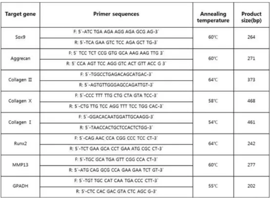
For reverse transcriptase polymerase chain reaction (RT-PCR), chondrogenic clusters in four different media were gathered at 7, and 14 days. Chondrogenic clusters were digested in liquid nitrogen. Then, total RNA was extracted from chondrogenic clusters using Trizol Reagent (Invitrogen Life Technologies, USA). To synthesize a complementary DNA (cDNA) used as a template in subsequent PCR reaction, 2 μg of total RNA was reverse-transcribed with cDNA Synthesis Kit (M-MLV RT, Invitrogen, USA) according to manufacturer’s instructions. In PCR reactions, the total reaction volume was 20 μl which include 1 μl of 10-fold dilution cDNA, 1 μl of each of primers (5 pmole), PCR premix (Accupower PCR premix, Bioneer, Korea) and sterilized 17 μl of water. PCR amplification was conducted with Mastercycler gradient (Eppendorf, Germany) under followed conditions: denaturation at 94 ℃ for 5 min, amplification at 94℃ for 30 sec, annealing at specific temperature for each primer pair (as shown in table 1) for 30 sec, extension at 72℃ for 20 sec, and total 28-37 cycles. A final extension step was performed at 72℃ for 5 min. RT-PCR primer sequences and each annealing temperature are shown in table 1. For analysis of PCR products, DNA gel electrophoresis was carried out in 1.2% agarose gel with 0.01 mg/mL ethidium bromide and quantified by Bio-profil X press zoom 2000 (Vilber Lourmat, Marne-la-Vallee, France).
Statistical analysis
Four independent experiments were performed in triplicate and data are presented as means ± SDs. Data analysis was preformed using Student’s t-test a significance level of P value.
Results
Characterization of hBM-MSCs
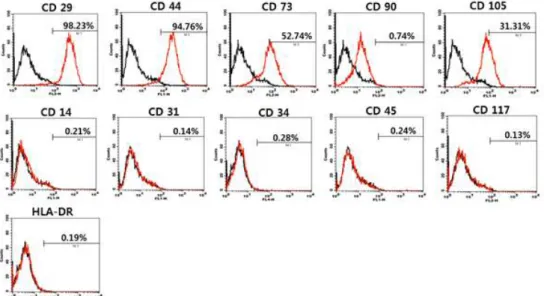
As shown in Figure 1, hBM-MSCs expressed the mesenchymal stem cell markers such as CD29 (98.23%), CD44 (94.76%), CD73 (52.74%), CD90 (0.74%), and CD105 (31.31%), whereas few cells expressed hematopoietic marker such as CD14 (0.21%), CD34 (0.28%), CD45 (0.24%), CD117 (0.13%), and HLA-DR (0.19%), or the endothelial marker CD31 (0.14%). The homogeneously high expression of CD29 (β1-integrin) and CD44 (hyaluronan receptor) indicates that the used hBM-MSCs are mainly mesenchymal stem cells. However, the lack of hematopoietic cell marker and endothelial cell marker reveals that hBM-MSCs were not contaminated by hematopoietic stem cells or endothelial cells. The relative low level of CD105 (endoglin), which is a specific marker for mesenchymal stem cell, implies that there exists osteogenic differentiation process.
The effect of GAG synthesis of small molecules with TGF-β3 on hBM-MSCs
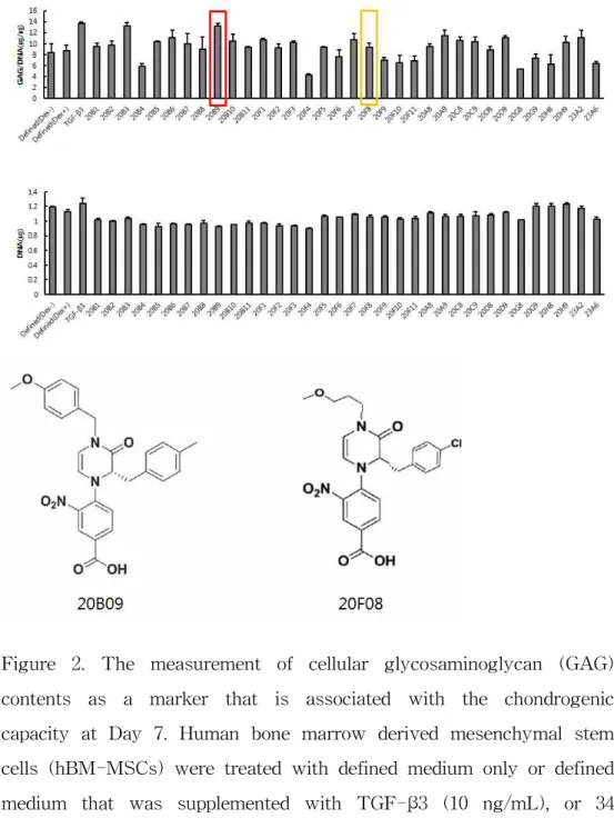
We selected 34 compounds from the small molecules library and measured cellular GAG contents, the biomarker of chondrogenic extracellular matrix (15), with this collection. As shown in Figure 2, the treatment of hBM-MSCs either with TGF-β3 (10 ng/mL), 20B03
(10 uM), 20B09 (10 μM) or 20F08 (10 μM) in the defined media made a significant difference to the total GAG contents normalized with DNA contents, compared to the defined medium only (defined medium only, 8.69; TGF-β3, 13.73; 20B03, 13.25; 20B09, 13.28; 20F08, 9.42 at 7 days). Due to the disadvantages of 20B03 in mass production, the small molecule was not used in the following experiments despite the significant effect on the total GAG contents. On the basis of these screening results with the small molecules, we identified 20B09 and 20F08 as a potential inducer of chondrogenic activity in hBM-MSCs and subjected it to further in vitro biological evaluation.
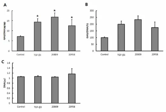
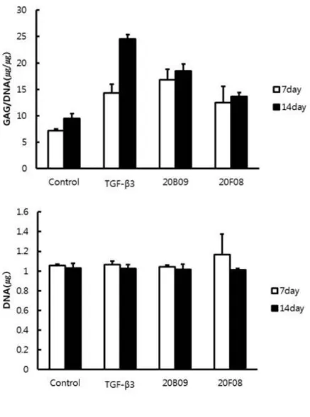
Confirmation of chondrogenic differentiation of small molecules Considering these initial results above, we further confirmed the chondrogenesis-inducing activity of 20B09 and 20F08 via the robust formation of chondrogenic clusters and the measurement of cellular GAG contents at days 7 and 14. As shown in Figure 3, chondrogenic clusters upon treatment with TGF-β3, 20B09 and 20F08 were larger than those prepared in defined medium only. The size of chondrogenic clusters is known to be related to the degree of chondrogenic differentiation due to the increased proteoglycan synthesis (16). In addition, we observed a significant difference in the GAG/DNA contents in the resulting chondrogenic clusters prepared in four different conditions. As shown in Figure 3A, Among TGF-β3, 20B09 and 20F08 supplemented in the defined media enhanced the cellular GAG/DNA contents, related to the degree of chondrogenic
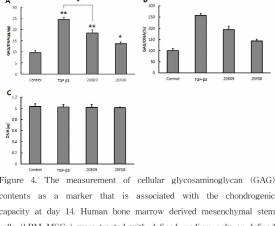
differentiation, compared to the defined medium only (defined medium only, 7.2; TGF-β3, 14.3; 20B09, 16.8; and 20F08, 12.5 at 7 days). As shown in Figure 3B, GAG/DNA synthesis were higher than those of the control by TGF-β3 200%, 20B09 234% and 20F08 174%. Total cellular DNA contents of all chondrogenic clusters were similar relative to the control shown in Figure 3C. As shown in Fig. 4A, Among TGF-β3, 20B09 and 20F08 supplemented in the defined media enhanced the cellular GAG/DNA contents, related to the degree of chondrogenic differentiation, compared to the defined medium only (defined medium only, 9.5; TGF-β3, 24.5; 20B09, 18.4; and 20F08, 13.6 at 14 days). As shown in Figure. 4B, GAG/DNA synthesis were higher than those of the control by TGF-β3 258%, 20B09 194% and 20F08 143%. Total cellular DNA contents of all chondrogenic clusters were similar relative to the control shown in Figure 4C. Our chondrogenic inducer, 20B09, shows the faster enrichment of GAG contents than TGF- β3 at 7 days, but this trend was reversed at 14 days shown in Figure. 5.
The characteristics of resulting chondrogenic clusters
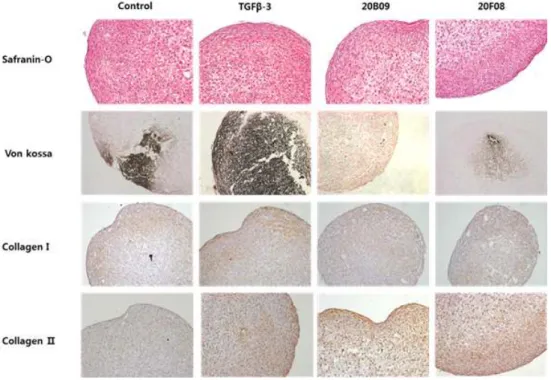
In order to examine the characteristics of resulting chondrogenic clusters, we further investigated them using histological staining and immunohistochemistry. As shown in Figure 6, the treatment with TGF-β3, 20B09 and 20F08 allowed the efficient expression of proteoglycan, which was visualized by Safranin-O staining. In order to measure the levels of calcium deposition, which is known as a key
biomarker of osteogenesis (17) - one of the competing routes of hBM-MSC differentiation - we visualized chondrogenic clusters with Von Kossa staining. TGF-β3-treated chondrogenic clusters showed higher levels of calcium deposition than small molecules-treated ones, which strongly supports that small molecules can selectively induce the directed chondrogenic differentiation of hBM-MSCs, compared to TGF-β3. On the basis of immunohistochemistry, we observed the higher expression levels of collagen II, a biomarker of chondrogenesis (3), than that of collagen I, a biomarker of osteogenesis (17), in small molecules-treated chondrogenic clusters, compared to the similar expression levels of collagen I and II in TGF-β3 - treated clusters, which strengthened our above statement.
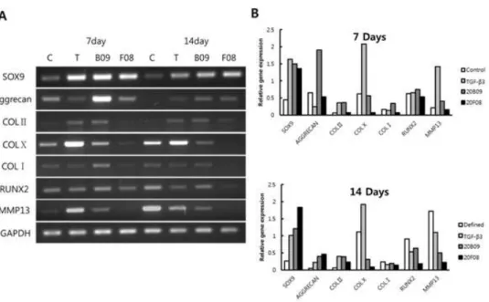
The confirmation of the directed chondrogenic differentiation For the confirmation of the directed chondrogenic differentiation of hBM-MSCs upon treatment with small molecules in contrast with TGF-β3, we measured the transcription levels of messenger RNAs (mRNAs) for the chondrogenic markers (SOX9, Aggrecan and collagen type II) (18), the hypertrophic chondrocyte specific marker (collagen type X) (19) and the osteogenic markers (collagen type I, RUNX2, and MMP-13) (20, 21) using reverse transcriptase polymerase chain reaction (RT-PCR). As shown in Figure 7, TGF-β3 drastically enhanced the mRNA levels of collagen type X and matrix metalloproteinase 13 (MMP-13) - specific osteogenic markers - at both day 7 and 14. Based on the fact that the hypertrophic
differentiation of chondrocytes is one of processes for endochondral ossification (22), this data confirm that TGF-β3 might induce the non-specific differentiation of hBM-MSCs via facilitating osteogenesis of MSCs along with chondrogenesis. In addition, TGF-β3 also increased the mRNA level of MMP-13 (collagenase-3), which plays important roles in regulating osteoblastic differentiation of osteogenic cells. However, we didn’t observe any significant enhancement of mRNA levels in collagen type X and MMP-13 upon treatment with small molecules compared to increases by TGF-β3 at both days 7 and 14. In the case of RUNX2 (one of the osteogenic transcription factors) and SOX9 (one of the chondrogenic markers), we observed their comparable mRNA levels both in TGF-β3 and 20B09 treated chondrogenic clusters at days 7 and 14. In addition, the mRNA level of aggrecan, an early expressed chondrogenesis specific proteoglycan, was dramatically increased by 20B09 and 20F08 at day 7, which are 7.9-fold and 2.2-fold higher than that by TGF-β3 and the mRNA level of collagen type X, the hypertrophic chondrocyte specific marker, dramatically increased by TGF-β3 at day 7 and 14, which are 3.7-fold and 6.1-fold higher than that by 20B09. Also, the mRNA level of MMP-13, specific osteogenic markers, dramatically increased by TGF-β3 at day 7 and 14, which are 3.5-fold and 2.2-fold higher than that by 20B09 shown in Figure 7B.
This observation strongly supports our statement that small molecules might specifically induce the chondrogenesis of hBM-MSCs at the early stage of their differentiation. The mRNA levels of Collagen type I (one of the osteogenic markers) and Collagen type II
(one of the chondrogenic markers) were not significantly different between TGF-β3 and small molecules treated chondrogenic clusters at days 7 and 14.
Discussion
Osteoarthritis is a highly prevalent and chronic joint disease. There is no effective disease-modifying treatment for osteoarthritis, necessitating surgical method as a therapeutic option. TGF-β is known for the maintenance of articular cartilage and structural integrity. However, TGF-βs have many clinical side effects. We report the discovery of the novel synthetic small-molecule inducers that can facilitate the differentiation of human bone marrow-derived MSCs (hBM-MSCs) into chondrocytes. To identify novel small-molecule modulators with chondrogenesis-inducing activity in hBM-MSCs, the pDOS-derived small-molecule collection containing over 3,000 members with 60 unique molecular frameworks was subjected to high throughput screening (HTS) (14, 23) using a cell viability assay and GAG content screening with primary hBM-MSCs.
As shown in Figure 1, homogeneously high expression of CD 29 (β 1-integrin) and CD44 (hyaluronic acid receptor) indicated that the hBM-MSCs used in this study were mesenchymal stem cells. The low levels of CD14, CD31, CD34, CD45, CD117, and HLA-DR demonstrated that the hBM-MSCs used in this study were not contained by hematopoietic and endothelial cells. We treated human bone marrow mesenchymal stem cells with TGF-β3 as positive control. To strengthen safety and reproducibility our experiment, GAGs analyses were performed in triplication. As shown in Figure 5, 20B09 shows the faster enrichment of GAG contents than TGF- β3
at 7 days, but this trend was reversed at 14 days. These observations led us to hypothesize that 20B09 might induce the directed chondrogenesis in the early stage of its differentiation, which is currently under mechanistic investigation and will be reported in due course.
The chondrogenic-related genes, SOX9, aggrecen, and collagen type
Ⅱ, were up-regulated by small-molecules. Specially, aggrecan, cartilage-specific proteoglycan core protein, increased expression, whereas collagen type Ⅹ, hypertrophic marker, decreased expression at day 7 and 14. In this study, we identified that hBM-MSCs underwent chondrogenic differentiation upon treatment small-molecules. Unlike the typical conditions for chondrogenesis of MSCs using TGF-β3, the selective chondrogenic differentiation of hBM-MSCs without osteogenic differentiation by small molecules was supported by our in vitro confirmation, including histological Von Kossa staining, immunohistological collagen type I and II staining and RT-PCR analyses of collagen type X, MMP-13 and aggrecan.
In summary, small-molecule inducers have key role in chondrogenic differentiation of hBM-MSCs. Furthermore, their potential and functional roles with hBM-MSCs were investigated. Our data suggested that small-molecules seemed to have the selective induction by way of hyaline cartilage in hBM-MSCs. A detailed mechanistic study and target identification are currently under way and will be reported in due course.
References
1. Lawrence, R. C., Felson, D. T., Helmick, C. G., Arnold, L. M., Choi, H., Deyo, R. A., Gabriel, S., Hirsch, R., Hochberg, M. C., Hunder, G. G., Jordan, J. M., Katz, J. N., Kremers, H. M., Wolfe, F., and National Arthritis Data, W. (2008) Estimates of the prevalence of arthritis and other rheumatic conditions in the United States. Part II. Arthritis and rheumatism 58, 26-35.
2. Brittberg, M., Lindahl, A., Nilsson, A., Ohlsson, C., Isaksson, O., and Peterson, L. (1994) Treatment of deep cartilage defects in the knee with autologous chondrocyte transplantation. The New England journal of medicine 331, 889-895.
3. Khan, W. S., Johnson, D. S., and Hardingham, T. E. (2010) The potential of stem cells in the treatment of knee cartilage defects. The Knee 17, 369-374.
4. Eble, J. A. (2001) The molecular basis of integrin-extracellular matrix interactions. Osteoarthritis and cartilage / OARS, Osteoarthritis Research Society 9 Suppl A, S131-140.
5. Pittenger, M. F., Mackay, A. M., Beck, S. C., Jaiswal, R. K., Douglas, R., Mosca, J. D., Moorman, M. A., Simonetti, D. W., Craig, S., and Marshak, D. R. (1999) Multilineage potential of adult human mesenchymal stem cells. Science 284, 143-147.
6. Terraciano, V., Hwang, N., Moroni, L., Park, H. B., Zhang, Z., Mizrahi, J., Seliktar, D., and Elisseeff, J. (2007) Differential response of adult and embryonic mesenchymal progenitor cells
to mechanical compression in hydrogels. Stem cells 25, 2730-2738.
7. Hardingham, T., Tew, S., and Murdoch, A. (2002) Tissue engineering: chondrocytes and cartilage. Arthritis research 4 Suppl 3, S63-68.
8. Tang, Q. O., Shakib, K., Heliotis, M., Tsiridis, E., Mantalaris, A., Ripamonti, U., and Tsiridis, E. (2009) TGF-beta3: A potential biological therapy for enhancing chondrogenesis.
Expert opinion on biological therapy 9, 689-701.
9. Maehara, Y., Kakeji, Y., Kabashima, A., Emi, Y., Watanabe, A., Akazawa, K., Baba, H., Kohnoe, S., and Sugimachi, K.
(1999) Role of transforming growth factor-beta 1 in invasion and metastasis in gastric carcinoma. Journal of clinical oncology : official journal of the American Society of Clinical Oncology 17, 607-614.
10. Johnstone, B., Hering, T. M., Caplan, A. I., Goldberg, V. M., and Yoo, J. U. (1998) In vitro chondrogenesis of bone marrow-derived mesenchymal progenitor cells. Experimental cell research 238, 265-272.
11. Erlebacher, A., Filvaroff, E. H., Ye, J. Q., and Derynck, R.
(1998) Osteoblastic responses to TGF-beta during bone remodeling. Molecular biology of the cell 9, 1903-1918.
12. Lyssiotis, C. A., Lairson, L. L., Boitano, A. E., Wurdak, H., Zhu, S., and Schultz, P. G. (2011) Chemical control of stem cell fate and developmental potential. Angewandte Chemie 50, 200-242.
13. Johnson, K., Zhu, S., Tremblay, M. S., Payette, J. N., Wang, J., Bouchez, L. C., Meeusen, S., Althage, A., Cho, C. Y., Wu, X., and Schultz, P. G. (2012) A stem cell-based approach to cartilage repair. Science 336, 717-721.
14. Kim, J., Lee, WS., Koo, J., Lee, J., and Park, SB. (2014) Synthesis and library construction of privileged tetra-substituted Δ5-2-oxopiperazine as β-turn structure mimetics. ACS Comb Sci. 16, 24-32.
15. Huang, A. H., Motlekar, N. A., Stein, A., Diamond, S. L., Shore, E. M., and Mauck, R. L. (2008) High-throughput screening for modulators of mesenchymal stem cell chondrogenesis. Annals of biomedical engineering 36, 1909-1921.
16. Sekiya, I., Colter, D. C., and Prockop, D. J. (2001) BMP-6 enhances chondrogenesis in a subpopulation of human marrow stromal cells. Biochemical and biophysical research communications 284, 411-418.
17. Shi, X., Wang, Y., Varshney, R. R., Ren, L., Zhang, F., and Wang, D. A. (2009) In-vitro osteogenesis of synovium stem cells induced by controlled release of bisphosphate additives from microspherical mesoporous silica composite. Biomaterials 30, 3996-4005.
18. Woods, A., Wang, G., Dupuis, H., Shao, Z., and Beier, F.
(2007) Rac1 signaling stimulates N-cadherin expression, mesenchymal condensation, and chondrogenesis. The Journal of biological chemistry 282, 23500-23508.
19. Goldring, M. B., Tsuchimochi, K., and Ijiri, K. (2006) The control of chondrogenesis. Journal of cellular biochemistry 97, 33-44.
20. Komori, T., Yagi, H., Nomura, S., Yamaguchi, A., Sasaki, K., Deguchi, K., Shimizu, Y., Bronson, R. T., Gao, Y. H., Inada, M., Sato, M., Okamoto, R., Kitamura, Y., Yoshiki, S., and Kishimoto, T. (1997) Targeted disruption of Cbfa1 results in a complete lack of bone formation owing to maturational arrest of osteoblasts. Cell 89, 755-764.
21. Smith, G. N., Jr. (2006) The role of collagenolytic matrix metalloproteinases in the loss of articular cartilage in osteoarthritis. Frontiers in bioscience : a journal and virtual library 11, 3081-3095.
22. Colnot, C. (2005) Cellular and molecular interactions regulating skeletogenesis. Journal of cellular biochemistry 95, 688-697.
23. Park, EG., Cho, TJ., Oh, KH., Kwon, SK., Lee, DS., Park, SB., and Cho, JJ. (2012) Establishment of high throughput screening system using human ubilical cord-derived mesenchymal stem cells. Int. J ournal of oral biology 37, 43-49.
Figure 1. Immunophenotypic characterization of hBM-MSCs at passage 6 (P6). Data are shown as an overlay plot with immunoglobulin isotype control (in black) and different specific cell-surface markers (in red).
Figure 2. The measurement of cellular glycosaminoglycan (GAG) contents as a marker that is associated with the chondrogenic capacity at Day 7. Human bone marrow derived mesenchymal stem cells (hBM-MSCs) were treated with defined medium only or defined medium that was supplemented with TGF-β3 (10 ng/mL), or 34 individual small molecules (10 μM) and the chemical structure of 20B09, 20F08. Total GAG contents were normalized by dividing with the total DNA contents (n=3).
Figure 3. The measurement of cellular glycosaminoglycan (GAG) contents as a marker that is associated with the chondrogenic capacity at day 7. Human bone marrow derived mesenchymal stem cells (hBM-MSCs) were treated with defined medium only or defined medium that was supplemented with TGF-β3 (10 ng/mL), 20B09 (10 μM), and 20F08 (10 μM). (A) Relative amount of GAG/DNA (ug/ug) contents in each chondrogenic clusters grow in the four different groups. Total GAG contents were normalized by dividing with the total DNA contents (n=3). (B) The percentage of GAG/DNA (%) in each chondrogenic clusters grow in the four different conditions. (C) Total amount of DNA in chondrogenic clusters, indicating that viability and proliferation of cells in four conditions were similar, which implies the results are reliable (n=3). *: p < 0.05, ** : p <
0.001
Figure 4. The measurement of cellular glycosaminoglycan (GAG) contents as a marker that is associated with the chondrogenic capacity at day 14. Human bone marrow derived mesenchymal stem cells (hBM-MSCs) were treated with defined medium only or defined medium that was supplemented with TGF-β3 (10 ng/mL), 20B09 (10 μM), and 20F08 (10 μM). (A) Relative amount of GAG/DNA (ug/ug) contents in each chondrogenic clusters grow in the four different groups. Total GAG contents were normalized by dividing with the total DNA contents (n=3). (B) The percentage of GAG/DNA (%) in each chondrogenic clusters grow in the four different conditions. (C) Total amount of DNA in chondrogenic clusters, indicating that viability and proliferation of cells in four conditions were similar, which implies the results are reliable (n=3). *: p < 0.05, ** : p <
0.001
Figure 5. The measurement of cellular glycosaminoglycan (GAG) contents as a marker that is associated with the chondrogenic capacity at Day 7, 14. Human bone marrow derived mesenchymal stem cells (hBM-MSCs) were treated with defined medium only or defined medium that was supplemented with TGF-β3 (10 ng/mL), 20B09 (10 μM), and 20F08 (10 μM). Total GAG contents were normalized by dividing with the total DNA contents (n=3).
Figure 6. Histological staining and immunohistochemistry of hBM-MSCs-derived chondrogenic clusters treated with defined medium only and defined medium that was supplemented with TGF- β3 (10 ng/mL), 20B09 (10 μM), and 20F08 (10 μM) for 14 days.
Safranin-O staining indicates the level of proteoglycan, and Von Kossa staining indicates the level of depoited calcium.
Immunohistochemical staining was performed with collagen type Ⅰ and Ⅱ specific antibodies.
Figure 7. (a) The levels of expression of mRNA of various biomarkers, including the chondrogenic markers (SOX9, Aggrecan Collagen type II), the hypertrophic chondrocyte-specific marker (Collagen type X) and the osteogenic markers (Collagen type I, RUNX2 and MMP13) were determined by RT-PCR analysis after the treatment of hBM-MSCs in chemically defined medium only or defined media supplemented with TGF-b3 (10 ng/mL), 20B09 (10 mM) and 20B09 (10 mM) for days 7 and 14. (b) RT-PCR analysis of the related gene expression normalized by GAPDH in chondrogenic clusters at days 7 and 14.
Table 1. Primer sequence and specific annealing temperature for each primer pair for RT-PCR.
요약(국문초록)
새로운 저분자 화합물에 의한 인간 골수 유래 줄기세포의 선택적 연골 분화
조 태 준 서울대학교 대학원
치의과학과 치의재생생명공학전공 (지도교수 조 재 진)
중배엽 줄기세포는 골관절염과 같은 연골 결함을 치료하기 위한 우수한
세포 자원으로 고려되고 있다. 특히 골수 유래의 줄기세포는
성체줄기세포와 다양한 연골 분화 연구에 이용되고 있다. 골수 유래 줄기세포의 연골화 과정은 defined media 에 TGF-β3 를 넣어 유도되어진다. 본 연구는 high throughput screening(HTS) 방법으로 세포에 영향을 주는 저분자 물질 20B09 를 발견하였다. 이 실험의
목적은 저분자 화합물이 TGF-β3 를 대체할 수 있는지를 확인하는
것이다. 원심분리로 물리적인 자극을 준 후에 20B09 의 연골 분화 능력을 측정하였다. 배양 7 일과 14 일 후에 3 차원적으로 연골화된 masses 는 GAG assay, RT-PCR, Safranin-O 와 Von Kossa 와 같은 특수염색으로 분석하였다. 또한 CollagenⅠ, Ⅱ로 면역조직염색법을 실시하였다. 육안으로의 결과는 14 일 동안 TGF-β3 와 20B09 의 차이가 없었다. 그렇지만 GAG assay 결과는 7 일에서 20B09 가 TGF-β3 보다
GAG 양이 더 풍부하였지만 14 일의 결과에서는 이러한 경향이 바뀌었다. Safranin-O 에 의한 조직염색 결과는 TGF-β3 와 20B09 에서 proteoglycan 의 발현이 효과적으로 관찰되었다. CollagenⅠ과 CollagenⅡ의 면역조직염색 결과에서도 20B09 가 TGF-β3 를 처리한 그룹과 비슷한 레벨의 발현량을 보여주었다. RT-PCR 의 결과도
TGF-β3 는 연골화 과정에서 강력하게 작용하는 것을 보여주지만
hyperthroph 와 osteogenesis 와 관계된 전사인자도 증가하는 것을 보여주었다. 반면에 실험에 선택된 저분자 화합물 20B09 는 적은 발현량을 보여주었고 골수유래줄기세포의 연골분화를 선택적으로 유도할 수 있었다. 자세한 작용 기작과 표적 식별은 현재 연구 중이며 저분자 물질을 이용한 실험은 연골화 과정의 신호전달 문제를 해결하기 위해 중요한 물질로 고려된다.
주요어 : 중배엽 줄기세포, 저분자 화합물, TGF-β3, 연골분화, 골수줄기 세포
학번 : 2009-23606