저작자표시-비영리-변경금지 2.0 대한민국 이용자는 아래의 조건을 따르는 경우에 한하여 자유롭게
l 이 저작물을 복제, 배포, 전송, 전시, 공연 및 방송할 수 있습니다. 다음과 같은 조건을 따라야 합니다:
l 귀하는, 이 저작물의 재이용이나 배포의 경우, 이 저작물에 적용된 이용허락조건 을 명확하게 나타내어야 합니다.
l 저작권자로부터 별도의 허가를 받으면 이러한 조건들은 적용되지 않습니다.
저작권법에 따른 이용자의 권리는 위의 내용에 의하여 영향을 받지 않습니다. 이것은 이용허락규약(Legal Code)을 이해하기 쉽게 요약한 것입니다.
Disclaimer
저작자표시. 귀하는 원저작자를 표시하여야 합니다.
비영리. 귀하는 이 저작물을 영리 목적으로 이용할 수 없습니다.
변경금지. 귀하는 이 저작물을 개작, 변형 또는 가공할 수 없습니다.
의학석사 학위논문
The study for characterization and integrinα V β 3 targeting of
64 Cu-cRGDyK-HSA
방사성구리-cRGDyK-알부민을 이용한 인테그린α V β 3 표적화 및
특성에 관한 연구
2016 년 2 월
서울대학교 대학원 협동과정 종양생물학 전공
박 초 롱
A thesis of the Master’s degree
방사성구리-cRGDyK-알부민을 이용한 인테그린α V β 3 표적화 및
특성에 관한 연구
The study for characterization and integrinα V β 3 targeting of
64 Cu-cRGDyK-HSA
February 2016
Interdisciplinary Program in Tumor Biology, Seoul National University
College of Medicine
Cho Rong Park
ABSTRACT
Cho Rong Park Interdisciplinary Program in Tumor Biology The Graduate School Seoul National University
Introduction: RGD is famous integrinαVβ3 targeting peptide, so integrinαVβ3
targeting for tumor imaging is useful for tumor imaging. However, RGD has
the short circulation time and the majority of the injected probes are cleared
through the renal system or hepatobiliary system. To enhance the half-lives of
RGD and tumor targeting, cRGDyK was conjugated to HSA via bioorthogonal
click reaction. To develop the best RGD conjugated HSA nanoparticles, two
types of cRGDyK conjugated HSA were synthesized and the probes (cRGDyK-
HSA) were characterized, radiolabeled and preliminarily tested in in vitro and
in vivo properties of integrinαVβ3 expressing cancer targeting.
i
Methods: HSA was conjugated with DBCO-NHS ester (molar ratio of HSA :
DBCO-NHS ester; 1 : 5.62 for reaction 1 and 1 : 11.24 for reaction 2) for click
reaction linker. And DBCO-HSA was conjugated with N3-cRGDyK (HSA-
DBCO : N3-cRGDyK; 1 : 3 for reaction 1 and 1 : 6 for reaction 2). For
radiolabeling, 64Cu-labeled 3-azidopropyl-NOTA was conjugated to DBCO-
HSA and cRGDyK-HSA via click reaction. At each conjugation step, the
conjugates were purified using PD-10 column, eluted with the PBS buffer. All
conjugation products were analyzed via MALDI-TOF-MS and radiolabeling
efficiencies were measured by instant thin layer chromatography (ITLC). The
stability of 64Cu-labeled HSA and -cRGDyK-HSA in serum were monitored
during 48 hrs. To certify cRGDyK-HSA binding to integrinαVβ3 in cell level,
FNR648-labeled cRGDyK-HSA was used for confocal microscopy imaging.
64Cu-labeled HSA and -cRGDyK-HSA were intravenously injected to SK-OV3
tumor bearing mice and the distribution of the probes in mice were imaged by
small animal PET at 10 min, 4 hours, 24 hours and 48 hours post injection (p.i.)..
ii
Results: DBCO-NHS ester and cRGDyK were successfully conjugated to HSA,
according to their molar ratio. In case of DBCO, the number of DBCO
conjugated to HSA; reaction 1 was 3.94 ± 0.70 and reaction 2 was 6.72 ±
0.41. In case of cRGDyK, the number of cRGDyK conjugated to DBCO-HSA;
reaction 1 was 2.07 ± 0.51 and reaction 2 was 5.29 ± 0.76. Radiolabeling
efficiencies of 64Cu-HSA and 64Cu-cRGDyK-HSA (reaction 2) was 100% and
after PD-10 purification, that of 64Cu-cRGDyK-HSA (reaction 1) was almost
100%. 64Cu-HSA and -cRGDyK-HSA were stable in serum after 48 hours
incubation. Confocal microscopy images showed that FNR648-labeled
cRGDyK-HSA were localized in cell membrane and intracellular regions, this
localization in cells was blocked when cells were pre-incubated with excess
cRGDyK. The cell uptake of 64Cu-labeled cRGDyK-HSA (reaction 1 and
reaction 2) was higher in SK-OV3 cells (integrinαVβ3 positive) than 22Rv1
cells (integrinαVβ3 negative, P < 0.05). PET images revealed that reaction 2
of cRGDyK-conjugated HSA had the highest uptake in tumor (5.37 ±
1.09 %ID/g).
iii
Conclusion: DBCO-HSA and cRGDyK-HSA were successfully synthesized.
As the molar ratio of DBCO or cRGDyK were different, the number of attached
DBCO or cRGDyK to HSA were consistently different. Using click reaction,
64Cu was successfully labeled to HSA and cRGDyK-HSA. cRGDyK-HSA
could bind integrinαVβ3 in tumor cells and in vivo PET imaging results probed
that the 64Cu-cRGDyK-HSA could target tumor after 4 hours p.i.. These results
demonstrate that 64Cu-labeled cRGDyK-HSA can be used as PET tumor
imaging probes.
---
Keywords: integrinαVβ3, RGD, Human serum albumin (HSA),
bioorthogonal click reaction, PET imaging, 64Cu
Student number: 2014-21152
iv
CONTENTS
Abstract ... i
Contents ... ⅴ List of tables and figures ... ⅵ List of abbreviations ... ⅶ Introduction ... 1
Material and Methods ... 5
Results ... 15
Discussion ... 40
v
LIST OF TABLES AND FIGURES
Figure1. Characterization of DBCO-HSA and cRGDyK- HSA... 16 Table 1. Investigation of molecular mass using MALDI-TOF and
analysis of MALDI-TOF data ... 18 Figure2. Labeling efficiency and stability of
64Cu-HSA and -
cRGDyK-HSA ... 22 Figure3. Screening for the
integrinαVβ3expressing cell line ... 25 Figure4. Cell binding and internalization of cRGDyK-HSA ... 30-31 Figure5. In vitro uptake test of
64Cu-HSA and –cRGDyK-HSA...35 Figure6. In vivo tumor imaging of
64Cu-HSA and -cRGDyK-HSA
... 38
vi
LIST OF ABBREVIATIONS
RGD, Arg-Gly-Asp
HSA, Human Serum Albumin
cRGDyK, cyclic Arg-Gly-Asp-D-Tyr-Lys DBCO-NHS, dibenzocyclooctyl-NHS
MALDI-TOF, Matrix Assisted Laser Desorption/Ionization-Time Of
FlightNOTA, 1,4,7-triaza-cyclononane-1,4,7-triacetic acid ITLC-SG, Instant Thin Layer Chromatography-Silica Gel FBS, Fetal Bovine Serum
RT-PCR, Reverse Transcriptase-Polymerase Chain Reaction HBSS, Hank’s Balanced Salt Solution
BSA, Bovine Serum Albumin
PET, Positron Emission Tomography
%ID/g, % Injected Dose/gram
vii
INTRODUCTION
Integrinα
Vβ
3targeting for tumor imaging
For the expansion of a tumor mass, new blood vessel formation is
important. Angiogenesis is regulated through the balance of pro-angiogenic and
anti-angiogenic processes and when pro-angiogenic processes are dominant,
angiogenesis occurs (1, 2, 4). Because angiogenesis depends on the adhesive
interaction of vascular cells, adhesive receptor integrinαVβ3 was identified and
known as angiogenic marker. RGD (Arg-Gly-Asp) is famous integrinαVβ3
targeting peptide, which was identified from finding the cell attachment activity
of fibronectin (5, 6). For selectively targeting to integrinαVβ3 and improving
affinity, many modified RGD peptides are found (7). Many research groups are
already using RGD or modified RGD peptides for tumor targeting (8-10).
Because integrinαVβ3 is already well known about the angiogenesis aspect
during tumor mass expansion and there are many molecules can target
1
integrinαVβ3, so integrinαVβ3 targeting for tumor imaging is very useful for
tumor imaging.
HSA as a carrier
Human serum albumin (HSA) is molecular weight of 65.5kDa and the
major soluble protein in circulating system, a blood concentration is about 50
mg/mL (11). Because HSA has long half-life (an average half-life is 19 days)
and already exists in body, many HSA-based nanoparticles are developed. The
most famous one is ABI 007 (Abraxane®), which was approved by the FDA in
2005, already used in clinical trials (12-14). Paclitaxel has bad solubility in
water, as the drug were incorporated in albumin, the solubility and toxicity
problem are improved.
As mentioned before, there are many kinds of RGD-based peptides.
But RGD has the short circulation time and the majority of the injected probes
are cleared through the renal system (9) or hepatobiliary system (15). As the
imaging moiety, fast clearance is a good aspect for tumor imaging, but as the 2
therapy aspects, particles should be incorporated to tumor for a long time to
cure the tumor. And when using with radionuclide labeled RGD sequences for
therapy, because the radionuclide for therapy emits high energy, high uptake in
kidney may cause kidney damages (16, 17). To solve this problem, HSA can
be an answer to improve longer circulation in body. With prolonged circulation
time in blood, we can also expect more accumulation of the probes to tumors.
Click reactions for conjugation
Click reactions have broad meaning, which meet being selective, high
yielding, and having good reaction kinetics. At the early time, copper-catalyzed
azide–alkyne cycloaddition was called “click” chemistry. But in these days,
researchers are started to focus on bioorthogonal click reaction, which are inert
to biological environments (18). Because this reaction doesn’t need exogenous
metal catalysts, which can cause mild to severe cytotoxic effects, it is good for
making nanoparticles to use in vivo (19).
3
Purpose of this study
RGD peptides have been used as tumor imaging to target angiogenesis
for a long times, but fast-clearance via kidney is problematic. Conjugating RGD
peptides to HSA via bioorthogonal click reaction, which can conduct the
conjugating reaction in favorable condition, I wanted to enhance the half-lives
of RGD in in vivo and the targeting capacity of RGD. To develop the best RGD
conjugated HSA nanoparticles, I synthesized two types of cRGDyK (cyclic
Arg-Gly-Asp-D-Tyr-Lys) conjugated HSA using click reaction. Then the
probes (cRGDyK-HSA) were characterized, radiolabeled with 64Cu and tested
in in vitro and in vivo level whether this probes can be used or not.
4
MATERIALS AND METHODS
Conjugation of HSA with DBCO and cRGDyK
First, HSA (5 mg) was conjugated with dibenzocyclooctyl-NHS ester
(DBCO-NHS ester, 40 mg/mL, dissolved in DMSO) in 1 mL of phosphate-
buffered saline (PBS, pH 7.4) in two molar ratio of 1 : 5.62 and 1 : 11.24 for 30
min at 37℃. Two types of DBCO-HSA conjugates were then purified using
PD-10 column (GE Healthcare, Little Chalfont, Buckinghamshire, UK) and
eluted with the same PBS buffer. The purified DBCO-HSA were then reacted
with azido cyclic RGDyK (N3-cRGDyK, 1.4 mg/mL, dissolved in PBS) in 1mL
of PBS in two molar ratio of 1 : 3 and 1 : 6 for 30 min at 37℃. Both of HSA-
cRGDyK conjugates were further purified using the PD-10 column and eluted
with PBS buffer. In each step, the protein concentration was measured by the
bicinchoninic acid (BCA) protein assay kit (Pierce Endogen, Rockford, IL,
U.S.A), and the samples were analyzed via MALDI TOF-TOF 5800 System
(AB SCIEX, Framingham, MA, U.S.A) in every conjugation step.
5
Radiolabeling of DBCO-HSA and cRGDyK-HSA
The total volume of 64Cu was reduced with N2 gas, the pH was
adjusted to 4.5 using 1 M sodium acetate buffer (NaOAc, pH 5.4). 3-
azidopropyl-NOTA (3-azidopropyl-1,4,7-triaza-cyclononane-1,4,7-trizcetic
acid, 16 nmole dissolved in 10µL D.W) was added pH adjusted 64CuCl2. The
reaction mixture was incubated at 50℃ for 10 minutes in heating block. After
64Cu labeling to 3-azidopropyl-NOTA, 3-azidopropyl-64Cu was added to
DBCO-HSA or cRGDyK-HSA at 37℃ for 30 minutes in heating block. To
improve the radiolabeling efficiency, 3-azidopropyl-64Cu added cRGDyK-
HSA were incubated in 4℃ for 16 hours at shaker. Two types of DBCO-HSA
and cRGDyK-HSA conjugates were purified using PD-10 column and eluted
with PBS buffer. In each step, the labeling efficiency of products (3-
azidopropyl-NOTA with 64Cu or 3-azidopropyl-64Cu with DBCO-HSA or
cRGDyK-HSA) was determined using Instant Thin Layer Chromatography
(ITLC-SG) with 0.1 M citric acid as the solvent. The strip was counted by Bio-
6
Scan AR-2000 System imaging scanner (Bio-Scan Inc., Washington D.C,
U.S.A).
Serum stability test
Human serum was filtered with Minisart syringe filter, hydrophilic
(0.2㎛, Sartorius stedim biotech., Bohemia, NY, U.S.A) and 5ml syringe (BD,
Franklin Lakes, NJ, U.S.A). Filtered human serum was mixed with 64Cu-
labeled HSA or -cRGDyK-HSA and incubated 37℃ shaking incubator. At
each time points (10 minutes, 4 hours, 24 hours, 48 hours after the probes mixed
with human serum), the mixture were analyzed using ITLC-SG with 0.1 M
citric acid as the solvent. The strip was counted by Bio-Scan AR-2000 System
imaging scanner.
Cell culture
The human cancer cell lines, SK-OV3(ovarian cancer), PC3, DU145,
22Rv1(prostate cancer) and KB(head and neck cancer) were grown in RPMI 7
medium (Welgene, Daegu, South Korea) containing 10% (v/v) fetal bovine
serum (FBS, Invitrogen, Grand Island, NY, U.S.A) and 1% antibiotics
(Invitrogen, Grand Island, NY, U.S.A). Cells were incubated in a 37℃
humidified incubator with 5% CO2 atmosphere.
Reverse Transcriptase-Polymerase Chain Reaction
(RT-PCR)
Total RNA were obtained from cells (SK-OV3, PC3, DU145, 22Rv1
and KB) with the Trizol reagent (Invitrogen, Carlsbad, CA, U.S.A). For cDNA
synthesis, amfiRivert Platinum cDNA synthesis Master Mix (GenDEPOT,
Barker, TX, U.S.A) was used with 2 ug of mRNA following the manufacturer’s
instructions. From synthesized cDNA, the mRNA expression level of
integrinαv, β3 and β-actin were detected. The sequences of the forward and
reverse primers of integrinαv were 5’-GAA AAG AAT GAC ACG GTT GC
and 5’-TAA CCA ATG TGG AGT TGG TG, which give product size of 318
8
base pairs. The sequences of the forward and reverse primers of integrinβ3
were 5’-CAG ACT TGG GCA GGG TAC AG and 5’-GAC CTT CAA GAC
TGG CTG CT, which give product size of 396 base pairs. The sequences of the
forward and reverse primers of β-actin were 5’-ACC AGG GCT GCT TTT
AAC TCT and 5’-GAG TCC TTC CAC GAT ACC AAA. The PCR was run
after an initial single cycle of 94℃ for 5 minutes. After initial single cycle,
there is 30 cycles of following procedure; 94℃ for 30 seconds, annealing
temperature for 1 minutes and 72℃ for 10 minutes. The annealing temperature
of integrin αv is 62℃, integrin β3 is 58℃ and β-actin is 58℃. In the last
step, 72℃ for 10 minutes. After amplification, PCR products were analyzed
by gel electrophoresis in 1.2% agarose gels and visualized with Loading star
(DyneBio Inc, Seoul, Korea) staining.
9
Western blotting
Total proteins were isolated from cells (SK-OV3, PC3, DU145,
22Rv1 and KB) using radio-immunoprecipitation assay (RIPA) buffer (Sigma,
St. Louis, MO, U.S.A) and protease inhibitor (Roche, Nutley, NJ, Switzerland).
Lysates were cleared by centrifugation at 15,000 rpm at 4°C. Total proteins
were quantified by BCA protein assay kit. Lysate of each cell sample (20㎍)
mixed with 4× polyacrylamide gel electrophoresis sample buffer (Invitrogen,
Grand Island, NY, U.S.A) was loaded onto 8% SDS-PAGE gel. After gel
electrophoresis, the gels were blotted onto PVDF membranes (Millipore,
Watford, UK). The PVDF membranes were blocked with 5% skim milk in Tris-
Buffered Saline – Tween-20 buffer (20mM Tris, 138mM NaCl and 0.1%
Tween-20, TBS-T) for 1 hour at room temperature. The membranes were
incubated overnight at 4℃ with primary antibodies for integrin αv (sc-9969,
Santa Cruz, Dallas, Texas, U.S.A; diluted 1:200), integrin β3 (#13166, Cell
signaling, Danvers, MA, U.S.A; diluted 1:1000) and β-actin (A5441, Sigma-
Aldrich, St. Louis, MO, U.S.A; diluted 1:5000). Membrane were then probed 10
with HRP conjugated anti-rabbit or anti-mouse IgG (Cell Signaling Technology,
Danvers, MA, U.S.A). Visualization was performed using the ECL reagents
(Roche, Nutley, NJ, U.S.A). The signal intensity was measured using an LAS-
3000 imaging system (Fujifilm, Tokyo, Japan).
Confocal microscopy
For immunofluorescence analysis in cancer cells, 1.5 × 10⁵ cells
(SK-OV3) were seeded in each well of 12-well plate (Nalge NUNC
International, Naperville, IL, U.S.A) since the day before. For staining, all
procedures did 1% BSA (g/v) containing Hank’s Balanced Salt Solution (HBSS,
Invitrogen, Grand Island, NY, U.S.A). For cold cRGDyK blocking group, the
cells were blocked for 30 minutes with 500-fold higher deprotected cRGDyK
(Futurechem, Seoul, South Korea) in 1% BSA containing HBSS. For cold HSA
blocking group, the cells were blocked for 30 minutes with 1000nmole HSA
(500 fold higher) in 1% BSA containing HBSS. Cells were incubated with 2
nmole of FNR648-labeled HSA or cRGDyK-HSA for 30 minutes. Cells were
11
washed three times with PBS. After staining, the slides were mounted with
Prolong Gold reagent (Invitrogen, Grand Island, NY, U.S.A). Fluorescence
images were taken by confocal laser scanning microscope (Leica TCS SP8,
Wetzlar, Hesse, Germany).
In vitro cell uptake assay
The 22Rv1 and SK-OV3 cells were split 4 × 10⁶ cellsper 5mL test
tube and cells were washed once with PBS. All cells in test tubes were treated
with the 64Cu-labeled HSA or cRGDyK-HSA (74000 Bq per tube) in 0.2 mL of
1% BSA (g/v) containing HBSS for 1 hours in 37℃ shaking incubator. The
cells were washed two times with cold PBS and lysed in 0.3 mL 1% SDS. The
radioactivity of the cells was counted using a PerkinElmer 1470 automatic γ-
counter (PerkinElmer, Waltham, Massachusetts, U.S.A).
12
Animal modeling and PET imaging
All animal studies were performed under approval from the Seoul
National University Institutional Animal Care and Use Committee (IACUC).
BALB/c nude mice (5 week-old, female) were purchased from the Orient Bio
Inc., (Seongnam, South Korea). Ovarian cancer cell line, SK-OV3 (4 × 10⁶)
was subcutaneously injected into right lower flanks. Tumors were grown to a
size of 5 – 10 mm in diameter (3 - 4 weeks). The tumor-bearing mice were
subjected to PET imaging studies.
Small animal PET imaging of tumor-bearing mice was performed
using PET box (SOFIE Bioscience, Culver, CA, U.S.A). SK-OV3 tumor
bearing mice were injected with 0.37–0.74 M㏃ (1.5-3 nmole for 64Cu-HSA;
3.5–3.8 nmole for 64Cu-cRGDyK-HSA) of 64Cu-HSA or 64Cu-cRGDyK-HSA
via tail vein. At various times after injection of probes (10 minutes, 4, 24, 48
hours), the mice were anesthetized with 2% isoflurane and placed in the prone
position. Static scans (10 minutes scans for 10 min, 4 hours after injection; 20
minutes scans for 24, 48 hours after injection) were obtained and the images 13
were reconstructed by AMIDE algorithm. Reconstructed data from PET box
were visualized and co-registered using InVivoScope (Bioscan, Washington
D.C, WA, U.S.A).
14
RESULTS
Characterization of DBCO-HSA and cRGDyK-HSA
Figure 1. shows the procedure to make DBCO-HSA and cRGDyK-
HSA. To confirm how many DBCO or cRGDyK were conjugated to HSA,
MALDI-TOF analysis was conducted in each conjugation steps. The molecular
weight of all reaction products shown in Table 1. The number of conjugated
DBCO in HSA was 3.94 ± 0.70 in reaction 1 (reaction ratio of HSA : DBCO-
NHS ester was 1 : 5.62) and 6.72 ± 0.41 in reaction 2 (reaction molar ration of
HSA : DBCO-NHS ester was 1 : 11.24). Each of DBCO-HSA (reaction 1 and
reaction 2) was further conjugated with N3-cRGDyK via copper-free azide-
alkyne cycloaddition. The number of conjugated cRGDyK in DBCO-HSA also
certified by MALDI-TOF analysis, which was 2.07 ± 0.51 in reaction 1
cRGDyK-HSA (reaction 1, reaction molar ratio of DBCO-HSA : N3-cRGDyK
was 1 : 3) and 5.29 ± 0.76 in reaction 2 cRGDyK-HSA(reaction 2, reaction
molar ratio of DBCO-HSA : N3-cRGDyK was 1 : 6).
15
A
B
16
Figure 1. Characterization of DBCO-HSA and cRGDyK-HSA
(A) Diagram of procedure to conjugate DBCO, cRGDyK or 64Cu to HSA. First,
DBCO was conjugated to HSA. Using this DBCO, N3-cRGDyK or 3-
azidopropyl-NOTA was conjugated to HSA via azide-alkyne cycloaddition.
The difference between reaction 1 and reaction 2 is the number of DBCO or
cRGDyK (The number of DBCO or cRGDyK is on Table 1). (B) The
representative MALDI-TOF analysis results. The number is molecular weight.
As HSA was conjugated to DBCO or cRGDyK, the molecular weight was
increased.
17
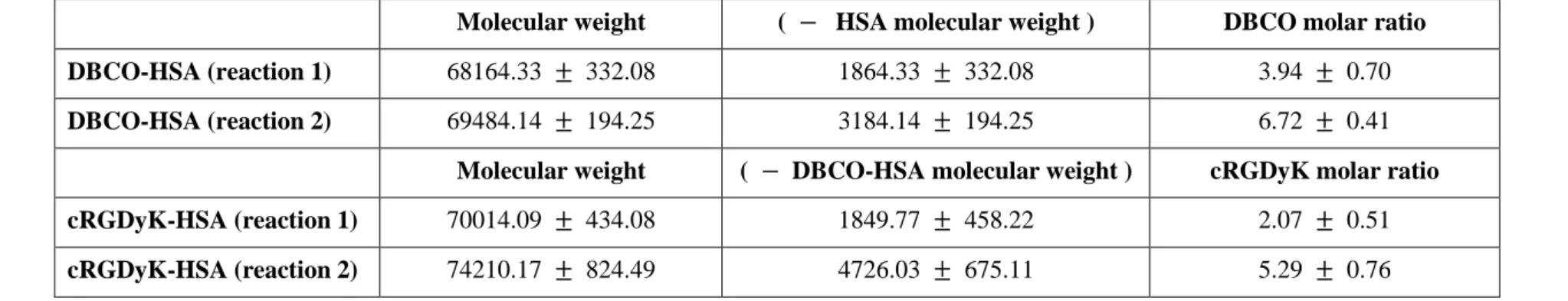
Table 1. Investigation of molecular mass using MALDI-TOF and analysis of MALDI-TOF data
Data are expressed as means ± SDs of three independent experiments.
Molecular mass was measured using MALDI-TOF and the number of DBCO or cRGDyK was calculated as follows;
(Molecular weight of HSA is 66300, DBCO-NHS-ester is 473.48, N3-cRGDyK is 892.69)
DBCO molar ratio = ( 𝑀𝑀𝑀𝑀𝑀𝑀𝑀𝑀𝑀𝑀𝑀𝑀𝑀𝑀𝑀𝑀𝑀𝑀 𝑤𝑤𝑀𝑀𝑤𝑤𝑤𝑤ℎ𝑡𝑡 𝑀𝑀𝑜𝑜 𝑀𝑀𝑀𝑀𝑀𝑀𝑀𝑀𝑀𝑀 − 𝑇𝑇𝑇𝑇𝑇𝑇 ) −( 𝑚𝑚𝑀𝑀𝑀𝑀𝑀𝑀𝑀𝑀𝑀𝑀𝑀𝑀𝑀𝑀𝑀𝑀 𝑤𝑤𝑀𝑀𝑤𝑤𝑤𝑤ℎ𝑡𝑡 𝑀𝑀𝑜𝑜 𝐻𝐻𝐻𝐻𝑀𝑀 ) ( 𝑚𝑚𝑀𝑀𝑀𝑀𝑀𝑀𝑀𝑀𝑀𝑀𝑀𝑀𝑀𝑀𝑀𝑀 𝑤𝑤𝑀𝑀𝑤𝑤𝑤𝑤ℎ𝑡𝑡 𝑀𝑀𝑜𝑜 𝑀𝑀𝐷𝐷𝐷𝐷𝑇𝑇 − 𝑁𝑁𝐻𝐻𝐻𝐻 𝑀𝑀𝑒𝑒𝑡𝑡𝑀𝑀𝑀𝑀 )
cRGDyK molar ratio = ( 𝑀𝑀𝑀𝑀𝑀𝑀𝑀𝑀𝑀𝑀𝑀𝑀𝑀𝑀𝑀𝑀𝑀𝑀 𝑤𝑤𝑀𝑀𝑤𝑤𝑤𝑤ℎ𝑡𝑡 𝑀𝑀𝑜𝑜 𝑀𝑀𝑀𝑀𝑀𝑀𝑀𝑀𝑀𝑀 − 𝑇𝑇𝑇𝑇𝑇𝑇 )−( 𝑚𝑚𝑀𝑀𝑀𝑀𝑀𝑀𝑀𝑀𝑀𝑀𝑀𝑀𝑀𝑀𝑀𝑀 𝑤𝑤𝑀𝑀𝑤𝑤𝑤𝑤ℎ𝑡𝑡 𝑀𝑀𝑜𝑜 𝐻𝐻𝐻𝐻𝑀𝑀 ) ( 𝑚𝑚𝑀𝑀𝑀𝑀𝑀𝑀𝑀𝑀𝑀𝑀𝑀𝑀𝑀𝑀𝑀𝑀 𝑤𝑤𝑀𝑀𝑤𝑤𝑤𝑤ℎ𝑡𝑡 𝑀𝑀𝑜𝑜 𝑁𝑁3− 𝑀𝑀𝑐𝑐𝑐𝑐𝑀𝑀𝑐𝑐𝑐𝑐 )
Molecular weight ( − HSA molecular weight ) DBCO molar ratio DBCO-HSA (reaction 1) 68164.33 ± 332.08 1864.33 ± 332.08 3.94 ± 0.70 DBCO-HSA (reaction 2) 69484.14 ± 194.25 3184.14 ± 194.25 6.72 ± 0.41
Molecular weight ( − DBCO-HSA molecular weight ) cRGDyK molar ratio cRGDyK-HSA (reaction 1) 70014.09 ± 434.08 1849.77 ± 458.22 2.07 ± 0.51 cRGDyK-HSA (reaction 2) 74210.17 ± 824.49 4726.03 ± 675.11 5.29 ± 0.76
18
Radiolabeling efficiency and stability of DBCO-HSA and
cRGDyK-HSA
For quantitative in vitro and in vivo study, DBCO-HSA (reaction 1
and reaction 2) and cRGDyK-HSA (reaction 1 and reaction 2) were
radiolabeled with 64Cu. Radiolabeling efficiencies were analyzed by ITLC-SG
with 0.1 M citric acid as a solvent at each step; free 64CuCl2 reached up to Rf
=1.0 (retention factor), 64Cu-3-azidopropyl goes up to Rf=0.7 and 64Cu-HSA or
64Cu-cRGDyK-HSA remained at the origin (Rf=0.0). For using azide-alkyne
cycloaddition, at first, 3-azidopropyl-NOTA was radiolabeled with 64Cu in 50℃
for 10 minutes at heat block. 64Cu-labeled 3-azidopropyl had only one peak at
Rf=0.7 in ITLC-SG, so 3-azidopropyl-NOTA was well radiolabeled with 64Cu
(Figure 2A). 64Cu-labeled 3-azidopropyl was then added to DBCO-HSA
(reaction 1 and reaction 2) and cRGDyK-HSA (reaction 1 and reaction 2) in
37℃, 30 minutes for azide-alkyne reaction. In Figure 2B, both of 64Cu-3-
azidopropyl added DBCO-HSA (reaction 1 and reaction 2) successfully
radiolabeled with 64Cu (Rf=0.0). In case of cRGDyK-HSA, radiolabeling 19
efficiency was 27% for reaction 1 and almost 100% for reaction 2 (Figure 2C).
So, cRGDyK-HSA in reaction 2 were successfully radiolabeled as DBCO-HSA.
For further enhancing the radiolabeling efficiency of cRGDyK-HSA in reaction
1, 64Cu-labeled 3-azidopropyl added cRGDyK-HSA (reaction 1) was incubated
in 4℃ for 16 hours at shaker. After the incubation at 4℃, the radiolabeling
efficiency of 64Cu-3-azidopropyl added cRGDyK-HSA (reaction 1) was
increased (78%, Figure 2C). To purify the 64Cu-HSA or -cRGDyK-HSA, all
products were purified by PD-10 column. After the purification procedure, all
of the radiolabeling efficiency of 64Cu-HSA or -cRGDyK-HSA (reaction 1 and
reaction 2) were 100% (Rf=0.0, Figure 2B, 2C). After the PD-10 purification,
64Cu-cRGDyK-HSA (reaction 1) can be used as the imaging probe for PET
imaging.
To certify the stability of 64Cu-HSA and 64Cu-cRGDyK-HSA (in each
type; reaction 1 and reaction 2) were mixed with filtered human serum in 37℃
shaking incubator. As time goes on, 64Cu-HSA or 64Cu-cRGDyK-HSA
(reaction 1 and reaction 2) remained at the origin (Rf=0.0) and there were no
20
64Cu-labeled 3-azidopropyl or free 64Cu. So, the 64Cu-labeled HSA or
cRGDyK-HSA were stable in human serum after 48 hours.
21
A
B
C
D
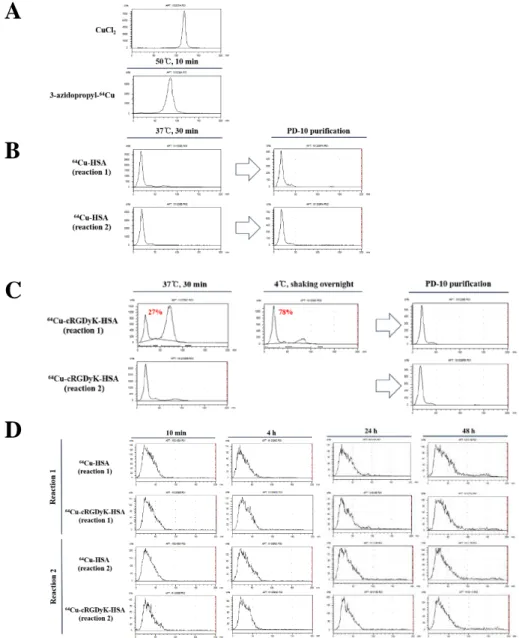
Figure 2. Labeling efficiency and stability of 64Cu-HSA and -cRGDyK-
HSA
(A) ITLC-SG data of 64CuCl2 and 3-azidopropyl-64Cu at 0.1 M citric acid.
64CuCl2 was at Rf=1.0 and 3-azidopropyl-64Cu was at Rf=0.7. (B) 64Cu-HSA
(reaction 1 and reaction 2) were labeled at 37℃ for 30 minutes in heating block.
22
Labeled 64Cu-HSA was at the origin (Rf=0.0). After reaction, 64Cu-HSA
(reaction 1 and reaction 2) were purified with PD-10 column. (C) 64Cu-
cRGDyK-HSA(reaction 1 and reaction 2) were also labeled at 37℃ for 30
minutes in heating block. Labeled 64Cu-cRGDyK-HSA was at Rf=0.0. For
further labeling reaction, after the 37℃ for 30 minutes reaction, 4℃ for
overnight (16 hours) shaking reaction was added to reaction 1. After reaction,
64Cu-cRGDyK-HSA (reaction 1 and reaction 2) were purified with PD-10
column. At each step, the labeling efficiency of 64Cu-HSA and -cRGDyK-HSA
were detected in ITLC-SG at 0.1 M citric acid as the solvent. (D) Serum
stability test of 64Cu-HSA and -cRGDyK-HSA (reaction 1 and reaction 2). The
stability of 64Cu-HSA and –cRGDyK-HSA were detected in ITLC-SG, eluted
with 0.1 M citric acid.
23
Integrinα
Vβ
3expression in cancer cell lines
To select a integrinαVβ3 positive cell lines, integrinαVβ3 mRNA and
protein expression levels in some cancer cell lines were confirmed using RT-
PCR and western blot (Figure 3A, B) RT-PCR data showed that the mRNA
expression levels of integrinαVwere very similar, but the integrin β3 mRNA
was only expressed in PC3, DU145, SK-OV3 cells (Figure 3A). In a case of
western blotting data, integrinαV was also expressed in all cell lines as protein
level, but integrin β3 was only expressed in DU145, SK-OV3 cells (Figure 3B).
According to the mRNA expression (RT-PCR) and protein expression (western
blot) results, SK-OV3 was the highest integrinαVβ3 expressing cell in all
compared cells (KB, 22Rv1, PC3, DU145, SK-OV3).
24
A
B
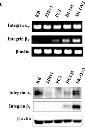
Figure 3. Screening for the integrinαVβ3 expressing cell line
(A) RT-PCR for mRNA expression level analysis. The 319-bp fragments of
αV mRNA were detected at all cell lines, and 396-bp fragments of β3 mRNA
were detected at PC3, DU145, SK-OV3 cells. β-actin was used as an internal
loading control. (B) Western blot for protein expression level analysis.
IntegrinαV proteins were detected at all cell lines and integrin β3 proteins were
25
detected at DU145 and SK-OV3 cells. β-actin was used as an internal loading
control. (Protein size; integrinαV is 135-145 kDa, integrin β3 is 100 kDa)
26
In vitro level integrinα
Vβ
3binding of cRGDyK-HSA
To confirm integrinαVβ3 specific binding of the cRGDyK-HSA in
cellular level, DBCO-HSA and cRGDyK-HSA were conjugated with
fluorescence, FNR648, and visualized using confocal microscopy. There are
two types of DBCO-HSA and cRGDyK-HSA in each case, the difference is the
number of DBCO and cRGDyK; in case of DBCO, reaction 1 is 3.94 ± 0.70
and reaction 2 is 6.72 ± 0.41; in case of cRGDyK, reaction 1 is 2.07 ± 0.51,
reaction 2 is 5.29 ± 0.76 (Table 1). After the fluorescence labeling to DBCO-
HSA and cRGDyK-HSA, FNR648 labeled HSA and cRGDyK-HSA were
purified with PD-10 column. At figure 4A, fluorescence labeled HSA were
accumulated in cytosol and membrane, but there were no difference in reaction
1 and reaction 2. And when pre-treated excess HSA (500-fold higher amount),
the accumulation in cells were decreased in all reactions. (Figure 4A). To
certify cRGDyK-HSA targeting integrinαVβ3, FNR648 labeled cRGDyK-
HSA (reaction 1 and reaction 2) were treated and there are two groups of
confocal images; one is cells treated with only FNR648 conjugated cRGDyK- 27
HSA, the other is cells pre-treated with excess cRGDyK and after then treated
with FNR648-cRGDyK-HSA. Reaction 1 and reaction 2 of the FNR648-
cRGDyK-HSA were localized in the cell membrane and the intracellular
regions. And in reaction 2 of FNR648-cRGDyK-HSA, the accumulation of
FNR648 labeled cRGDyK-HSA were higher than that of in reaction 1 (Figure
4B). And in excess cRGDyK (500-fold higher) pre-treated groups, compared
to FNR648-cRGDyK-HSA only treated groups, very small amount of FNR648-
labeled particles were found in the cell membrane or the intracellular regions
(Figure 4B). Also, the decreased amounts of cRGDyK-HSA were higher in
reaction 2. Compared fluorescence labeled HSA treated groups with cRGDyK-
HSA treated groups, in reaction 1, there were almost no difference in
accumulation of probes. But in reaction 2, as cRGDyK were conjugated to HSA,
more probes were accumulated in cells. This results demonstrate that the
cRGDyK-HSA bound to integrinαVβ3 and entered the cells via integrinαVβ3-
receptor-mediated endocytosis and when cRGDyK were more conjugated to
28
HSA, the accumulation of probes were increased by cRGDyK targeting
integrinαVβ3.
29
A
30
B
31
Figure 4. Cell binding and internalization of cRGDyK-HSA
(A) SK-OV3 cells were treated with fluorescence (FNR648) labeled HSA (2
nmole) for 30 minutes. To certify FNR648 labeled HSA were bound to cells by
HSA, excess HSA (500-fold, 1000 nmole) were pre-treated for blocking.
(B) SK-OV3 cells were treated with FNR648 labeled cRGDyK-HSA (2 nmole)
for 30 minutes. For certifying that cRGDyK-HSA is targeting integrinαVβ3,
excess cRGDyK (500-fold) were pre-treated for blocking groups. In case of
cRGDyK-HSA only treated groups, FNR648-labeled cRGDyK-HSA were
located in cell membrane and intracellular regions. On the other hand, in
cRGDyK pre-treated groups, there were very small amount of cRGDyK-HSA
were observed in cells. And compared reaction 1 and reaction 2 in cRGDyK
blocking, accumulation were more decreased in reaction 2. (Scale bar is 75㎛)
32
In vitro uptake of
64Cu-HSA and
64Cu-cRGDyK-HSA
To investigate the radiolabeled cRGDyK-HSA also specifically bind
to cells, cell uptake test were conducted. 64Cu labeled HSA and cRGDyK-HSA
were treated to two cell lines, one is 22Rv1, which is integrinαVβ3 negative
cell line, the other is SK-OV3, which is integrinαVβ3 positive cell line (Figure
3). Cells were treated with 64Cu-HSA or –cRGDyK-HSA (reaction 1 and
reaction 2). Cell uptake values were divided to treated source counts per minute
(CPM) and changed as % values for comparing all different probes. And all
values were expressed as means ± SDs of four samples. The uptake
percentage of 64Cu-cRGDyK-HSA in reaction 1 was 0.086 ± 0.01 % for
22Rv1 and 0.45 ± 0.06 % for SK-OV3. The uptake percentage of 64Cu-
cRGDyK-HSA in reaction 2 was 0.065 ± 0.017 % for 22Rv1 and 0.38 ±
0.04 % for SK-OV3. In all reaction, 64Cu-cRGDyK-HSA showed higher uptake
in SK-OV3 cells, integrin αVβ3 specific uptake (reaction 1, P = 0.0159 and
reaction 2, P = 0.0357, Figure 5). In 64Cu-HSA, two cell lines had very similar
uptake percentage values (reaction 1; 22Rv1 was 0.23 ± 0.06 %, SK-OV3 was 33
0.24 ± 0.08 %. Reaction 2; 22Rv1 was 0.20 ± 0.12 %, SK-OV3 was 0.33 ±
0.05 %. In each reaction, compared the value of 22Rv1 and SK-OV3, P > 0.1),
so there were no differences in 64Cu-HSA uptake in these two cell lines. From
this results, 64Cu-labeled cRGDyK-HSA were integrinαVβ3 specifically taken
up into the cells.
34
Figure 5. In vitro uptake test of 64Cu-HSA and -cRGDyK-HSA
Uptake percentage of 22Rv1 and SK-OV3 cells. Cells were treated with 64Cu
labeled HSA or cRGDyK-HSA for 1 h at 37℃. The values are expressed as
uptake %, each CPM values were divided by the CPM of applied radioactivity
and changed as percentage value. Each values are means of four samples ±
SD (N = 4). Each values (uptake percentage) were written in results paragraph.
35
Small animal PET imaging of
64Cu-HSA and
64Cu-
cRGDyK-HSA
There are differences in reaction 1 and reaction 2, for example, in case
of HSA, each reaction has different number of DBCO (DBCO-HSA; reaction
1 was 3.94 ± 0.70 and reaction 2 was 6.72 ± 0.41, Table 1). DBCO is linker for
cRGDyK conjugation in HSA, reaction 2 have more cRGDyK than reaction 1
(cRGDyK-HSA; reaction 1 was 2.07 ± 0.51 and reaction 2 was 5.29 ± 0.76,
Table 1). So, to investigate the difference in biodistribution in case of DBCO-
HSA and cRGDyK-HSA or the biodistribution pattern were affected by the
number of DBCO or cRGDyK conjugated to HSA, 64Cu-labeled HSA and -
cRGDyK-HSA (each had reaction 1 and reaction 2) were injected to tumor
bearing mice (SK-OV3, integrinαVβ3 positive) via tail vein. PET images were
acquired at 10 min, 4 h, 24 h, 48 h post injection (p.i.) and shown in Figure 5A.
Compared to reaction 1 and reaction 2 of 64Cu-HSA, blood vessels were visible
to 24 h p.i. in reaction 1 and were to 4 h p.i. in reaction 2. When compared
reaction 1 of 64Cu-HSA and -cRGDyK-HSA, at 24 h p.i., 64Cu-HSA could 36
recognize blood vessels but 64Cu-cRGDyK-HSA could not. In case of reaction
2, blood vessels were well visible in 64Cu-HSA, but not visible in 64Cu-
cRGDyK-HSA at 4 h p.i.. In the aspect of tumor targeting, all 64Cu-HSA and –
cRGDyK-HSA (reaction 1 and reaction 2), the highest accumulation in tumor
were observed at 4 h p.i. and after that time point, tumor accumulation were
decreased. When compared 64Cu-HSA and -cRGDyK-HSA, because cRGDyK-
HSA have targeting moiety, cRGDyK, 64Cu-cRGDyK-HSA were more
accumulated in tumor than 64Cu-HSA. And the highest accumulation was
observed in reaction 2 of 64Cu-cRGDyK-HSA at all time points (Figure 6A and
6B). Quantification analysis in tumor also revealed reaction 2 of 64Cu-
cRGDyK-HSA had the highest %ID/g (% injected dose per gram) value, 5.37
± 1.09 %ID/g, at 4 h p.i.. And at all time points, %ID/g value of 64Cu-
cRGDyK-HSA in reaction 2 was higher than that of others. From this results,
when HSA conjugated with cRGDyK, the blood circulation time were
decreased, but reaction 2 of cRGDyK-HSA can use to target integrinαVβ3 high
expressing tumor.
37
A
B
38
Figure 6. In vivo tumor imaging of 64Cu-HSA and 64Cu-cRGDyK-HSA
(A) Representative small animal PET images in SK-OV3 tumor bearing nude
mice. Maximum intensity projection (MIP) images at 10 min, 4 h, 24 h, 48 h
after tail vein injection of 64Cu-HSA and -cRGDyK-HSA. Yellow circle
indicates tumor region. (B) Time versus %ID/g curves from PET images at
tumor site. Each time points %ID/g values are mean of two values ± SD (N =
2). The tumor %ID/g of 64Cu-cRGDyK-HSA (reaction 2) was the highest at 4
h p.i. and was about 1.6-fold higher than that of 64Cu-HSA (reaction 2).
39
DISCUSSION
RGD has been used in many tumor targeting imaging, to target
integrinαVβ3 (7). For tumor growth more than 1-2 mm in diameter, pro-
angiogenic switch on, integrinαVβ3 is overexpressed in tumors (20). But RGD
is small peptides, so fast clearance through the kidney system is a problem for
targeting the tumor. Aim of this study was to develop HSA-loaded cRGDyK
nanoparticles for targeting tumor. By using HSA, I expected to improve the
half-lives of RGD and the ability of tumor targeting. To conjugate RGD to HSA,
click reaction was used in favorable condition (pH 7, 37℃).
In radiolabeling process at representative radiolabeling data (Figure
2B), cRGDyK-HSA in reaction 1 showed relatively low radiolabeling
efficiencies. The 3-azidopropyl was conjugated to cRGDyK-HSA via click
reaction, but the number of DBCO left to click reaction for radiolabeling was
low (approximately reaction 1; 1-2, calculated from Table 1). If the number of
DBCO will be increased, then DBCO will be left more for conjugating 3-
azidopropyl-64Cu after N3-cRGDyK conjugation. Consider the structure of 40
cRGDyK, this peptide is cyclic form, cRGDyK may conceal DBCO which
were left. Using the linker, such as polyethylene glycol (PEG), linker
conjugated cRGDyK can have some distance from the HSA and DBCO, then
radiolabeling efficiencies can be increased. Some researchers using RGD
peptides with linkers, tumor targeting of RGD peptides were enhanced (21, 22,
23). Although those studies were not conjugated RGD to a carriers, using the
linker conjugated cRGDyK may enhance the targeting efficiencies of
cRGDyK-HSA.
There is a study about changing the number of RGD (24). In this study,
there were two probes, one is dimer-RGD and the other is tetramer-RGD. When
compared with dimer- and tetramer-RGD in in vivo, because these probes have
different number of RGD, tetramer-RGD probes were more accumulated in
tumors. Also, this research group also accomplished tetramer- and octamer-
RGD probes imaging, to compare tumor targeting efficiencies (25). In this
study, the tumor accumulation at initial time points were very similar, but when
compared at later time points, octamer-RGD probes were retain their tumor
41
accumulation percentage, but in case of tetramer-RGD, tumor accumulation
were decreased. From these papers, the number of RGD can affect the
characteristics of probes and also change tumor targeting efficiency. In my
paper, from PET images, reaction 2 of cRGDyK-HSA had the highest %ID/g
value to tumor compared to the others (Figure 6A). So, using cRGDyK-HSA
with more cRGDyK can enhance tumor targeting more efficiently or retain the
accumulation of the probes at tumor. For more cRGDyK-HSA additions,
conjugating more cRGDyK to HSA (for this, it maybe need more DBCO should
be conjugated to HSA) and certifying there is any difference more cRGDyK in
HSA for targeting integrinαVβ3 positive tumor should be accomplished.
In this study, DBCO-HSA and cRGDyK-HSA were successfully
synthesized. And as the molar ratio of DBCO or cRGDyK were different, the
number of conjugated DBCO or cRGDyK to HSA were consistently different.
Using click reaction, 64Cu was successfully labeled to HSA and cRGDyK-HSA.
cRGDyK-HSA could bind to integrinαVβ3 in tumor cells and the probes were
internalized to cells. And the 64Cu-labeled cRGDyK-HSA were certainly
42
accumulated in integrinαVβ3 positive tumor cells. In vivo PET imaging results
probed that the 64Cu-cRGDyK-HSA could target tumor after 4 h p.i.. These
promising results demonstrate that 64Cu-labeled cRGDyK-HSA can be used as
PET tumor imaging probes, which will be useful for various diagnostic
applications.
43
REFERENCES
(1-25)
1. Carmeliet P, Jain RK. Angiogenesis in cancer and other diseases.
Nature. 2000;407(6801):249-57.
2. Brooks PC, Clark RA, Cheresh DA. Requirement of vascular integrin
alpha v beta 3 for angiogenesis. Science. 1994;264(5158):569-71.
3. Costouros NG, Diehn FE, Libutti SK. Molecular imaging of tumor
angiogenesis. Journal of cellular biochemistry Supplement. 2002;39:72-8.
4. Desgrosellier JS, Cheresh DA. Integrins in cancer: biological
implications and therapeutic opportunities. Nature reviews Cancer.
2010;10(1):9-22.
5. Pierschbacher MD, Ruoslahti E. Variants of the cell recognition site
of fibronectin that retain attachment-promoting activity. Proceedings of the
National Academy of Sciences of the United States of America.
1984;81(19):5985-8.
6. Pierschbacher MD, Ruoslahti E. Cell Attachment Activity of
Fibronectin Can Be Duplicated by Small Synthetic Fragments of the Molecule.
44
Nature. 1984;309(5963):30-3.
7. D'Andrea LD, Del Gatto A, Pedone C, Benedetti E. Peptide-based
molecules in angiogenesis. Chemical biology & drug design. 2006;67(2):115-
26.
8. Yang G, Nie P, Kong Y, Sun H, Hou G, Han J. MicroPET imaging of
tumor angiogenesis and monitoring on antiangiogenic therapy with an (18)F
labeled RGD-based probe in SKOV-3 xenograft-bearing mice. Tumour
biology : the journal of the International Society for Oncodevelopmental
Biology and Medicine. 2015;36(5):3285-91.
9. Liu Z, Jia B, Shi J, Jin X, Zhao H, Li F, et al. Tumor uptake of the
RGD dimeric probe (99m)Tc-G3-2P4-RGD2 is correlated with integrin
alphavbeta3 expressed on both tumor cells and neovasculature. Bioconjugate
chemistry. 2010;21(3):548-55.
10. Wu Y, Zhang XZ, Xiong ZM, Cheng Z, Fisher DR, Liu S, et al.
microPET imaging of glioma integrin alpha(V)beta(3) expression using Cu-64-
labeled tetrameric RGD peptide. J Nucl Med. 2005;46(10):1707-18.
11. He XM, Carter DC. Atomic structure and chemistry of human serum 45
albumin. Nature. 1992;358(6383):209-15.
12. Langer K, Anhorn MG, Steinhauser I, Dreis S, Celebi D, Schrickel I,
et al. Human serum albumin (HSA) nanoparticles: Reproducibility of
preparation process and kinetics of enzymatic degradation. Int J Pharm.
2008;347(1-2):109-17.
13. Gradishar WJ, Tjulandin S, Davidson N, Shaw H, Desai N, Bhar P, et
al. Phase III trial of nanoparticle albumin-bound paclitaxel compared with
polyethylated castor oil-based paclitaxel in women with breast cancer. Journal
of clinical oncology : official journal of the American Society of Clinical
Oncology. 2005;23(31):7794-803.
14. Kottschade LA, Suman VJ, Amatruda T, 3rd, McWilliams RR, Mattar
BI, Nikcevich DA, et al. A phase II trial of nab-paclitaxel (ABI-007) and
carboplatin in patients with unresectable stage IV melanoma : a North Central
Cancer Treatment Group Study, N057E(1). Cancer. 2011;117(8):1704-10.
15. Dijkgraaf I, Liu S, Kruijtzer JA, Soede AC, Oyen WJ, Liskamp RM,
et al. Effects of linker variation on the in vitro and in vivo characteristics of an
111In-labeled RGD peptide. Nuclear medicine and biology. 2007;34(1):29-35.
46
16. Vegt E, de Jong M, Wetzels JF, Masereeuw R, Melis M, Oyen WJ, et
al. Renal toxicity of radiolabeled peptides and antibody fragments: mechanisms,
impact on radionuclide therapy, and strategies for prevention. J Nucl Med.
2010;51(7):1049-58.
17. Gaertner FC, Kessler H, Wester HJ, Schwaiger M, Beer AJ.
Radiolabelled RGD peptides for imaging and therapy. European journal of
nuclear medicine and molecular imaging. 2012;39 Suppl 1:S126-38.
18. Jewett JC, Bertozzi CR. Cu-free click cycloaddition reactions in
chemical biology. Chemical Society reviews. 2010;39(4):1272-9.
19. Sletten EM, Bertozzi CR. Bioorthogonal chemistry: fishing for
selectivity in a sea of functionality. Angewandte Chemie. 2009;48(38):6974-
98.
20. Folkman J. Tumor angiogenesis: therapeutic implications. The New
England journal of medicine. 1971;285(21):1182-6.
21. Liu Z, Niu G, Shi J, Liu S, Wang F, Liu S, et al. (68)Ga-labeled cyclic
RGD dimers with Gly3 and PEG4 linkers: promising agents for tumor integrin
alphavbeta3 PET imaging. European journal of nuclear medicine and molecular 47
imaging. 2009;36(6):947-57.
22. Liu Z, Liu S, Wang F, Liu S, Chen X. Noninvasive imaging of tumor
integrin expression using (18)F-labeled RGD dimer peptide with PEG (4)
linkers. European journal of nuclear medicine and molecular imaging.
2009;36(8):1296-307.
23. Shi J, Kim YS, Zhai S, Liu Z, Chen X, Liu S. Improving tumor uptake
and pharmacokinetics of (64)Cu-labeled cyclic RGD peptide dimers with Gly(3)
and PEG(4) linkers. Bioconjugate chemistry. 2009;20(4):750-9.
24. Wu Y, Zhang X, Xiong Z, Cheng Z, Fisher DR, Liu S, et al. microPET
imaging of glioma integrin {alpha}v{beta}3 expression using (64)Cu-labeled
tetrameric RGD peptide. J Nucl Med. 2005;46(10):1707-18.
25. Li ZB, Cai W, Cao Q, Chen K, Wu Z, He L, et al. (64)Cu-labeled
tetrameric and octameric RGD peptides for small-animal PET of tumor
alpha(v)beta(3) integrin expression. J Nucl Med. 2007;48(7):1162-71.
48
국문 초록
서론: RGD는 인테그린 αVβ3를 표적으로 하는 잘 알려진 펩타 이드이며, 이를 이용한 종양의 영상화는 매우 효과적이다. 그 러나, RGD는 매우 짧은 체내 순환시간을 가지며 주입된 대부 분이 신장이나 간담도계를 통해 빠져나간다. 본 연구에서는 RGD의 생체 내 반감기 및 종양 표적을 향상시키기 위해서, 클릭 반응을 통해 cRGDyK를 알부민에 접합시켰다. 또한 가 장 적합한 RGD 접합 알부민 나노입자를 개발하기 위해서 두 가지 종류의 cRGDyK가 접합된 알부민을 합성하였고, 이 cRGDyK-알부민의 특징을 확인하였으며, 세포수준과 생체내 에서 인테그린 αVβ3를 발현하는 종양을 표적화 할 수 있는지 를 확인하였다.
방법: 알부민 : DBCO-NHS 에스터 물수 비율을 기준으로 반 응 1번은 1 : 5.62, 반응 2번은 1: 11.24로 알부민과 DBCO- NHS 에스터를 클릭 반응을 이용해 결합시켰다. 그리고 DBCO-알부민 : N3-cRGDyK 몰수 비율을 기준으로 반응
49
1번은 1 : 3, 반응 2번은 1 : 6으로 DBCO-알부민과 N3- cRGDyK의 결합반응을 진행했다. 동위원소 표지를 위해, 방사 성 구리-64가 붙은 3-azidopropyl-NOTA를 DBCO-알부 민과 cRGDyK-알부민에 클릭반응을 통해 결합시켰다. 각각의 결합 반응 후에는 반응물을 인산완충생리식염수를 용매로 다 공성컬럼인 PD-10을 이용해 분리했다. 모든 반응물은 질량분 석기를 이용해 분석하였으며, 방사성 표지 효율은 크로마토그 래피 방법으로 확인하였다. 방사성 구리-64가 붙은 알부민과 cRGDyK-알부민의 안정성을 확인하기위해, 이들 각각을 혈청 과 섞어주고 시간별로 크로마토그래피로 분석하였다.
cRGDyK-알부민이 인테그린αVβ3를 세포 수준에서 표적할 수 있는지 확인하기 위해 FNR648을 붙인 cRGDyK-알부민 을 공초점 현미경을 통해 영상화하였고, 구리-64가 붙은 cRGDyK-HSA로 세포 업테이크를 확인하였다. 이후 방사성 구리-64가 붙은 알부민과 cRGDyK-알부민을 종양을 가지고 있는 마우스에 정맥을 통해 투여하였으며, 소동물용 양전자단 층촬영 기기를 이용하여 정맥 투여 후 10 분, 4 시간, 24 시간,
50
48 시간에 마우스를 영상화 하였다.
결과: DBCO-NHS 에스터와 cRGDyK는 반응시킨 몰수에 따 라 성공적으로 알부민에 결합하였다. DBCO의 경우, 알부민에
붙은 DBCO의 개수는 반응 1의 경우 3.94 ± 0.70개, 반응 2 의 경우 6.72 ± 0.41개였다. cRGDyK의 경우, 알부민에 붙은 cRGDyK의 개수는 반응 1의 경우 2.07 ± 0.51개, 반응 2의 경우 5.29 ± 0.76개였다. 방사성 구리-64-알부민과 cRGDyK-알부민 중 반응 2번의 표지효율은 100%였으며, cRGDyK-알부민 중 반응 1번의 경우에는 PD-10으로 분리 해 낸 이후 거의 100% 표지효율을 보였다. 구리-64-알부민 과 cRGDyK-알부민은 혈청과 섞어준 이후 48 시간이 지나도 안정적으로 결합하여 있는 것을 확인할 수 있었다. 공초점현미 경 영상에서는, FNR648이 붙은 cRGDyK-알부민은 세포막과 세포 내 영역에 존재하였으며, cRGDyK-알부민의 500배의 cRGDyK를 우선 세포에 처리해주었을 때 cRGDyK-알부민의 이런 분포는 보이지 않았다. 구리-64가 붙은 cRGDyK-알부 민의 세포내 섭취는 22Rv1 세포 (인테그린αVβ3 비발현)에
51
비해 SK-OV3 세포(인테그린αVβ3 과발현)에서 확연히 높았 다. 양전자단층촬영 영상에서는, 구리64-알부민과 cRGDyK- 알부민의 종양 섭취가 정맥 주입 후 4 시간까지 증가하였으며, 이 시간대에 cRGDyK-알부민 반응 2번이 가장 높은 종양 내 섭취를 보였다.
결론: 이 연구에서 DBCO-알부민과 cRGDyK-알부민을 성공 적으로 합성하였고, 클릭 반응을 이용하여 방사성 구리-64를 알부민과 cRGDyK-알부민에 성공적으로 표지하였다.
cRGDyK-알부민은 인테그린 αVβ3를 종양 세포에서 결합할 뿐만 아니라 생체 수준에서도 효과적으로 종양을 표적하는 것 을 영상적으로 확인할 수 있었다. 따라서, 방사성 구리-64가 표지된 cRGDyK-알부민은 종양 영상 표적제로 사용할 수 있 을 것이다.
--- 주요어: 인테그린αVβ3, RGD, 알부민, 생물직교 클릭 반응, 양전자 단 층 촬영 영상, 구리-64
학 번: 2014-21152
52