저작자표시-비영리-변경금지 2.0 대한민국 이용자는 아래의 조건을 따르는 경우에 한하여 자유롭게
l 이 저작물을 복제, 배포, 전송, 전시, 공연 및 방송할 수 있습니다. 다음과 같은 조건을 따라야 합니다:
l 귀하는, 이 저작물의 재이용이나 배포의 경우, 이 저작물에 적용된 이용허락조건 을 명확하게 나타내어야 합니다.
l 저작권자로부터 별도의 허가를 받으면 이러한 조건들은 적용되지 않습니다.
저작권법에 따른 이용자의 권리는 위의 내용에 의하여 영향을 받지 않습니다. 이것은 이용허락규약(Legal Code)을 이해하기 쉽게 요약한 것입니다.
Disclaimer
저작자표시. 귀하는 원저작자를 표시하여야 합니다.
비영리. 귀하는 이 저작물을 영리 목적으로 이용할 수 없습니다.
변경금지. 귀하는 이 저작물을 개작, 변형 또는 가공할 수 없습니다.
이학박사학위논문
HAC-NPR1-TGA 복합체에 의한 애기장대 면역의 후성유전학적 조절
Epigenetic regulation of Arabidopsis immunity by the HAC-NPR1-TGA Complex
2018년 02월
서울대학교 대학원 생명과학부
김 홍 실
i
Abstract
Epigenetic regulation of Arabidopsis immunity by the HAC-NPR1-TGA Complex
HongShi Jin Department of Biological Sciences The Graduate School
Seoul National University
Unlike animals, plants lack specialized immune cells. Instead, plants have developed multiple layers of sophisticated immune responses through massive expression of immune-related genes including the pathogenesis related (PR) genes to respond to bacterial or viral pathogenic attacks. Upon pathogenic attack, plants turn on the innate immune system as the first line of defense. The immune response is initiated at the site of infection by accumulating salicylic acid (SA). Then, as a major signaling molecule in plant immunity, SA is accumulated at distal tissues to protect the entire plant against successive attacks by various pathogen. This “whole plant” resistance response is referred as systemic acquired resistance (SAR). SA
ii
signal results in the activation of the master immune regulator, NPR1, which is recruited by TGACG SEQUENCE-SPECIFIC BINDING PROTEIN (TGA) transcription factors to numerous downstream PR genes. However, despite the critical role of NPR1 in SA-triggered immunity, the biochemical mechanism of NPR1 as a transcriptional co-activator remain largely unknown.
Epigenetics is the study of heritable changes in gene expression which are not based on changes in DNA sequence. Histone acetylation is an epigenetic modification that occurs at the lysine residue of N-terminal histone tail. Histone acetyltransferases (HATs) transfer the acetyl group (COCH3) from acetyl coenzyme A (acetyl-CoA) to the NH3+ amino group of histones. HATs are also known as transcription co-activators, leading to transcriptional activation.
In Arabidopsis, recent studies reported that CBP/p300 HAC family proteins possess histone acetyltransferase activities. The two types of zinc finger domains ZnF-TAZ and ZnF-ZZ in CBP/p300 families are known to be important for mediating protein–protein interactions. Epigenetic regulation through several HACs is known to play crucial roles in flowering, various developmental processes, and ethylene signaling pathway.
In this study, I showed that the CBP/p300-family histone acetyltransferases, HAC1 and HAC5 (HAC1/5) are required for SA-triggered immunity and PR induction in Arabidopsis. During SA-triggered immune response, HAC1 form a complex with NPR1 and TGAs to activate PR genes by histone acetylation. Thus, this study reveals the function of HAC1 as a co-activator of NPR1
iii
and the precise biochemical mechanism of NPR1-mediated transcriptional activation.
Furthermore, this study also proposes epigenetic reprogramming acts as an essential part of plant immune system which allows plants to efficiently switch their regular developmental program to a defense program upon pathogenic attack.
Key words: CBP/p300, Histone acetyltransferase, HAC1, NPR1, SA, Immune system, Pathogen.
Student Number: 2008-30703
iv
Contents
Abstract ··· i
Contents ··· iv
List of tables ··· vii
List of figures ··· viii
Abbreviations ··· x
1. General Introduction ··· 1
1.1 Epigenetic control and gene expression ··· 1
1.1.1 Histone modification ··· 1
1.1.1.1 Histone acetylation ··· 4
1.1.1.2 Histone deacetylation··· 7
1.1.2 DNA methylation ··· 10
1.1.3 ATP-dependent chromatin remodeling ··· 12
1.1.4 Noncoding RNA-mediated silencing ··· 13
1.2 The plant immune system ··· 14
1.2.1 The innate immune system of plant ··· 15
1.2.2 PAMP-triggered immunity in Arabidopsis ··· 16
1.2.3 Systemic acquired resistance (SAR) and salicylic acid (SA)
v
signaling ··· 16
1.2.4 NON-EXPRESSER OF PR GENES1 (NPR1) ··· 18
1.2.5
PATHOGENESIS-RELATED (PR) GENES··· 21
1.2.6 TGA transcription factors ··· 22
1.2.7 SA-JA-ET crosstalk in plant immune response ··· 23
1.2.8 Epigenetic control of the SA-dependent defense ··· 25
2. Epigenetic reprogramming by the HAC-NPR1-TGA complex confers immunity in Arabidopsis ··· 30
2.1 Materials and methods ··· 31
2.1.1 Plant materials and growth conditions ··· 31
2.1.2 Pathogen infection ··· 31
2.1.3 Plasmid construction ··· 31
2.1.4 Protein purification and immunoblotting ··· 32
2.1.5 Co-IP assay ··· 33
2.1.6 Yeast-Two-Hybrid assay ··· 33
2.1.7 Gel filtration assay ··· 34
2.1.8 RNA extraction and RT-qPCR analysis ··· 35
2.1.9 ChIP assay ··· 35
vi
2.1.10 RNA sequencing analysis ··· 35
2.1.11 ChIP sequencing analysis ··· 36
2.1.12 Sequential ChIP assay··· 36
2.1.13 Confocal Microscopy ··· 37
2.2 Text ··· 51
2.2.1 The H3Ac increase at PR1 is undermined by the loss of either NPR1 or the three related Class II TGAs ··· 52
2.2.2 HACs activate SA-dependent plant immunity by promoting PR transcription through histone acetylation ··· 53
2.2.3 HAC1, NPR1, and TGA2 form a complex in Arabidopsis ··· 53
2.2.4 HAC1 and TGA2/5 do not interact directly but indirectly through NPR1 ··· 55
2.2.5 HAC1, NPR1, and TGA2/5 may form a multi-protein complex ··· 56
2.2.6 HAC-NPR1-TGA complex constitutes part of the genome-wide transcriptional activator system acting in plant immunity ··· 58
2.3 Figures ··· 59
References ··· 91
Abstract in Korean ··· 109
vii
List of tables
Table 1-1 HAT and HDAC family in Arabidopsis thaliana ··· 27
Table 2-1 List of all transgenic or multiple-mutant plants used in this study ··· 38
Table 2-2 Primers used for HAC1:HA, NPR1:GFP, and TGA2:FLAG constructs ··· 40
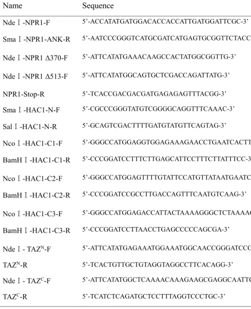
Table 2-3 Primers used for Yeast-Two-Hybrid constructs ··· 41
Table 2-4 Primers used for RT-qPCR analyses ··· 42
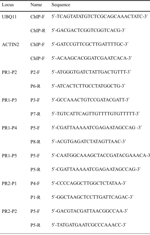
Table 2-5 Primers used for ChIP assays ··· 48
viii
1.
List of figures
2.
Figure 1-1 CBP/p300 HAC family in Arabidopsis thaliana ··· 28 Figure 1-2 The family of TGA transcription factors in Arabidopsis thaliana ··· 29
Figure 2-1 HAC1 and HAC5 are essential for PR1 transcription and plant immunity ··· 59 Figure 2-2 HAC1/5 regulate pathogen-induced PR2 transcription and
histone acetylation ··· 61 Figure 2-3 SA signal induces concurrent targeting of HAC1 and
NPR1 to PR1 ··· 63
Figure 2-4 NPR1 enrichment within PR1 chromatin in35S::NPR1:GFP
npr1-1plants either treated with INA or not ··· 65
Figure 2-5 Subcellular localization of HAC1, NPR1, and TGA2/5 ···· 67
Figure 2-6
In vivo interaction of HAC1 with NPR1 and TGA2/5 ··· 69Figure 2-7
In vivo interactions among HAC1, NPR1, and TGA2/5 ···· 72Figure 2-8 Interaction between HAC1 and NPR1 in yeast ··· 74
ix
Figure 2-9 Targeting of HAC1 and NPR1 to PR1 chromatin requires TGA2/5/6 ··· 76 Figure 2-10 Fractionation of the HAC-NPR1-TGA complex and its role
in the regulation of SA-induced transcriptome and
epigenome dynamics ··· 78 Figure 2-11 RT-qPCR analysis of randomly selected 22 Group 1-gene expression in Col, npr1-1, and hac1-2 hac5-2 treated with INA or not ··· 81 Figure 2-12 RT-qPCR analysis of randomly selected 21 Group 2-gene
expression in Col, npr1-1, and hac1-2 hac5-2 treated
with INA or not ··· 84
Figure 2-13 Visualization and confirmation of H3Ac ChIP-seq data ·· 87
Figure 2-14 Model for the epigenetic reprogramming of PR genes by
the HAC-NPR1-TGA complex ··· 89
x
3.
Abbreviations
CBP CREB-binding protein
ChIP chromatin immunoprecipitation
ChIP-seq chromatin immunoprecipitation
followed by sequencing Co-IP assay Co-immunoprecipitation assay
DNA deoxyribonucleic acid
EDTA ethylene diaminetetraacetic acid
FDR false discovery rate
GFP green fluorescence protein
HA hemagglutinin
HACs histone acetyltransferase
HAG1 histone acetyltransferase GCN5
HAT histone acetyltransferase
INA 2,6-Dichloroisonicotinic acid
KD kilo dalton
LD long day
xi
MG132 carbobenzoxy-Leu-Leu-leucinal
mRNA messenger ribonucleic acid
NPR1 nonexpresser of PR genes 1
OD optical density
PAGE polyacrylamide gel
PCR polymerase chain reaction
PI propidium iodide
PR
pathogenesis-related gene
Pst DC3000
P. syringae pathovar tomato DC3000
qPCR quantitative polymerase chain reaction
RNA-seq RNA sequencing
RT reverse-transcription
SA salicylic acid
SD short day
SDS sodium dodecyl sulfate
SE standard error
TAZ domains transcriptional adapter zinc binding
domains
xii
T-DNA transfer DNA
TGA TGACG sequence-specific binding
protein
UBQ ubiquitin
WT wild type
1
1.
1. General introduction
2.
1.1
Epigenetic control and gene expression
Epigenetics is the study of mitotically and/or meiotically heritable changes in gene expression which are not based on changes in the underlying DNA sequence.
The activity and/or function of various genes can be regulated epigenetically through covalent modifications of DNA, histone proteins, DNA-binding proteins, as well as noncoding RNAs. These epigenetic mechanisms are crucial for nearly all biological processes in eukaryotes.
In eukaryotic cells, genetic information is carried in the chromosomal DNA.
The DNA is associated with various proteins and RNAs to form a structure called
“chromatin". Epigenetic modifications control the structure of chromatin to regulate transcription. The main mechanisms underlying epigenetic regulation include histone modification, DNA methylation, ATP-dependent chromatin remodeling and noncoding RNA-mediated silencing. Epigenetic variations sensitively respond to the environmental and intercellular signals and regulate cue-specific gene expression and thus cause morphological and functional changes in cells. Epigenetic factors are well conserved in eukaryotic organisms and various homologs are identified among plants and animals (Istvan et al., 2013).
1.1.1 Histone modification
The structure of the chromatin undergoes various dynamic changes
2
according to the needs of the cell. The eukaryotic chromatin is comprised of 147 bp of DNA wrapped around histone proteins to form a fundamental structural unit referred as nucleosomes. The core histone protein consists of two H3, H4 histone dimers and two H2A, H2B dimers while histones H1 and H5 act as linker histones (Handy et al., 2011). The core histones are packaged into a globular structure whereas the N-terminal tail of each core histone is exposed from the core histone and loosely extended. Various enzymes modify multiple sites within the histone N- terminal tails resulting in an “open” or “closed” chromatin structure. The open chromatin structure also referred as euchromatin, is the loose chromatin state that is accessible to RNA polymerase and transcription factors resulting in gene activation.
On the other hand, heterochromatin is the condensed chromatin state consisting transcriptionally inactive genes and repetitive sequences. Thus, gene expression can be regulated at the transcriptional level depending on the chromatin structure itself.
Histones are covalently modified in different residues such as lysine, arginine, serine, and threonine. Among them, lysine residues are important substrates for various modifications such as acetylation, methylation, phosphorylation, ubiquitination, and sumoylation. Acetylation and methylation are major histone modifications which occur in the lysine residues of histones H3, H4, H2A, and H2B tails. Histone H3 can be acetylated at several lysine residues including K9, 14, 18, 23, and 5. Histone H3 can also be methylated on K4, 9, 27, 36, 79, and arginine residue (H3R2). In histone H4, K5, 8, 12, and 16 can be acetylated and K20 and R3 residues can be methylated (zhang et al., 2010).
3
Four types of correlations are established based on analyses of genome- wide profiles of histone modifications and gene expression: suppressed, active, poised, and bivalent (Weng et al., 2012). In the suppressed state, the closed chromatin structure results in suppression of gene transcription. In contrast, gene transcription is activated in open chromatin structure. However, even if the chromatin is at open state with high levels of active marks such as H3K4me3, gene transcription can still be low. This chromatin state is referred as the poised state.
When the chromatin is poised, gene transcription can be rapidly activated upon activation signal. The bivalent chromatin state was first identified in developmental gene promoters of embryonic stem cells (Bluma and David, 2014). In bivalent chromatin, both active and repressive marks are present at high levels. The bivalent chromatin state enables the chromatin to change to an open or closed conformation through cell differentiation and upon activation signals (Bluma and David, 2014;
Azuara et al., 2006; Bernstein et al., 2006; Mikkelsen et al., 2007). Among various histone modifications, H3K9me2/3 and H3K27me3 are major repressive markers. In contrast, H3Ac, H4Ac, H3K4me3, and H3K36me3 are active markers. The repressive mark H3K27me3 and the active mark H3K4me3 are the most common bivalent marks found in stem cells. H3K27me3 suppresses gene expression while H3K4me3 mark reactivates them when needed. Various histone modifications can make crosstalks. They affect each other negatively or positively and regulate gene expression.
4
1.1.1.1. Histone acetylation
Histone acetylation was first reported by Allfrey et al in 1964. Histone acetylation occurs at the lysine residue of the N-terminal histone tail. The modifying enzymes that catalyze histone acetylation are called histone acetyltransferases (HATs). HATs transfer the acetyl group (COCH3) from acetyl-coenzyme A (acetyl- CoA) to the NH3+ amino group of histones, mainly H3 and H4. HATs are also known as transcription co-activators.
Histone acetylation reduces the interaction between nucleosomes and DNA.
The decondensed chromatin structure allows access to transcription factors, DNA binding activators, and transcriptional co-activators. Thus, histone acetylation is involved in gene activation. Histone acetylation is involved in the regulation of various cellular processes including chromatin dynamics, differentiation, cell cycle progression, DNA replication, and DNA repair. Furthermore, histone acetylation is also involved transcriptional initiation and elongation (Barski et al., 2007).
HATs are divided into two classes according to their subcellular localization, Type-A and Type-B. Type-A HATs are localized in the nucleus and many of them possess a bromodomain, a specific domain that can recognize acetylated histones. Type-A HATs acts on histone H2A, H2B, H3, and H4. They are further classified into five families based on their sequence homology of their catalytic domains: the CREB-binding protein (CBP/p300) family, Gcn5-related N- acetyltransferases (GNATs), the more recently reported MOZ, Ybf2, Sas2, and Tip60 (MYST) family, TATA-binding protein-associated factor (TAFII250), and the
5
nuclear receptor co-activators (ACTR)/steroid receptor co-activators (SRC) family (Yasuto et al., 2017; Wang et al., 2009). The CBP/p300 family HATs generally act as transcriptional co-activators in the regulation of gene expression (Goodman and Smolik, 2000; Das et al., 2009; Wang et al., 2010). They are involved in a wide range of cellular activities such as DNA repair, cell growth, and cell differentiation (Zhang et al., 1998). GNATs are involved in many cellular processes such as cellular growth and cell differentiation. MYST HATs are involved in cell proliferation and transcriptional regulation (Michael et al., 2003). Type-B HATs are localized in the cytoplasm and lack the bromodomain. They acetylate synthesized histone H4K5 and H4K12 as well as the specific site within H3. Type B HATs are also known to acetylate newly synthesized histones H3 and H4 (Mackay et al., 1992).
Four groups HAT family proteins are identified in the Arabidopsis genome (Table 1-1) (Pandey et al., 2002; Liu et al., 2012). Among them, there are five CBP/p300 HAC family members, named HAC1, HAC2, HAC4, HAC5, and HAC12 (Figure 1-1). Arabidopsis HAC1, HAC4, HAC5, and HAC12 possess HAT activities but unlike in animals, HAC2 does not have HAT activity (Li et al., 2014). Previous studies have reported that CBP/p300 HAC proteins can specifically acetylate histones H3 and H4. For instance, HAC1 can specifically acetylate H4K14 and HAC1, HAC5 and HAC12 can acetylate H3K9 (Earley et al., 2007). HAC family proteins also share the CBP-type HAT domain, the partially conserved KIX domain, as well as the partially conserved PHD finger motif. Moreover, two types of zinc finger domains ZnF-TAZ and ZnF-ZZ in HAC family proteins are important for mediating protein-protein interactions (Li et al., 2014).
6
In Arabidopsis,
CBP/p300 HAC proteins are reported to regulate flowering time, ethylene (ET) signaling, and environmental stress-dependent pattern-triggered immunity (PTI) (Han et al., 2006; Li et al., 2014; Singh et al., 2014). HAC1, HAC5, and HAC12 are reported to be functionally redundant in the regulation of flowering. They promote flowering by negatively regulating the expression of FLOWERING LOCUS C (FLC) which is a major floral repressor (Deng et al., 2007; Han et al., 2007). Accordingly, mutation of HAC1 cause late flowering phenotypes, and the late flowering phenotype is enhanced in the hac1/hac5 and hac1/hac12 mutant (Han et al., 2007). HACs are also known to be involved in ET signaling pathway. In hac1hac5 double mutant the transcription level of several ethylene-reactive genes including ERF1, ERF4, ERF6, and ERF11 are significantly increased (Li et al., 2014). HAC1 also plays an important role in UV-B signaling (Fina et al., 2017). Additionally, HAC1 is known to be responsible for bacterial resistance and PTI priming after exposure to environmental stress such as cold, heat, and salt stress (Singh et al., 2014).The GNAT family HATs are named HAG1/GCN5, HAG2/HAT1, and HAG3/ELP3. Arabidopsis HAG1 is involved in floral development, cold tolerance as well as root and shoot development (Benhamed et al., 2006; Bertrand et al., 2003;
Long et al., 2006; Vlachonasios et al., 2003). Two MYST HAT family proteins are named as HAM1/HAG4 and HAM2/HAG5, respectively. Arabidopsis TAFII250 homologs were identified and named as HAF1 and HAF2. HAF2 plays a role in the regulation of light-induced gene expression. Typically, HAF2 is known to function in the regulation of both red/far-red and blue light signaling pathways (Benhamed et
7 al., 2006; Bertrand et al., 2005).
1.1.1.2. Histone deacetylation
Acetylated histones can be reversibly deacetylated by histone deacetylases (HDACs). HDACs alter the compaction of chromatin by removing the acetyl group of lysine residues from both histone and non-histone proteins. The compacted chromatin structure prevents access of transcription factors and RNA polymerases to the target DNA leading to transcriptional repression. Histone acetylation is mainly catalyzed at histone H3K9, 14, 18, and 23 and histone H4K8, 12, 16, and 20 residues (Fuchs et al., 2006).
There are 18 HDACs divided into four classes in higher eukaryotes: Class I (HDAC1, 2, 3, and 8), Class II (HDAC4, 5, 6, 7, 9, and 10), Class III (SIRT1, 2, 3, 4, 5, 6, and 7), and Class IV (HDAC11) (Table 1-1) (Pandey et al., 2002).
In Arabidopsis, HDACs can be classified into three groups based on the sequence similarity (Yang and Seto, 2007). Among 18 Arabidopsis HDAC proteins, 12 HDACs belong to the yeast reduced potassium deficiency (RPD3/HDA1) superfamily, which is called as HDA. The other 2 sirtuins (SRTs) share similarity to the yeast silent information regulator 2 (SIR2). Additionally, 4 members belong to plant-specific histone deacetylase 2 (HD2) family, known as HD-tuins (HDT).
Members of RPD3/HDA1 superfamily are further divided into three classes: Class I (HDA6, 7, 9, 10, 17, 19), Class II (HDA5, 14, 15, 18), and Class III (HDA2) which
8
possess an incomplete HDAC domain (Pandey et al., 2002).
HDACs play important roles in the regulation of various aspects of Arabidopsis life cycle including plant growth, flowering, circadian regulation, seed development, germination, as well as in ET, jasmonic acid (JA), salicylic acid (SA) signaling pathway and basal defense to pathogens (Wang et al., 2014). Among the Class I proteins, HISTONE DEACETYLASE 6 (HDA6), HISTONE DEACETYLASE 9 (HDA9), and HISTONE DEACETYLASE 19 (HDA19) are the most intensely investigated for their biological function. HDA6 is involved in the regulation of flowering time by directly interacting with lysine-specific demethylase 1 type histone demethylase FLOWERING LOCUS D (FLD), FVE/MSI4, and MSI5 (Jiang et al., 2007; Gu et al., 2011).
Additionally, it has been reported that the crosstalk between histone deacetylation and demethylation is mediated by the physical association of HDA6 and FLD (Yu et al., 2011).HDA6 also acts as a negative regulator of the JA signaling pathway. HDA6 is recruited by JASMONATE ZIM-DOMAIN 1 (JAZ1) to repress the expression of ETHYLENE INSENSITIVE 3 (EIN3), thereby inhibiting JA signaling (Zhu et al., 2011). Additionally, HDA6 and HDA19 redundantly co-repress the expression of embryogenesis-related genes such as LEAFY COTYLEDON 1 (LEC1), FUSCA 3 (FUS3), and ABA INSENSITIVE 3 (ABI3) by forming multi- functional complexes with other co-factors (Tanaka et al., 2008). Besides its role in embryogenesis, HDA19 plays a crucial role in plant development and the loss of HDA19 result in various developmental abnormalities (Tanaka et al., 2008). HDA9
9
is involved in the regulation of flowering time by directly targeting AGAMOUS- LIKE 19 (AGL19) and repressing its expression (Kim et al., 2013; Kang et al., 2015).
Moreover, HDA19 functions in SA biosynthesis and regulates expression of SA- regulated defense genes through histone deacetylation. It is also known to be involved in the repression of pathogenesis related 1 (PR1) expression (Choi et al., 2012).
HDA19
is known to be apositive regulator of basal disease resistance in plants and represses the expression of the transcription
factors WRKY DNA- BINDING PROTEIN 38 (WRKY38) and WRKY DNA-BINDING PROTEIN 62 (WRKY62). WRKY38 and WRKY62 are both known to negatively regulate the expression of pathogenesis related (PR)
genes. Accordingly, mutation in
HDA19 abolishes the resistance to Pseudomonas syringae pv. tomato DC3000 (Pst DC3000)(Kim et al., 2008).
HDA15 is involved in the repression of chlorophyll biosynthesis and photosynthesis. HDA15 interacts with PHYTOCHROME INTERACTING FACTOR 3 (PIF3) which act as a negative regulator in light responses. Together, they repress chlorophyll biosynthesis and chlorophyll biosynthesis-dependent gene expression in dark condition (Liu et al., 2013). HDA18 plays a key role in the cell fate control of Arabidopsis root epidermis (Xu et al., 2005; Liu et al., 2013). HD2C associates with HDA6 and enhances the transcription level of abiotic stress- responsive genes, such as ABI1, ABI2, and ERF4. Moreover, it has been demonstrated that HD2C and HDA6 regulate rRNA gene expression through histone modifications (Luo et al., 2012).
10
1.1.2 DNA methylation.
DNA methylation is a widely-studied epigenetic mechanism that is typically involved in transcriptional repression. DNA methylation is involved in a variety of biological processes and is conserved in higher eukaryotic organisms including plants, animals, fungi, budding yeast Saccharomyces cerevisiae, and the nematode worm Caenorhabditis elegans (Colot et al., 1999).
DNA methylation occurs at cytosine residues. The methyl group (-CH3) is added to cytosine bases of the DNA resulting in 5-methylcytosine (5-mC). The methylated status is stable and inherited to the next generation. DNA methylation is usually distributed in CpG islands in animals and plants.
In mammals, DNA methylation mainly occurs in symmetric CG context, although non-CG methylation is ubiquitously distributed in embryonic stem cells. In plants, DNA methylation occurs in the contexts of CG, CHG, and CHH (where H = A, C, or T) (Ramsahoye et al., 2000; Lister et al., 2009). The methyltransferases which catalyze DNA methylation are DNA METHYLTRANSFERASE 1 (DNMT1), DNA (CYTOSINE-5)-METHYLTRANSFERASE 3A (DNMT3A), and DNA (CYTOSINE-5)-METHYLTRANSFERASE 3B (DNMT3B). DNMT3A and DNMT3B are involved in establishing de novo DNA methylation patterns during germ cell development (Zhao and Chen, 2014).
The Arabidopsis genome encodes four classes of DNA methyltransferases,
11
METHYLTRANSFERASE 1 (MET1), CHROMOMETHYLASE 3 (CMT3), DOMAINS REARRANGED METHYLTRANSFERASE 2 (DRM2), and DNA METHYLTRANSFERASE 2 (DNMT2). MET1 is the main CG methyltransferase in Arabidopsis. MET1 is orthologous to the mammalian DNMT1 which contain the bromo-adjacent homology(BAH) domain and function to maintain CG methylation (Simon et al., 2005). In addition to MET1, DECREASE IN DNA METHYLATION 1 (DDM1), a factor involved in SWI2/SNF2-like chromatin remodeling, is also known to regulate CG methylation. Mutation in DDM1 resulted in the loss of CG methylation and H3K9 methylation (Gendrel et al., 2002; Johnson et al., 2002). The histone deacetylase HDA6 is also known to be required to maintain DNA methylation. Mutation in HDA6 resulted in reduced cytosine methylation at CG as well as CHG sites. Moreover, the expression of the targets of RNA-directed DNA methylation (RdDM) pathway, a main siRNA-mediated epigenetic pathway, were derepressed suggesting that HDA6 also function in RdDM (Aufsatz et al., 2002;
Probst et al., 2004; Matzeke and Mosher, 2014). De novo DNA methylation is established by DRM2, an orthologue of the mammalian DNMT3 which is regulated by the RdDM pathway (Cao et al., 2003; Pontes et al., 2006). CMT3 is a plant- specific DNA methyltransferase and is required for the maintenance of DNA methylation at CHG sites (Simon et al., 2005).
Importantly, H3K9 and DNA methylation are known to be closely related.
KRYPTONITE (KYP) and its homologs SU (VAR) 3-9 HOMOLOG 5 (SUVH5) and SU (VAR) 3-9 HOMOLOG 6 (SUVH6) are known as typical H3K9 histone methyltransferases and are required for maintaining CMT3-dependent CHG
12 methylation (Hume et al., 2017).
The removal of the methylated state of DNA, or DNA demethylation, is catalyzed by DNA glycosylases. In Arabidopsis, DEMETER (DME), REPRESSOR OF SILENCING 1 (ROS1), DEMETER-LIKE 2 (DML2), and DEMETER-LIKE 3 (DML3) are known to possess DNA glycosylase activity. They recognize and remove methylated cytosine leading to DNA demethylation (Pilar et al., 2008).
1.1.3 ATP-dependent chromatin remodeling
Various types of ATP-dependent chromatin remodeling complexes play different roles in
eukaryotic cells
. During ATP-dependent remodeling, the position and structure of the nucleosome is altered by sliding, ejecting, or restructuring the nucleosome using the energy obtained from ATP hydrolysis. In this manner, the conformational changes in histone-DNA interaction allow access for transcription factors or recruitment of transcription machinery to the genomic region. In addition, ATP-dependent chromatin remodeling complexes associate with histone chaperones to alter histone H2A-H2B or remove the octameric core from the DNA. In eukaryotes, the ATP-dependent chromatin remodeling complexes are divided into four classes: switching defective/sucrose non-fermenting (SWI/SNF), imitation SWI (ISWI), chromodomain (CHD), and the INO80 groups, respectively (Eisen et al., 1995; Vignali et al., 2000; Weisz et al., 2001; Jerzmanowski et al., 2007).42 SNF2 family ATPases are annotated and categorized into 24 distinct
13
subfamilies in Arabidopsis (Flaus et al., 2006). Among them, PHOTOPERIOD INDEPENDENT EARLY FLOWERING 1 (PIE1) is homologous to SWR1 and is known to play a key role in the repression of floral transition (Noh and Amasino, 2003; Kumar and Wigge, 2010). SPLAYED (SYP) and BRAHMA (BRM) are identified as SNF2 subfamily involved in development and immunity in Arabidopsis (Wagner and Meyerowitz, 2002; Bezhani et al., 2007). DECREASED DNA METHYLATION 1 (DDM1) of the LYMPHOID SPECIFIC HELICASE (LSH) subfamily are also known to function as immune regulators. SYD and BRM have functional redundancy in regulating some defense-related genes (Bezhani et al., 2007; Walley et al., 2008). In addition, the expression of the SA-responsive gene PR1 is increased in syd-2 mutant upon Pst DC3000 infection indicating that SYD negatively regulates SA pathway (Walley et al., 2008).
1.1.4
Noncoding RNA-mediated silencing
Recent studies using genome-wide analysis have shown that 90% of the eukaryotic genome is transcribed. However, only 1-2% of the genome encodes proteins. This implies that a very large number of RNAs do not have the potential to encode protein. This type of RNAs is called noncoding RNAs (ncRNAs). NcRNAs are transcribed from the intergenic region or antisense strand of protein-coding genes.
They regulate the expression of their target genes in an epigenetic manner at the transcriptional or post transcriptional level.
NcRNAs can be divided into two groups: the short ncRNAs (snRNAs) and long
14
ncRNAs (lncRNAs). First, short ncRNAs are less than 200 nucleotides long. Among various short ncRNAs, micro RNAs (miRNAs) and small interfering RNAs (siRNAs) are the most studied. Both animals and plant miRNAs are about 20-22 nt long. They are cleaved by RNase III-like DICER enzymes. In Arabidopsis,
DICER-LIKE 1 (DCL1) proteins are known to be involved in the biogenesis of miRNAs
(Speth et al., 2013). SiRNAs are cleaved by endoribonuclease DICER-LIKE 3 (DCL3) and loaded to ARGONAUTE 4 (AGO4). The siRNA-AGO4 complex associates with DRM2 and regulates target gene transcription (Ramachndran and Chen, 2009). Unlike short ncRNAs, the long ncRNAs (lncRNAs) are greater than 200 nucleotides long. According to a previous report two lncRNAs, COLD INDUCED LONG ANTISENSE INTRAGENIC RNA (COOLAIR) and COLD ASSISTED INTRONIC (COLDAIR) function in the repression of FLC expression during vernalization through the association with POLYCOMB REPRESSIVE COMPLEX 2 (PRC2) (Heo et al., 2011).1.2 The plant immune system
In nature, plants live in a dynamic environment and are constantly threatened by different types of attackers including fungi, bacteria, viruses, and microbial pathogens. However, unlike animals, plants cannot flee from danger. In turn, plants are equipped with a highly sophisticated immune system that can respond to their attackers and withstand challenges. The ability of the plants to defend themselves against various environmental, abiotic stresses is critical for their
15 survival and reproductive success.
Plant pathogens can be divided into two types based on their lifestyle, the necrotrophs and biotrophs (Corné et al., 2009). Necrotrophs invade and rapidly destroy the host cell to obtain nutrients from dead cells. They are inhibited by JA and ET-related defense system. In contrast, biotroph pathogens absorb nutrients from living cells. They are sensitive to SA-dependent defenses (Corné et al., 2009).
1.2.1 The innate immune system of plants
Unlike animals, plants are in lack of the adaptive immune system. However, plants can compensate this weakness by sensitively recognizing pathogens and responding to their attack by activating specific defense mechanisms through the innate immune system (Chisholm et al., 2006; Dodds et al., 2010). Upon pathogen attack, the primary immune response allows plants recognize common features of various microbial pathogens. These microbial determinants are named as pathogen- associated molecular patterns (PAMPs). Flagellin from gram-negative bacteria represents a typical PAMPs recognized by the immune system of Arabidopsis (Corné et al., 2009).
PAMP-triggered immunity (PTI) and effector-triggered immunity (ETI) are the two main systems of the plant immune response. In PTI, the pattern-recognition receptors (PRR) of the host plant recognizes the PAMPs upon pathogen infection.
Then PRRs initiate downstream signaling cascades and activate the immune response to protect the host. However, the co-evolution of pathogens and their host
16
plants allowed pathogens to acquire effector molecules which can suppress PTI and promote virulence in the host cell leading to effector-triggered susceptibility (ETS).
To cope with ETS, plants acquired resistance (R) proteins that recognize these attacker-specific effectors, resulting in a sophisticated immune response referred as effector-triggered immunity (ETI) (Corné et al., 2009).
1.2.2
PAMP-triggered immunity in Arabidopsis
During PTI, the leu-rich repeat transmembrane receptor kinase (LRR-RLK) FLAGELLIN SENSITIVE2 (FLS2) plays a crucial role in flagellin perception. FLS2 contains the extracellular LRR domain and has similar characteristics with the Toll receptors in Drosophila and Toll-like receptors in mammals. Generally, LRR domains are important for protein-protein interactions (Delphin et al., 2006). FLS2 initiate PTI through the association with other proteins such as BRI1-ASSOCIATED KINASE 1 (BAK1) and other LRR-RLKs (Delphine et al., 2006).
1.2.3 Systemic acquired resistance (SAR) and salicylic acid (SA) signaling
Following pathogen attack, the plant defense response is induced at the site of infection by accumulating SA. Then a systemic defense response is triggered in the whole plant to protect the undamaged tissues against successive attack by various pathogens. This “whole-plant” resistance response is referred as systemic acquired resistance (SAR). SAR is a sophisticated defense strategy that allows plants to17
protect themselves in long-term against a broad range of pathogens. SAR can be activated by PTI and ETI-mediated pathogen recognition and is related to increased levels of SA (Corné et al., 2009).
SA is an important phenolic metabolite that regulates many biological processes in prokaryotic and eukaryotic organisms. In plants, SA plays essential roles in plant development, cell growth, leaf senescence, seed germination, fruit production as well as disease resistance. Plants produce SA as a key defense signal after pathogen attack. SA plays crucial roles in delivering the extracellular PAMP signal into the cell. Cellular accumulation of SA elevates the expression of PR genes which contribute to the onset of SAR. The SA-dependent immune response pathway is typically triggered against microbial biotrophic pathogens (Glazebrook et al., 2005).
SA is synthesized from two different biosynthetic pathways: from cinnamate via PHENYLALANINE AMMONIA LYASE (PAL) pathway, and the other from isochorismate via ISOCHORISMATE SYNTHASE (ICS/SID2) pathway (Garcion and M´etraux, 2006). The Arabidopsis genome encodes two ICS genes, ICS1/SID2 and ICS2. Previous studies have reported that mutation in ICS1 impairs SA biosynthesis (Dewdney et al., 2000; Wildermuth et al., 2001). Four PAL genes are encoded in Arabidopsis: PAL1, PAL2, PAL3, and PAL4. In pal1pal2pal3pal4 quadruple mutants, basal and pathogen-induced SA levels are highly reduced (Huang et al., 2010).
In Arabidopsis, SA accumulation is promoted by PHYTOALEXIN
18
DEFICIENT 4 (PAD4) and ENHANCED DISEASE SUSCEPTIBILITY 1 (EDS1). In addition, the transcription level of PAD4 and EDS1 is upregulated by SA treatment (Cui et al., 2017). The feedback loop involving SA accumulation and the upregulation of PAD4 and EDS1 expression amplifies the defense output.
Both PAD4 and EDS1 encode lipase-like proteins and function in basal and R protein-mediated immune response (Bart et al., 2001). The TIR-NBS-LRR-type R proteins in plants are determinants of the immune response which recognize effector molecules and initiate ETI. The interaction of EDS1 and PAD4 proteins in pathogen-challenged or healthy plant cells were demonstrated through in vivo co- immunoprecipitation assays (Co-IP) (Bart et al., 2001).
In Arabidopsis, pathogen-activated expression of ICS1 and ENHANCED DISEASE SUSCEPTIBILITY 5 (EDS5/SID1) is blocked in EDS1 and PAD4 loss of function mutants. This result indicates that EDS1 and PAD4 play roles upstream of ICS1 and EDS5 in the production of SA (Glazebrook et al., 2003). The CALMODULIN BINDING PROTEIN 60g (CBP60g) and its homolog SYSTEMIC ACQUIRED RESISTANCE DEFICIENT 1 (SARD1), two CaM-binding transcription factors bind to the ICS1 promoter and activate its transcription during calcium signaling-dependent SA biosynthesis. Accordingly, cbp60g sard1 double mutant shows partial deficiency in pathogen-induced SA biosynthesis (Wang et al., 2011).
1.2.4 NON-EXPRESSER OF PR GENES1 (NPR1)
19
NON-EXPRESSER OF PR GENES 1 (NPR1) is a transcription co- activator and master regulator of SA-induced immune response required for the activation of SA-regulated defense genes (Delaney et al., 1995; Glazebrook et al., 1996; Shah et al., 1997; Beckers and Spoel, 2006; Loake and Grant, 2007; Spoel et al., 2009). NPR1 was first identified through a mutant screening (Cao et al., 1994;
Wang et al., 2006). In npr1mutant, the SA-mediated PR gene expression and SA signaling are blocked in the presence of SA or 2, 6-dichloroisonicotinic acid (INA:
synthetic SA analog) leading to defective pathogen resistance. The Cys521/Cys529 transactivation domain of NPR1 is critical for its binding with SA and for its function as a co-activator (Yue et al., 2012).
Following pathogen attack, the accumulation of SA leads to a cellular redox change. This leads to the monomerization of NPR1 oligomers by thioredoxins (TRXh3 and TRXh5) facilitating its translocation into the nucleus for the regulation of SA-response genes including the PR genes (Carolin and Kenichi, 2014). Mutation in NPR1 results in decreased transcription of defense-responsive genes and pathogen resistance (Cao et al., 1994; Shah et al., 1997). A previous study has shown that 193 SA and NPR1-dependent genes are significantly induced through complete Arabidopsis transcriptome microarray (CATMAv2) analysis (Blanco et al., 2009).
Furthermore, NPR1 and its paralogues NON-EXPRESSER OF PR GENES 3 (NPR3) and NON-EXPRESSER OF PR GENES 4 (NPR4) are proposed to act as SA receptors (Zheng et al., 2012; Yue et al., 2012).
NPR1 contains the nuclear localization sequence (NLS) yet the endogenous
20
NPR1 proteins localize both in the cytosol and nucleus (Despre´ s et al., 2000). NPR1 has two protein-protein interaction motifs: Ankyrin repeat domain and broad- complex, tramtrack, and bric-a-brac/pox virus and zinc finger (BTB/POZ) domain (Aravind and Koonin, 1999; Bardwell and Treisman, 1994).
In Arabidopsis, the transcription factor TGACG SEQUENCE-SPECIFIC BINDING PROTEIN (TGA2) functions as a transcriptional repressor for basal repression of PR genes. Upon activation of SAR, TGA2 recruits NPR1 and activates PR genes. The ankyrin repeat domain is essential for the interaction of TGA2 with NPR1 and mutation of the ankyrin repeat abolishes NPR1-TGA complex formation and PR gene expression (Cao et al., 1997; Ryals et al., 1997; Zhang et al., 1999;
Despres et al., 2003). A previous study demonstrated that BTB/POZ domain in NPR1 is important for the transactivation function of the TGA2-NPR1 enhanceosome. Stoichiometry analysis demonstrated that TGA2-NPR1 enhanceosome may form a stoichiometry of 2:2 (TGA2 and NPR1) (Patrick et al., 2009).
NPR1 is also known to function upstream of the WRKY transcription factor.
In Arabidopsis, 74 WRKY transcription factors are involved in the defense response.
WRKY18, WRKY38, WRKY53, WRKY54, WRKY58, and WRKY70 are known as direct targets of NPR1 (Wang et al., 2006).
Upon SA induction, NPR1 is sumoylated by small SMALL UBIQUITIN- LIKE MODIFIER 3 (SUMO3). NPR1 contains three putative SIM sequence [VIL]- x-[VIL]-[VIL] or [VIL]-[VIL]-x-[VIL], but only SIM3 is involved in the NPR1-
21
SUMO3 interaction (Kerscher et al., 2007; Wang et al., 2011). In addition, NPR1- SUMO3 interaction is necessary for Ser11/Ser15 phosphorylation to produce active forms of NPR1 through the signal amplification loop. The activated form of NPR1 interacts with TGA transcription factor to activate the transcription of PR1 (Abdelaty et al., 2015).Moreover, upon SA accumulation, sumoylation of NPR1 causes its dissociation with the transcriptional repressor, WRKY70 (Saleha et al., 2015).
1.2.5 PATHOGENESIS-RELATED (PR) GENES
PR proteins were first identified in tobacco by Gianinazzi and Martin in 1970. They were shown to be hypersensitive to tobacco mosaic virus (TMV) in tobacco leaves (Kauffmann et al., 1987). PR proteins are classified into 17 families and they accumulate upon pathogen infections (Ichiro et al., 2008). Among various PR genes, PR1 is the most abundantly induced and generally used as a marker gene for SAR in various plant species.
The Arabidopsis genome encodes 22 PR1-like genes (Tamara et al., 2017).
However, PR1 (At2g14610) is the only pathogen-inducible gene and other PR1-like genes do not respond to pathogens (Loon et al., 2006). Moreover, PR1 is the most abundantly induced gene among other immune genes that are induced by pathogen attack.
Besides PR1, pathogenesis related 2 (PR2) and pathogenesis related 5 (PR5) are also known as major defense genes that are activated by SA-dependent
22
signaling and pathogen infection in Arabidopsis. In contrast, PR3 and PR4 are increased by JA and ET-dependent signaling pathways (Thomma et al., 1998; Seo et al., 2008). PR genes are also involved in abiotic stress responses and plant development (Seo et al., 2008).
The PR1 promoter contains three cis-regulatory elements LS5, LS7, and LS10. LS5 and LS7 have the TGA binding sequence, TGACG. TGA2 was shown to bind to the LS5 element of the PR1 promoter (Lebel et al., 1998).
1.2.6 TGA transcription factor
TGACG SEQUENCE-SPECIFIC BINDING PROTEIN (TGA) are bZIP family transcription factors crucial for the regulation of PR genes through the physical interaction with the positive regulator, NPR1. In Arabidopsis, 10 genes are identified to encode TGA transcription factors and they are classified into five classes as demonstrated in Figure 1-2. Clade I, II, and III are mainly involved in plant defense whereas clade IV and V are reported to be involved in flower development (Gutsche et al., 2016).
The DNA binding activity of TGA is crucial for the NPR1-TGA complex to activate PR1 and PR2 transcription upon pathogen attack as NPR1 lacks the DNA binding domain. Accordingly, tga2-1tga5-1tga6-1 triple-knockout mutant result in altered PR genes expression and defective SAR. TGA2, TGA3, TGA5, TGA6, and TGA7 are known to interact with NPR1 in yeast. Their interaction was also observed
23
through transient assay in plants (Kesarwani et al., 2007). In Arabidopsis, NPR1 was observed to interact specifically with TGA2 and TGA5 through Co-IP assay. This interaction was enhanced by SA or INA treatment (Weihua and Dong, 2002).
TGA2, TGA5, and TGA6 act as transcriptional repressors of PR1 in uninfected conditions. However, upon pathogen infection or SA treatment, they act as transcriptional activators (Zhang et al., 2003; Choi et al., 2012). Upon pathogen infection or SA treatment, TGA2 and TGA5 interact with NPR1 and is recruited to the PR locus to activate PR gene transcription (Zhang et al., 2003; Rochon et al., 2006; Kesarwani et al., 2007; Boyle et al., 2009). In the absence of SA, TGA2 forms an oligomer state and acts as a transcriptional repressor of the target gene. In particular, the N-terminal suppression domain of TGA2 plays an important role in the binding of TGA2 oligomers to DNA. Both TGA1 and TGA4 regulate plant defense in NPR1-independent manners (Shearer et al., 2012). A recent study demonstrated that TGA1 and TGA4 are involved in SA biosynthesis by modulating plant immune transcription factors, such as SARD1 and CBP60g (Sun et al., 2017).
TGA3 is known to interact with the cytokinin-activated transcription factor ARABIDOPSIS RESPONSE REGULATOR 2 (ARR2) to induce PR1 gene activation in a cytokinin-dependent manner. Thus, the resistance of Arabidopsis to Pst DC3000 is enhanced by cytokinin (Choi et al., 2010).
1.2.7 SA-JA-ET crosstalk in plant immune response
Plant hormones play important roles in plant growth and development.
24
Among various plant hormones, SA, JA, and ET are known to act as important signal molecules and critical regulators in the plant defense pathway (Corné et al., 2009).
In Arabidopsis, activation of SA pathway is known to suppress JA- mediated defenses. NPR1, a key regulator and a transcriptional co-activator in SA- mediated defense pathway is also required for the SA-JA crosstalk. SA-mediated suppression of JA-responsive gene expression is blocked in the NPR1 mutant (Spoel et al., 2003). By contrast, in wild-type Col-0 plant, the expression of JA-responsive marker gene PLANT DEFENSIN 1.2 (PDF1.2) and VEGETATIVE STORAGE PROTEIN 2 (VSP2) are suppressed by exogenous SA treatment (Spoel et al., 2003).
TGA is also required for SA-mediated suppression of the JA-responsive gene expression. The interaction between TGA2 with the SA-mediated gene, GLUTAREDOXIN 48 (GRX480) suppresses JA-responsive marker gene PDF1.2 expression (Ndamukong et al., 2007). Moreover, Clade II TGAs are important activators of JA-ET induced defense response (Zander et al., 2010). WRKY50 and WRKY51, two WRKY transcription factors which are known to be important for SA-dependent defense responses, are also required for SA-mediated suppression of JA signaling (Gao et al., 2011). In addition, WRKY62 is induced by SA and JA signals, but not in the npr1 mutant indicating that WRKY62 functions downstream of NPR1 and may be involved in the SA-JA crosstalk (Mao et al., 2007). WRKY8, WRKY11, WRKY17, WRKY18, WRKY40, WRKY60, and WRKY41 are also reported to be involved in SA-JA crosstalk (Corné et al., 2012).
ETHYLENE STABILIZED TRANSCRIPTION FACTORS 3 (EIN3/EIL1)
25
is involved in the inhibition of PAMP-responsive gene expression including ICS/SID2 and reduces the accumulation of SA (Corné et al., 2012; Chen et al., 2009).
1.2.8 Epigenetic control of the SA-dependent defense
Recent studies have revealed the importance of epigenetic control of SA- dependent genes in plant defense system. For instance, ARABIDOPSIS TRITHORAX 1 (ATX1), a SET domain protein comprising histone methyltransferase activity, is known to regulate the transcription of WRKY70, which is a key regulator of Arabidopsis immunity. WRKY70 transcription is activated by ATX1-mediated H3K4me3 at the WRKY70 promoter (Venegas et al., 2007). In addition, another epigenetic regulator PHOTOPERIOD-INDEPENDENT EARLY FLOWERING 1 (PIE1) is also involved in regulating several defense-related genes.
Moreover, histone variant H2A.Z is also reported to regulate the priming process in SAR through the interaction with PIE1 (March et al., 2007).
Various HDACs are also known to be involved in the transcriptional repression of defense-related genes in Arabidopsis. First, HDA19 (AtHD1), an RPD3/HDA1 family gene, is known to be involved in plant defense system. The overexpression of HDA19 activates several JA and ET responsive gene expression and displays increased resistance to necrotrophic pathogen Alternaria brassicicola (Zhou et al., 2005). Also, the expression of SA biosynthetic genes and SA levels are increased in hda19 mutants. HDA19 directly deacetylates histones at PR1 and PR2 promoters to repress SA biosynthesis and SA acid-mediated defense responses (Choi
26 et al., 2012).
A member of the SIRT family of HDACs, SRT2, is involved in repressing SA biosynthesis genes, such as PAD4, EDS5, and ICS1 (Wang et al., 2010).
ELONGATOR COMPLEX SUBUNIT 2 (ELP2) is also known to regulate histone acetylation levels at several defense gene loci and functions in the pathogen-induced dynamic DNA methylation. Accordingly, loss of function mutants of ELP2 result in reduced histone acetylation levels in the coding region of defense genes (Wang et al., 2013).
27
Table 1-1.
HAT and HDAC family in Arabidopsis thaliana.
HAT and HDAC family
Arabidopsis gene
name MIPS
HAT CBP HAC1 At1g79000
HAC2 At1g67220
HAC4 At1g55970
HAC5 At3g12980
HAC12 At1g16710
GNAT HAG1 (atGCN5) At3g54610
HAG2 At5g56740
HAG3 At5g50320
MYST HAG4 At5g64610
HAG5 At5g09740
TAFII250 HAF1 At1g32750
HAF2 At3g19040
HDAC RPD3/HDA1 Class I HDA6 At5g63110
HDA7 At5g35600
HDA9 At3g44680
HDA10 At3g44660
HDA17 At3g44490
HDA19 At4g38130
Class II HDA2 At5g26040
Class III HDA5 At5g61060
HDA8 At1g08460
HDA14 At4g33470
HDA15 At3g18520
HDA18 At5g61070
HD2 HDT1(AtHD2A) At3g44750
HDT2(AtHD2B) At5g22650
HDT3 At5g03740
HDT4 At2g27840
SIR2 SRT1 At5g55760
SRT2 At5g09230
28
Han et al., 2007
Figure 1-1. CBP/p300 HAC family in Arabidopsis thaliana.
29
Figure 1-2. The family of TGA transcription factors in Arabidopsis
thaliana.30
4.
Epigenetic reprogramming by the
5.
HAC-NPR1-TGA complex confers immunity in Arabidopsis
This part has been submitted to Nature Communication as “Hongshi Jin1†, Sun- Mee Choi1†, Min-Jeong Kang1, Se-Hun Yun1, Dong-Jin Kwon1, Yoo-Sun Noh1,2*, and Bosl Noh3* (2017). Epigenetic reprogramming by the HAC-NPR1-TGA complex confers immunity in Arabidopsis” and is currently under revision.
31
2.1 Materials and methods
2.1.1 Plant materials and growth conditions
All the Arabidopsis mutants (hac1-1, hac1-2 hac5-2, hac1-1 hac12-1, haf1-2 haf2- 5, hag1-6, ham1-1 ham2-1, npr1-1, tga2 tga5 tga6) and transgenic plants used in this study are in the Columbia-0 (Col) background. Details of the HAT mutants (Kim, J.-Y et al., 2015), npr1-1(Cao, H et al., 1994) and tga2 tga5 tga6 (Table. 2-1) are described elsewhere. List of all the transgenic or multiple-mutant plants used in this study and the ways to generate them are summarized in Table S3. All plants were grown under 100 μE m-2 sec-1 cool white fluorescence light (8 hr light/16 hr dark photoperiod; short days (SD)) at 22℃. For INA treatment, 4-week-old plants were sprayed with water or INA (Sigma-Aldrich) as previously described (Choi et al., 2012).
2.1.2 Pathogen infection
Pathogen inoculation was performed as described (Choi et al., 2012). Three days after inoculation, three inoculated leaf discs each from different plants were combined and homogenized in sterile H2O, with at least three times of replication.
Leaf extracts were plated on King’s B medium and incubated at 28℃ for 2 days, and then bacterial growth was determined by counting the colony-forming units.
2.1.3 Plasmid construction
32
A HAC1 genomic DNA including the HAC1 ORF was generated by PCR with HAC1-gate-F/HAC1-R7 (Table. 2-2), cloned into pENTR/SD/D-TOPO (Invitrogen), and then recombined into pGWB511, resulting in 35S::HAC1:FLAG-DES. For the construction of pNPR1::NPR1c:GFP-DES, an NPR1 cDNA amplified by PCR with NPR1 ORF-F (NdeI)/NPR1 ORF-R (Table. 2-2) was cloned into pENTR/SD/D- TOPO, and then an NPR1 promoter covering 1.7 kb upstream of the start codon generated by PCR with NPR1 P-F (NotI) / NPR1 P-R (NdeI) (Extended Data Table.
2) was inserted into pENTR/SD/D-TOPO in front of the NPR1 ORF. Finally, the resulting pNPR1::NPR1-ENTR was integrated into pEarlyGate301-GFP in which the HA tag of pEarleyGate301 (Earley et al., 2006) was replaced by the GFP:6xHis tag from pEarleyGate103 (Earley et al., 2006) For the construction of pTGA2::TGA2c:FLAG-DES, a TGA2 cDNA generated by PCR with TGA2 ORF-F (NdeI)/TGA2-R (w/o stop) (Table. 2-2) was cloned into pENTR/SD/D-TOPO, and then a TGA2 promoter covering 1.5 kb upstream of the start codon generated by PCR with TGA2 P-F (NotI)/TGA2 P-R (NdeI) (Table. 2-2) was inserted into pENTR/SD/D-TOPO in front of the TGA2 ORF. Subsequently, pTGA2::TGA2c:FLAG-ENTR was integrated into ImpGWB 510 (Nakagawa et al., 2007) resulting in pTGA2::TGA2c:FLAG-DES. All the constructs were introduced into plants by floral dip method (Clough et al., 1988) via Agrobacterium tumefaciens strain GV301 or C58C1.
2.1.4 Protein purification and immunoblotting
Proteins from nuclear and non-nuclear fractions were prepared as previously
33
described (Kang et al., 2015; Kinkema et al., 2000) Proteins were quantified using the Protein Assay Kit (Bio-Rad), separated on sodium dodecyl sulfate polyacrylamide gel electrophoresis (SDS-PAGE), and subsequently transferred onto nitrocellulose membranes (Millipore). For the detection of proteins, the following antibodies were used with indicated dilutions: α-HA (1:3,000; Abcam ab9110), α- GFP (1:4,000; Roche 11814460001), α-TGA2/5 antiserum (1:3,000; gift from C.
Gatz), α-FLAG (1:3,000; Sigma-Aldrich A8592), α-H3 (1:10,000; Abcam ab1791), and α-Tubulin (Sigma-Aldrich T9026).
2.1.5 Co-IP assay
Co-IP assay was performed as previously described (Mou, Z et al., 2003) with minor modifications. Briefly, total proteins were extracted from 4-week-old plants by grinding in liquid N2 and homogenizing in extraction buffer (50 mM Tris-HCl pH 7.5, 150 mM NaCl, 10 Mm MgCl2, 5 mM EDTA, 10% glycerol, 60 µM MG132, 100 mM β-glycerophosphate, 20 mM sodium fluoride, protease inhibitors, and phenylmethylsulfonyl fluoride (PMSF). After preclearing with protein-A agarose beads, proteins were incubated with α-HA agarose beads (Sigma-Aldrich A2095) or α-GFP (Roche) coupled to protein-A agarose (Santa Cruz) at 4°C for 3 hr. For protein elution, the beads were boiled in 2 x SDS sample buffer, and the supernatant obtained after centrifugation was saved and used for protein detection.
2.1.6 Yeast-Two-Hybrid assay
NPR1 cDNA fragments encoding NPR1 BTB/POZ-ANK (1-369 aa), NPR1 Δ370
34
(370-593 aa), and NPR1 Δ513 (513-593 aa) were amplified by PCR with primers NdeI-NPR-F/SmaI-NPR1-ANK-R, NdeI-NPR1 Δ370-F/NPR1-Stop-R, and NdeI- NPR1 Δ513-F/ NPR1-Stop-R, respectively (Extended Data Table. 3). HAC1 cDNA fragments encoding HAC1-N (7-896 aa), HAC1-C1 (875-1,335 aa), HAC1-C2 (991- 1,536 aa), HAC1-C3 (1,356-1,697 aa), TAZN (624 ~ 716 aa), and TAZC (1,575-1,667 aa) were also generated by PCR with primers SmaI-HAC1-N-F/SalI-HAC1-N-R, NcoI-HAC1-C1-F/BamHI-HAC1-C1-R, NcoI-HAC1-C2-F/BamHI-HAC1-C2-R, NdeI- TAZN-F/TAZN –R, and NdeI- TAZC-F/TAZC -R, respectively (Extended Data Table. 3). NPR1 and HAC1 cDNA fragments were cloned into pGADT7 and pGBKT7 vectors (Clontech), respectively, and introduced into yeast strain AH109 by lithium acetate method as described in the Clontech yeast protocol handbook.
Interactions were assessed by yeast growth on synthetic drop-out medium lacking leucine, tryptophan, adenine, and histidine in the presence of 1 or 3 mM 3-AT.
2.1.7 Gel filtration assay
Proteins were prepared by homogenizing 4-week-old plant tissues in extraction buffer (20 mM Tris-HCl pH 7.5, 200 mM NaCl, 10% glycerol, 60 µM MG132, 100 mM β-glycerophosphate, 20 mM sodium fluoride, protease inhibitors, and PMSF) followed by 20 min of incubation at 4°C. After centrifugation at 13,000 rpm for 5 min, the supernatant was saved and filtered through a 0.45 µm filter (Millipore). 1.5 mg of total proteins were injected on the Superdex 200 10/300GL column (GE Healthcare Life Sciences) and fractionated by the AKTA fast protein liquid chromatography system (Amersham Biosciences). Proteins in each fraction were
35
concentrated using acetone (Junsei), separated by SDS-PAGE, and transferred onto nitrocellulose membranes (Millipore) for immunoblot analysis.
2.1.8 RNA extraction and RT-qPCR analysis
Total RNA extraction and reverse transcription were performed as described previously (Choi et al., 2012). The sequences of primers used for reverse transcription followed by quantitative real-time PCR (RT-qPCR) are provided in Table. 2-4.
2.1.9 ChIP assay
ChIP assay was performed as previously described (Choi, S.-M et al., 2012, Han, S.K et al., 2007) using 4-week-old plants grown in SD. Antibodies used for ChIP were α-H3Ac (Millipore 06-599), α-FLAG (Sigma-Aldrich F1804), α-HA (Abcam ab9110), and α-GFP (Life technologies A6455). The amount of immunoprecipitated chromatin was measured by qPCR using primers listed in Table. 2-5. The 2-ΔΔCT method (Livak and Schmittgen, 2015) was used to calculate the relative amount of amplified products in samples.
2.1.10 RNA sequencing analysis
Total RNA was isolated using Tri Reagent (MRC) and further purified with RNeasy MiniKit (Qiagen) to have OD260/280 ratio of 1.8 to 2.2. RNA-seq library was constructed and sequenced on the Illumina HiSeqTM 2000 at Beijing Genomics Institute (Hong Kong). Reads were aligned to the Arabidopsis reference genome
36
using SOAPaligner/soap2 allowing mismatches of no more than 2 bases. Gene- expression level was calculated by using RPKM method (Reads per kb per Million reads). Differentially expressed genes (DEGs) were selected with FDR ≤ 0.01 and
|log2 Ratio| ≥ 1 as thresholds.
2.1.11 ChIP sequencing analysis
ChIP was performed as previously described (Choi et al., 2012; Han et al., 2007) with minor modifications. Protein-DNA immune-complex was precipitated using agarose A beads (Santa Cruz 2001) instead of salmon sperm DNA/Protein A agarose beads to avoid the contamination of ChIPed DNA with salmon sperm DNA. 12-20 ng of DNA pooled from 6 independent ChIPs was used for library construction after quality check with 2100 Bioanalyzer (Agilent). Library construction and sequencing on Illumina HiSeqTM 2000 were performed at Beijing Genomics Institute (Hong Kong). Reads were aligned to the TAIR10 Arabidopsis genome by using SOAP2 aligner and BWA, and uniquely mapped reads were used for further analysis. Using MACS2 version 2.1.0 normalized signals respective to Col input were obtained and H3Ac-enriched peaks were identified (P value<1.00e-02). The wiggle files obtained from peak scanning was visualized and analyzed by using Integrative Genomics Viewer (IGV).
2.1.12 Sequential ChIP assay
Two-week-old seedlings were treated with water or 300 µM INA for 12 hr before
37
harvest. Sequential ChIP was performed as previously described (Bernstein, B. E et al., 2006) with minor modifications. Chromatin was isolated form cross-linked seedlings by using 450 ml of nuclei lysis buffer (50mM Tris-HCl pH 8.0, 10 mM EDTA, 1% SDS, 0.1 mM phenylmethylsulfonyl fluoride (PMSF), and protease inhibitors), sonicated and subjected to immunoprecipitation with α-HA (Abcam ab9110) antibody. Immune complexes were eluted by gentle agitation in 300 µl of elution buffer (30 mM DTT, 500 mM NaCl, and 0.1% SDS) at 37℃ for 30 min and purified using ZebaTM Spin Desalting Column (Thermo Fisher Scientific 89882).
Eluted chromatin was diluted with 600 µl of ChIP dilution buffer (50mM Tris-HCl pH 8.0 and 0.1% SDS), subjected to the second immunoprecipitation with α-GFP (Life Technologies A6455) antibody, and then eluted with elution buffer (1% SDS and 0.1 M NaHCO3). DNA was isolated by reverse-crosslinking and proteinase K treatment and purified using QIAquick PCR Purification Kit (Qiagen).
Quantification of immunoprecipitated DNA and the evaluation of the relative amount of amplified products in samples were performed as described in the ChIP assay section.
2.1.13 Confocal Microscopy
Images of GFP fluorescence were observed with confocal laser microscope Carl Zeiss LSM700.
38
Table 2-1. List of all transgenic or multiple-mutant plants used in this study.
Transgenic plant How to generate
HAC1:HA Kim et al, 2015 (28)
35S::HAC1:FLAG By introducing 35S::HAC1:FLAG-DES into Col
35S::NPR1:GFP Obtained from X. Dong (Duke University, Durham, North Carolina, USA)
NPR1:GFP in npr1-1 By introducing pNPR1::NPR1c:GFP-DES into npr1-1
TGA2:FLAG By introducing pTGA2::TGA2c:FLAG-DES into Col
HAC1:HA in npr1-1 By crossing HAC1:HA with npr1-1
HAC1:HA in tga2 tga5 tga6 By crossing HAC1:HA with tga2 tga5 tga6
HAC1:HA 35S::NPR1:GFP By crossing HAC1:HA with 35S::NPR1:GFP in npr1-1
HAC1:HA 35S::NPR1:GFP in
tga2 tga5 tga6
By crossing HAC1:HA 35S::NPR1:GFP with tga2 tga5 tga6
HAC1:HA NPR1:GFP By crossing HAC1:HA with NPR1:GFP in npr1- 1
35S::NPR1:GFP in hac1-2 hac5-2
By crossing 35S::NPR1:GFP with hac1-2 (+/-) hac5-2 (-/-) and PCR-based genotyping in the following generations
39 35S::NPR1:GFP
TGA2g:FLAG
in hac1-2 (+/-) hac5-2 (-/-)
By crossing 35S::NPR1:GFP