저작자표시-비영리-변경금지 2.0 대한민국 이용자는 아래의 조건을 따르는 경우에 한하여 자유롭게
l 이 저작물을 복제, 배포, 전송, 전시, 공연 및 방송할 수 있습니다. 다음과 같은 조건을 따라야 합니다:
l 귀하는, 이 저작물의 재이용이나 배포의 경우, 이 저작물에 적용된 이용허락조건 을 명확하게 나타내어야 합니다.
l 저작권자로부터 별도의 허가를 받으면 이러한 조건들은 적용되지 않습니다.
저작권법에 따른 이용자의 권리는 위의 내용에 의하여 영향을 받지 않습니다. 이것은 이용허락규약(Legal Code)을 이해하기 쉽게 요약한 것입니다.
Disclaimer
저작자표시. 귀하는 원저작자를 표시하여야 합니다.
비영리. 귀하는 이 저작물을 영리 목적으로 이용할 수 없습니다.
변경금지. 귀하는 이 저작물을 개작, 변형 또는 가공할 수 없습니다.
의학박사 학위논문
Microvascular reactivity and endothelial glycocalyx degradation
when administering
hydroxyethyl starch or crystalloid during off-pump coronary artery
bypass graft surgery
– a randomized trial –
체외순환 없는 관상동맥 우회술에서 하이드록시에틸
스타치와 정질액
사용에 따른 미세혈관 반응성 및 내피당질층의 분해
– 무작위 연구 –
2017 년 2 월
서울대학교 대학원
의학과 마취통증의학 전공
김 태 경
A thesis of the Degree of Doctor of Philosophy
체외순환 없는 관상동맥 우회술에서
하이드록시에틸 스타치와 정질액 사용에 따른 미세혈관 반응성 및
내피당질층의 분해
– 무작위 연구 –
Microvascular reactivity and endothelial glycocalyx degradation
when administering
hydroxyethyl starch or crystalloid during off-pump coronary artery
bypass graft surgery
– a randomized trial –
February 2017
The Department of Medicine, Seoul National University
College of Medicine
Tae Kyong Kim
Microvascular reactivity and endothelial glycocalyx degradation
when administering
hydroxyethyl starch or crystalloid during off-pump coronary artery
bypass graft surgery
– a randomized trial –
by
Tae Kyong Kim
A thesis submitted to the Department of Medicine in partial fulfillment of the requirements
for the Degree of Doctor of Philosophy in Medicine ( Anesthesiology and Pain Medicine) at
Seoul National University College of Medicine
February 2017
Approved by Thesis Committee:
Professor Chairman
Professor Vice chairman Professor
Professor
Professor
체외순환 없는 관상동맥 우회술에서 하이드록시에틸
스타치와 정질액
사용에 따른 미세혈관 반응성 및 내피당질층의 분해
– 무작위 연구 –
지도교수 전 윤 석
이 논문을 의학박사 학위논문으로 제출함
2017 년 2 월
서울대학교 대학원
의학과 마취통증의학 전공
김 태 경
김태경의 박사학위논문을 인준함
2017 년 1 월
위 원 장 ( 인 )
부 위 원 장 ( 인 )
위 원 ( 인 )
위 원 ( 인 )
위 원 ( 인 )
ABSTRACT
Introduction: Fluid infusion may affect tissue microcirculation and endothelial glycocalyx. However, the effects of hydroxyethyl starch and crystalloid on endothelial glycocalyx degradation and microvascular reactivity have not been evaluated in detail. We hypothesized that hydroxyethyl starch may cause less endothelial glycocalyx degradation and better microvascular reactivity than that caused by crystalloid.
Methods: We randomly allocated 120 patients undergoing off-pump coronary artery bypass graft surgery to receive up to 20 ml/kg of either hydroxyethyl starch 670/0.75 or crystalloid for intra-operative fluid resuscitation. Crystalloid was then infused to meet ongoing fluid requirements. During the peri-operative period, vascular occlusion tests were performed to assess microvascular reactivity, and serum syndecan-1 was measured as an index of endothelial glycocalyx degradation.
Results: The median (IQR [range]) fluid infused during surgery was significantly less in the hydroxyethyl starch group than the crystalloid group; 2800 (2150-3550 [1400-7300]) vs. 3925 (3100-4725 [1900-6700]) ml, respectively, p < 0.001. Vascular occlusion test parameters, including tissue oxygen saturation, occlusion and recovery slope did not differ significantly between the groups.
Peri-operative changes in syndecan-1 were not significantly different between the groups.
Conclusions: We conclude that, in patients undergoing off-pump coronary artery bypass graft surgery, compared with crystalloid, the use of hydroxyethyl starch 670/0.75 did not result in significant differences in microvascular reactivity or endothelial glycocalyx degradation.
* This work is published in Anaesthesia Journal (Kim TK, Nam K, Cho YJ, Min JJ, Hong YJ, Park KU, et al. Microvascular reactivity and endothelial glycocalyx degradation when administering hydroxyethyl starch or crystalloid during off-pump coronary artery bypass graft surgery: a randomised trial.
Anaesthesia. 2016 Sep 26. doi: 10.1111/anae.13642.).
--- Keywords: off-pump coronary artery bypass graft; crystalloid;
glycocalyx; hydroxyethyl starch; microcirculation
Student Number : 2014-30644
CONTENTS
Abstract ...i
Contents ... iii
List of tables ...iv
List of figures ...v
List of abbreviations ...vi
Introduction ...1
Methods ...3
Results ...8
Discussion ...24
References ... 28
Abstract in Korean ... 34
LIST OF TABLES
Table 1 Baseline characteristics of patients receiving
hydroxyethyl starch or crystalloid during off-pump coronary artery bypass surgery ... 11
Table 2 Fluid balance of patients receiving hydroxyethyl starch or crystalloid during and after during off-pump
coronary artery bypass surgery... 13
Table 3 Rotational thromboelastometry parameters of patients receiving hydroxyethyl starch or crystalloid at
study time points ... 15
Table 4. Study outcomes by treatment group ... 18
LIST OF FIGURES
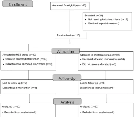
Figure 1 Consort flow diagram ...20
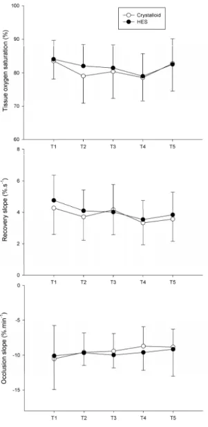
Figure 2 Vascular occlusion test parameters of patients receiving hydroxyethyl starch (●) or crystalloid (○) during the surgery ...21
Figure 3 Serum syndecan-1 of patients receiving
hydroxyethyl starch (dark grey) or crystalloid (light grey) during the surgery ...23
LIST OF ABBREVIATIONS
hydroxyethyl starch (HES) intensive care unit (ICU)
off-pump coronary artery bypass graft surgery (OPCAB) vascular occlusion test (VOT)
mixed venous oxygen saturation (SvO2) near-infrared spectroscopy (NIRS) tissue oxygen saturation (StO2)
rotational thromboelastometry (ROTEM)
INTRODUCTION
Fluid therapy is one of the most important elements in maintaining an effective circulating volume and tissue perfusion.
Appropriate fluid resuscitation improves tissue microcirculation, which plays a pivotal role in oxygen delivery to the tissues (1).
However, infused fluid may impair microcirculation by leading to tissue edema. Moreover, fluid infusion may increase the degradation of endothelial glycocalyx and may further aggravate fluid loss to the extravascular compartment (2, 3). Consequently, fluid resuscitation may affect clinical outcomes, and what constitutes the “ideal” fluid resuscitation strategy has long been debated.
Recent trials have raised concerns regarding the use of hydroxyethyl starch (HES) for fluid resuscitation in critically ill patients, suggesting that it may increase the risk of acute kidney injury and mortality (4, 5). These trials were performed on patients in the intensive care unit (ICU), including patients with severe sepsis, and it is unclear whether the results of these trials can be extrapolated to the peri-operative setting. In patients undergoing elective surgery, baseline organ function and vascular endothelial function differ from those in patients with sepsis.
HES has several characteristics that may theoretically improve microcirculation. First, it may maintain intravascular volume more effectively and requires infusion of less volume than crystalloid (6). Thus, it may cause less tissue edema, which compromises microvascular perfusion by increasing interstitial pressure. Moreover, previous studies reported that HES has advantages over crystalloid in improving microcirculation (7, 8).
Compared with crystalloid, HES inhibits platelet function, decreases blood viscosity and increases fibrinolysis, which may lead to an improved microcirculation (9-11). However, comparison of the microcirculatory effects of HES and crystalloid during surgery and the resulting clinical outcomes have not been evaluated in detail. Endothelial glycocalyx degradation coincides with microcirculatory dysfunction (12), thus we hypothesized that HES may cause less endothelial glycocalyx degradation and improved microvascular reactivity than those caused by crystalloid. The aim of this study was therefore to compare the effects of HES and crystalloid on endothelial glycocalyx degradation and microvascular reactivity in patients undergoing off-pump coronary artery bypass graft surgery (OPCAB).
METHODS
Following approval from the local research ethics committee, written informed consent was obtained from adult patients scheduled for OPCAB surgery. Exclusion criteria were pre-operative use of steroids, infectious disease, chronic liver disease, chronic kidney disease requiring dialysis, left ventricular ejection fraction < 30%, pre-operative administration of vasopressors or inotropes and pregnancy. Patients who could not tolerate the vascular occlusion test (VOT) due to arm deformity, burns, arteriovenous shunts, or peripheral vascular disease were also excluded.
Eligible patients were randomly allocated to the HES group or the crystalloid group using a computer-generated random number table. The randomization sequence was generated with a 1:1 allocation using a random block size of 4 by a blinded statistician. Group allocations were placed in sealed, opaque envelopes following randomization. An independent nurse prepared the study fluids for the respective groups. The study fluids were supplied in identical bags and were indistinguishable.
Five-lead electrocardiography, pulse oximetry and non-invasive blood pressure monitoring was commenced upon arrival in the operating room. Other monitoring included bispectral index, cerebral oximetry, pulmonary artery flotation catheter, and transesophageal echocardiography. Anesthesia was induced using intravenous midazolam 0.15 mg/kg, sufentanil 1 μg/kg and vecuronium 0.15 mg/kg. Anesthesia was maintained with continuous infusions of remifentanil 6–12 ng/ml and propofol 1.5–2.5 µg/ml target-controlled infusion, maintaining
bispectral index values between 40 and 60. After tracheal intubation, the patients’ lungs were ventilated in volume-controlled mode, with a tidal volume of 8 ml/kg predicted body weight; ventilation frequency was adjusted to maintain normocarbia throughout surgery. Normothermia was maintained during the peri-operative period using forced-air warming devices or warm water blankets and a heated humidifier circuit. Heparin was administered before coronary anastomoses and was neutralized with protamine after completion of anastomoses. The target activated clotting time during surgery was > 300 seconds. At the end of surgery, patients were transferred to the ICU.
In the HES group, 6% Hetastarch (670 kD/0.75 Hextend, CJ HealthCare, Seoul, Korea) was used for intravenous volume replacement during surgery and was administered in a volume of up to 20 ml/kg followed by plasmalyte solution if extra fluid was needed. In the crystalloid group, a plasmalyte solution (Plasma Solution A; CJ HealthCare, Seoul, Korea) was used for intravenous volume replacement during surgery. In both groups, fluids were infused to maintain a cardiac index > 2 l/min/m2, mean arterial pressure of 60–80 mmHg, and mixed venous oxygen saturation (SvO2) > 60% during surgery. A cell salvage system was routinely used for all patients. Packed red cells were transfused according to international transfusion guidelines (13). A transfusion hematocrit trigger of 25% was used during surgery.
Microvascular reactivity was evaluated using near-infrared spectroscopy (NIRS) and the VOT. Tissue oxygen saturation (StO2) was automatically recorded every 2 seconds
using a NIRS sensor (InSpectra StO2 tissue oxygenation monitor model 650; Hutchinson Technology Inc., Hutchinson, MN, USA) on the thenar eminence, as described in previous reports (14, 15).
VOT was conducted using an inflatable cuff with an adjustable air pressure source placed around the upper arm. After confirming a StO2 variation < 2% over 30 seconds, the cuff was inflated rapidly to 50 mmHg above systolic blood pressure and kept inflated until StO2 decreased to 40%. Then the cuff was deflated rapidly (< 0.5 seconds). If VOT was not performed correctly, the test was repeated 5 minutes later. VOT was performed five times during the perioperative period; immediately before induction of anesthesia (T1), 1 hour after the start of coronary artery anastomosis (T2), upon infusion of 20 ml/kg HES /crystalloid (T3), at the time of skin closure (T4) and 12 hours after ICU admission (T5). StO2, occlusion, and recovery slope, derived from changes in StO2 during the VOT, were calculated automatically by InSpectra Analysis (version 4.03;
Hutchinson Technology Inc.) software for every VOT.
Hemodynamic and ventilation variables were also recorded during every test.
Blood (6 ml) was sampled from the radial artery at the five time points stated above and stored in serum separator tubes. Blood samples were centrifuged (1000 × g, 10 minutes), and the serum was separated and frozen. Concentrations of syndecan-1, the main component of endothelial glycocalyx, were measured using an enzyme-linked immunosorbent assay kit (Human sCD138/syndecan-1 enzyme-linked immunosorbent assay kit, MyBioSource, CA, USA).
Rotational thromboelastometry (ROTEM, TEM
International, Munich, Germany) was performed after induction of anesthesia, after completion of the coronary anastomoses, at the time of skin closure and 12 hours after ICU admission. Blood samples were collected at these same five time points.
Major in-hospital complications, including myocardial infarction, coronary revascularization, stroke, re-operation for bleeding, postoperative atrial fibrillation, and acute kidney injury and mortality were recorded. In addition, PaO2/FIO2 ratio, lung mechanical ventilation time, and length of stay in the ICU and hospital were assessed. Myocardial infarction was defined as an elevation of cardiac biomarker values (> 10 × 99th percentile upper reference limit) in patients with normal baseline troponin values (< 99th percentile upper reference limit). In addition, new pathological Q waves, new left bundle branch block, angiographically documented new graft or new native coronary artery occlusion, or imaging evidence of new loss of viable myocardium or new regional wall motion abnormalities were recorded (16). Acute kidney injury was defined according to the Acute Kidney Injury Network staging system (17).
The primary outcome measure was the VOT recovery slope at the end of surgery. Secondary outcomes were serum syndecan-1 levels, VOT-derived variables, including StO2, occlusion, and recovery slope, fluid balance, ROTEM parameters during and after surgery, and in-hospital complications after surgery. Based on our pilot study performed in five OPCAB patients with infusion of plasmalyte solution, the mean (SD) VOT recovery slope at the end of surgery was 3.9 (1.2) %/s.
Assuming that a difference of 20% in the recovery slope was clinically significant, 50 patients would be needed in each group
to detect a difference with a type I error of 0.05 and a power of 0.9. To allow for a 20% dropout, 60 patients were recruited in each group. Continuous variables were compared using Student’s t test or the Mann–Whitney U-test after testing for normality.
Categorical variables were analyzed using the χ2 test or Fisher’s exact test, as appropriate. Student’s t test was used to compare continuous data, such as StO2-derived variables, cytokine levels and lactate levels at the different time points. These variables, which were recorded repeatedly in the same individuals, were further analyzed using repeated-measures ANOVA followed by post hoc tests with Bonferroni’s correction for multiple testing.
Data were analyzed using SPSS (version 22.0; IBM Corp., Armonk, NY, USA) software and a p value <0.05 was considered statistically significant.
RESULTS
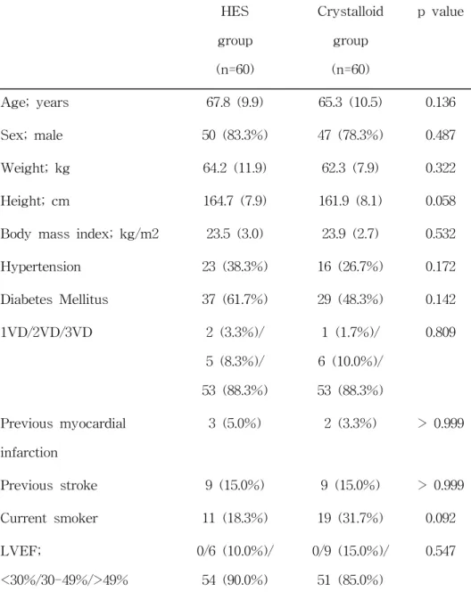
Of the 140 patients screened, 20 were excluded: nine because they had a left ventricular ejection fraction < 30%, nine because they had renal impairment requiring renal replacement therapy, one who required preoperative administration of inotropes, and one who declined to participate (Figure 1). A total of 120 patients were randomized, received the allocated interventions and were included in the final analysis. There were no cases of conversion to cardiopulmonary bypass from OPCAB. All operations were performed by a single surgeon. Baseline patient characteristics are shown in Table 1. Duration of surgery were comparable between the HES and crystalloid groups (369.6 (51.0) vs. 376.6 (67.3) min, respectively, p = 0.523).
The median (IQR [range]) volume of fluid infused during surgery were significantly less in the HES group compared with the crystalloid group; 2800 (2150-3550 [1400-7300] vs. 3925 (3100-4725 [1900-6700]) ml, respectively, p < 0.001, Table 2. Red cell transfusion during surgery were similar between the HES and crystalloid groups (1.5 (0-3 [0-9]) vs. 1 (0-2 [0-4]) units, respectively, p = 0.258). More blood loss occurred through the chest drains in the HES group during the first 8 hours (316 (220-433 [100-1019] vs. 504 (395-699 [70-2021] ml, respectively, p
< 0.001) and 24 hours (614 (485-700 [270-1765]) vs. 849 (677-1056 [198-2735]) ml, respectively, p < 0.001) after surgery.
In addition, more red cells were transfused in the HES group during the first 8 hours (0 (0-1 [0-3]) vs. 0 (0-0 [0-2]), p = 0.020) and 24 h (0 (0-2 [0-3]) vs. 0 (0-0 [0-2]), respectively, p = 0.011) after surgery. Perioperative hemodynamics, including heart
rate, mean arterial pressure, central venous pressure, cardiac index, and SvO2, did not differ significantly between the groups.
Baseline StO2 did not differ between the groups (Figure 2). StO2 decreased at the end of surgery in both the HES group (from 84.1 (5.5) to 78.5 (7.1) %, p < 0.001) and crystalloid group (from 83.6 (5.5) to 78.6 (7.0) %, p < 0.001). In addition, the recovery slope decreased at the end of surgery in both the HES groups (from 4.8 (1.6) to 3.6 (1.3) %/s, p < 0.001) and crystalloid group (from 4.3 (1.7) to 3.3 (1.4) %/s, p = 0.001). However, there were no significant differences in StO2, occlusion or recovery slope between the HES and crystalloid groups.
Baseline serum syndecan-1 levels were the same for both groups (Figure 3). Median (IQR [range]) levels were highest 1 hour after the start of coronary artery anastomosis in both the HES group (94.0 (55.7-208.1 [5.7-366.8]) ng/ml) and the crystalloid group (110.4 (67.9-169.3 [14.0-367.7]) ng/ml). However, peak serum syndecan-1 levels were not significantly different between the groups. Following infusion of 20 ml/kg of the study fluids, median (IQR [range]) syndecan-1 was higher in the HES group than the crystalloid group (79.9 (46.6-176.6 [5.1-398.4]) vs.
62.7 (30.1-103.0 [6.0-218.8]) ng/ml, respectively, p = 0.030).
However, overall perioperative changes in syndecan-1 were not significantly different between the groups.
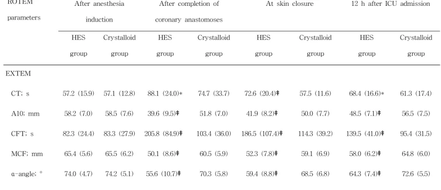
Baseline ROTEM parameters were comparable between the groups (Table 3). After completion of the coronary anastomoses, the HES group had a higher EXTEM clotting time (p < 0.035) and lower EXTEM and FIBTEM maximum clot firmness (both p < 0.001). EXTEM clotting time was consistently higher in the HES group at the end of surgery (p <
0.001) and 12 hours after ICU admission (p = 0.029). In addition, EXTEM and FIBTEM maximum clot firmness were consistently lower in the HES group at the end of surgery (both p < 0.001) and 12 hours after ICU admission (both p < 0.001) (Table 3).
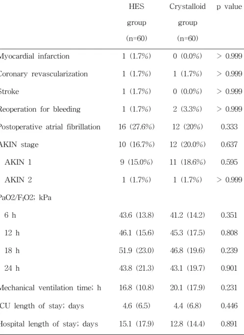
In the HES group versus the crystalloid group, postoperative in-hospital complications included mortality (0 vs.
0), myocardial infarction (1 vs. 0), coronary revascularization (relative risk 1.00, 95% CI 0.06 – 15.62), stroke (1 vs. 0), reoperation for bleeding (relative risk 0.50, 95% CI 0.05 – 5.37), postoperative atrial fibrillation (relative risk 1.38, 95% CI 0.72 – 2.66), and acute kidney injury (relative risk 0.83, 95% CI 0.39 – 1.78) respectively, with no statistically significant differences between the groups (Table 4). Mechanical ventilation time (mean difference -3.27, 95% CI -8.66 – 2.11), ICU stay (mean difference 0.17, 95% CI -2.24 – 2.57) and the lengths of hospital stay (mean difference 2.27, 95% CI -3.60 – 8.14) were similar for both groups.
Table 1. Baseline characteristics of patients receiving
hydroxyethyl starch or crystalloid during off-pump coronary artery bypass surgery
HES group (n=60)
Crystalloid group (n=60)
p value
Age; years 67.8 (9.9) 65.3 (10.5) 0.136
Sex; male 50 (83.3%) 47 (78.3%) 0.487
Weight; kg 64.2 (11.9) 62.3 (7.9) 0.322
Height; cm 164.7 (7.9) 161.9 (8.1) 0.058
Body mass index; kg/m2 23.5 (3.0) 23.9 (2.7) 0.532
Hypertension 23 (38.3%) 16 (26.7%) 0.172
Diabetes Mellitus 37 (61.7%) 29 (48.3%) 0.142
1VD/2VD/3VD 2 (3.3%)/
5 (8.3%)/
53 (88.3%)
1 (1.7%)/
6 (10.0%)/
53 (88.3%)
0.809
Previous myocardial infarction
3 (5.0%) 2 (3.3%) > 0.999
Previous stroke 9 (15.0%) 9 (15.0%) > 0.999
Current smoker 11 (18.3%) 19 (31.7%) 0.092
LVEF;
<30%/30-49%/>49%
0/6 (10.0%)/
54 (90.0%)
0/9 (15.0%)/
51 (85.0%)
0.547
Values are mean (SD) or number (proportion). HES, hydroxyethyl starch; 1VD, one-vessel disease; 2VD, two-vessel disease; 3VD, three-vessel disease; LVEF, left ventricle ejection fraction;
EuroSCORE, European System for Cardiac Operative Risk Evaluation.
Unstable angina 28 (46.7%) 25 (41.7%) 0.581
Previous cardiac surgery 1 (1.7%) 3 (5.0%) 0.619
EuroSCORE II 1.6 (1.0) 1.7 (1.4) 0.429
Serum creatinine; umol/l 0.96 (0.30) 0.93 (0.34) 0.588 Serum lactate; mmol/l 1.12 (0.39) 1.19 (0.65) 0.467 Platelet count; x109/l 143.9 (85.1) 152.1 (53.4) 0.527 Fibrinogen; mg/dl 237.4 (73.4) 255.18 (76.2) 0.196
Table 2. Fluid balance of patients receiving hydroxyethyl starch or crystalloid during and after during off-pump coronary artery bypass surgery
HES group (n=60)
Crystalloid group (n=60)
p value
Intraoperative parameters
Crystalloid; ml/kg 23.1 (15.0-37.9 [3.7-90.0])
62.6 (52.8-74.0 [27.8-128.3])
< 0.001
HES; ml/kg 20.0 (20.0-20.0 [20.0-20.0])
0 < 0.001
Total infused fluid;
ml/kg
43.4 (34.7-57.5 [21.8-110.2])
62.6 (52.8-74.0 [27.8-128.3])
< 0.001
Receiving red cells 38 (63.3%) 40 (66.7%) 0.702
Receiving FFP 2 (3.3%) 2 (3.3%) > 0.999
Receiving platelets 5 (8.3%) 1 (1.7%) 0.094
Diuresis; ml 1244.4 (596.5) 1419.5 (755.6) 0.161 Estimated blood
loss; ml
902.2 (448.1) 800 (769.7) 0.716
Postoperative parameters Chest tube drainage; ml
Values are means (SD), median (IQR [range]), or number
(proportion). FFP, fresh frozen plasma; HES, hydroxyethyl starch.
0-8 h 504 (395-699 [70-2021])
316 (220-433 [100-1019])
< 0.001
8-24 h 302 (236-417 [128-924])
250 (193-323 [140-746])
0.006
24-48 h 280 (178-397 [60-774])
184 (137-290 [35-1485])
0.012
Urine output; ml
0-8 h 1050 (685-1595 [215-3900])
1198 (715-1795 [250-4160])
0.395
8-24 h 1520 (1120-1943 [515-3890])
1885 (1168-2450 [665-4380])
0.049
24-48 h 1555 (1243-1990 [350-6060])
1803 (1315-2373 [385-5225])
0.139
Table 3. Rotational thromboelastometry parameters of patients receiving hydroxyethyl starch or crystalloid at study time points
ROTEM parameters
After anesthesia induction
After completion of coronary anastomoses
At skin closure 12 h after ICU admission
HES group
Crystalloid group
HES group
Crystalloid group
HES group
Crystalloid group
HES group
Crystalloid group
EXTEM
CT; s 57.2 (15.9) 57.1 (12.8) 88.1 (24.0)* 74.7 (33.7) 72.6 (20.4)‡ 57.5 (11.6) 68.4 (16.6)* 61.3 (17.4)
A10; mm 58.2 (7.0) 58.5 (7.6) 39.6 (9.5)‡ 51.8 (7.0) 41.9 (8.2)‡ 50.0 (7.7) 48.5 (7.1)‡ 56.5 (7.5)
CFT; s 82.3 (24.4) 83.3 (27.9) 205.8 (84.9)‡ 103.4 (36.0) 186.5 (107.4)‡ 114.3 (39.2) 139.5 (41.0)‡ 95.4 (31.5)
MCF; mm 65.4 (5.6) 65.5 (6.2) 50.1 (8.6)‡ 60.5 (5.9) 52.3 (7.8)‡ 59.1 (6.9) 58.0 (6.2)‡ 64.8 (6.0)
α-angle; ° 74.0 (4.7) 74.2 (5.1) 55.6 (10.7)‡ 70.3 (5.8) 59.4 (8.8)‡ 68.5 (6.8) 64.3 (7.4)‡ 72.6 (5.5)
FIBTEM
A10; mm 19.9 (8.7) 19.7 (8.6) 9.2 (6.6)† 13.6 (5.8) 8.6 (4.2)‡ 13.1 (4.8) 11.8 (4.4)‡ 18.6 (5.5)
MCF; mm 21.5 (10.3) 21.4 (9.0) 10.1 (7.0)† 15.2 (6.2) 9.2 (4.6)‡ 14.6 (5.1) 13.9 (6.6)‡ 20.9 (6.1)
INTEM
CT; s 173.3 (33.6) 174.6 (38.2) 244.3 (71.2) 220.9 (49.5) 220.4 (48.2)† 194.5 (41.3) 181.2 (33.1) 175.1 (5.1)
A10; mm 57.1 (6.8) 56.4 (8.3) 35.8 (9.7)‡ 48.8 (7.2) 39.0 (8.1)‡ 48.1 (8.4) 47.5 (7.5)‡ 55.3 (6.9)
CFT; s 69.9 (19.6) 78.9 (66.3) 238.2 (118.0)‡ 104.9 (50.4) 183.9 (81.0)‡ 109.9 (72.8) 123.4 (41.2)‡ 81.7 (27.7)
MCF; mm 63.3 (6.1) 62.5 (8.1) 44.8 (9.3)‡ 56.6 (6.7) 48.2 (7.5)‡ 56.2 (7.5) 55.8 (6.6)‡ 61.9 (5.7) α-angle; ° 76.3 (3.5) 76.0 (4.5) 55.9 (11.8)‡ 71.0 (6.3) 60.0 (10.1)‡ 70.5 (8.4) 67.3 (6.5)‡ 74.5 (4.4)
Values are mean (SD). *p < 0.05, †p < 0.01, ‡p < 0.001. ROTEM, rotational thromboelastometry; CT, clotting time; A10, amplitude of clot firmness after 10 min; CFT, clot formation time; MCF, maximum clot firmness; ICU, intensive care unit; HES, hydroxyethyl starch.
Table 4. Study outcomes by treatment group
HES group (n=60)
Crystalloid group (n=60)
p value
Myocardial infarction 1 (1.7%) 0 (0.0%) > 0.999 Coronary revascularization 1 (1.7%) 1 (1.7%) > 0.999
Stroke 1 (1.7%) 0 (0.0%) > 0.999
Reoperation for bleeding 1 (1.7%) 2 (3.3%) > 0.999 Postoperative atrial fibrillation 16 (27.6%) 12 (20%) 0.333
AKIN stage 10 (16.7%) 12 (20.0%) 0.637
AKIN 1 9 (15.0%) 11 (18.6%) 0.595
AKIN 2 1 (1.7%) 1 (1.7%) > 0.999
PaO2/FIO2; kPa
6 h 43.6 (13.8) 41.2 (14.2) 0.351
12 h 46.1 (15.6) 45.3 (17.5) 0.808
18 h 51.9 (23.0) 46.8 (19.6) 0.239
24 h 43.8 (21.3) 43.1 (19.7) 0.901
Mechanical ventilation time; h 16.8 (10.8) 20.1 (17.9) 0.231 ICU length of stay; days 4.6 (6.5) 4.4 (6.8) 0.446 Hospital length of stay; days 15.1 (17.9) 12.8 (14.4) 0.891
Values are number (proportion) or mean (SD). AKIN, Acute Kidney Injury Network; ICU, intensive care unit; HES, hydroxyethyl starch.
Figure 1. Consort flow diagram.
HES, hydroxyethyl starch.
Figure 2. Vascular occlusion test parameters of patients receiving hydroxyethyl starch (●) or crystalloid (○) during the surgery.
There were no significant differences in tissue oxygen saturation, occlusion and recovery slopes between hydroxyethyl starch and crystalloid groups.
Values are shown as mean ± SD. T1, immediately before induction of anesthesia; T2, 1 hour after the start of the coronary artery anastomosis; T3, upon infusion of 20 ml/kg of HES/crystalloids; T4, at skin closure; T5, 12 hours after intensive care unit admission; HES, hydroxyethyl starch.
Figure 3. Serum syndecan-1 of patients receiving hydroxyethyl starch (dark grey) or crystalloid (light grey) during the surgery.
Overall perioperative changes of syndecan-1 were not significantly different between hydroxyethyl starch and crystalloid groups.
Boxes represent the interquartile range and the lines dividing the boxes represent the median. The whiskers indicate the range of the data. T1, immediately before induction of anesthesia; T2, 1 hour after the start of the coronary artery anastomosis; T3, upon infusion of 20 ml/kg of HES/crystalloids; T4, at skin closure; T5, 12 hours after intensive care unit admission; HES, hydroxyethyl starch.
DISCUSSION
In the present study, the VOT recovery slope, an index of microvascular reactivity, and serum syndecan-1, a marker of endothelial glycocalyx, did not differ significantly between patients given HES and those given crystalloid fluids during OPCAB surgery. Although HES decreased the total amount of fluid infused, it impaired coagulation, as measured by ROTEM, and increased chest tube drainage and red cell transfusion.
Decreased microcirculation is observed in critically ill patients (18, 19) and during cardiac surgery (20, 21) and is associated with a poor prognosis (19, 22). These alterations may be explained by various factors, such as blood dilution, hypothermia, extracorporeal circulation, inflammatory response or surgical stimulation (20, 21). Microcirculatory dysfunction is recognized as one of the earliest manifestations of cardiovascular disease and, in particular, inflammatory processes (23). Thus, close observation of microcirculatory changes during cardiac surgery and methods to reduce microcirculatory alterations may have clinically important implications. A previous study indicated that microcirculatory changes occurred during hypovolemic periods and recovered after appropriate fluid therapy in patients undergoing abdominal surgery. If HES restores hypovolemia more effectively than crystalloid with less tissue edema, then it would be better for the microcirculation. HES may theoretically improve the microcirculation by several mechanisms, such as reduction in
platelet adhesion, a decrease in blood viscosity and enhanced fibrinolysis (9-11). However, in the present study, microvascular reactivity during OPCAB did not differ between patients given HES and those given crystalloids.
VOT recovery slope represents a capacity to recruit microvessels in response to an hypoxic stimulus (24). It has been reported that microvascular reactivity is related to worse clinical outcomes in patients with sepsis (25, 26). Thus, monitoring microvascular reactivity could be a useful method for guiding fluid therapy (27). In a study by Moerman et al. (28), the authors evaluated HES and gelatin as prime solutions of the cardiopulmonary bypass circuit. They concluded that HES 130/0.4 maintains better microvascular reactivity than gelatin during acute hemodilution. However, in their study, crystalloid was not compared with HES and changes to the glycocalyx were not evaluated.
Components of endothelial glycocalyx, including syndecan-1 and heparan sulfate, are released during cardiac surgery (29). Possible mechanisms of glycocalyx degradation during cardiac surgery include ischemia/reperfusion injury, release of pro-inflammatory cytokines, and mechanical stress (2, 29, 30).
In addition, hypervolemia triggers the release of atrial natriuretic peptide and increases the shedding of endothelial glycocalyx (2).
In our study, fluid resuscitation was guided by hemodynamic parameters, including the cardiac index and SvO2, which did not differ significantly between the groups. Overall, peri-operative changes in syndecan-1 were not significantly different between the groups. Syndecan-1 was higher in the HES group than the crystalloid group only after infusion of 20 ml/kg of the study
fluids and the difference was not maintained at the end of surgery, possibly because both groups received considerable volumes of crystalloid. However, these finding suggest that HES may cause more damage to endothelial glycocalyx, which contributes to vascular barrier function.
In our study, peri-operative hemostasis was evaluated by ROTEM, which was impaired to a greater extent in the HES group than in the crystalloid group. This is consistent with previous reports of potential hemostatic alterations associated with HES resuscitation (31). Consequently, more blood was lost via the chest drains in the HES group, and this group also received more allogeneic blood products. However clinical outcomes, including acute kidney injury, lengths of stay in the ICU and hospital and mortality, did not differ between the groups. Current FDA recommendations in the USA state that there are increased risks of bleeding with the use of HES in patients undergoing open heart surgery on cardiopulmonary bypass (32). Unfortunately our study demonstrated that HES was also associated with an increased risk of bleeding in OPCAB.
Intraoperative fluid therapy is often required for acute resuscitation due to intravascular hypovolemia. In OPCAB, the heart is frequently lifted and tilted into a vertical position, resulting in a marked increase in atrial pressure and decreased cardiac output, making timely fluid resuscitation essential. It has been suggested that HES allows faster hemodynamic stabilization, because it is more effective in expanding plasma due to its high oncotic pressure compared with crystalloid. In the present study, larger amounts of fluid were needed to achieve similar systemic endpoints in the crystalloid group versus the HES group. In
addition, intraoperative fluid balance was more positive in the crystalloid group than in the HES group, which raises concerns, because previous studies have indicated that a 10% fluid overload in cardiac surgical patients is independently associated with increased mortality (33), and patients with excess intraoperative fluid balance have more ICU complications and higher rates of in-hospital mortality (34).
Our study had several limitations. Firstly, it was limited to patients undergoing OPCAB, so the applicability of our conclusions to other procedures, especially surgery involving cardiopulmonary bypass, is limited. Secondly, we used high molecular weight HES, and the findings are not directly transferable to other types of HES. Thirdly, the NIRS technique is limited in the presence of swelling or thick adipose tissue, and VOT has not been standardized with regard to the site of measurement, ischemic threshold, probe size and time interval between tests (35). Fourthly, monitoring of endothelial glycocalyx biomarkers, including syndecan-1, is a surrogate marker of glycocalyx integrity.
We conclude that the use of HES 670/0.75 did not result in significant differences in microvascular reactivity or endothelial glycocalyx degradation compared with crystalloid in patients undergoing OPCAB. Although, HES 670/0.75 decreased the total volume of fluid infused during OPCAB, it resulted in more postoperative bleeding and impaired hemostasis.
REFERENCES
1. Ince C. The microcirculation is the motor of sepsis. Crit Care. 2005; 9 Suppl 4: S13-9.
2. Chappell D, Bruegger D, Potzel J, Jacob M, Brettner F, Vogeser M, et al. Hypervolemia increases release of atrial natriuretic peptide and shedding of the endothelial glycocalyx. Crit Care. 2014; 18(5): 538.
3. Rehm M, Zahler S, Lotsch M, Welsch U, Conzen P, Jacob M, et al. Endothelial glycocalyx as an additional barrier determining extravasation of 6% hydroxyethyl starch or 5% albumin solutions in the coronary vascular bed.
Anesthesiology. 2004; 100(5): 1211-23.
4. Perner A, Haase N, Guttormsen AB, Tenhunen J, Klemenzson G, Åneman A, et al. Hydroxyethyl starch 130/0.42 versus Ringer's acetate in severe sepsis. N Engl J Med. 2012; 367(2): 124-34.
5. Myburgh JA, Finfer S, Bellomo R, Billot L, Cass A, Gattas D, et al. Hydroxyethyl starch or saline for fluid resuscitation in intensive care. N Engl J Med. 2012;
367(20): 1901-11.
6. McIlroy DR, Kharasch ED. Acute intravascular volume expansion with rapidly administered crystalloid or colloid in the setting of moderate hypovolemia. Anesth Analg.
2003; 96(6): 1572-7.
7. Dubin A, Pozo MO, Casabella CA, Murias G, Pálizas F Jr, Moseinco MC, et al. Comparison of 6% hydroxyethyl starch 130/0.4 and saline solution for resuscitation of the microcirculation during the early goal-directed therapy of septic patients. J Crit Care. 2010; 25(4): 659.e1-8.
8. Strunden MS, Bornscheuer A, Schuster A, Kiefmann R, Goetz AE, Heckel K. Glycocalyx degradation causes microvascular perfusion failure in the ex vivo perfused mouse lung: hydroxyethyl starch 130/0.4 pretreatment attenuates this response. Shock. 2012; 38(5): 559-66.
9. Kupper S, Mees ST, Gassmann P, Brodde MF, Kehrel B, Haier J. Hydroxyethyl starch normalizes platelet and leukocyte adhesion within pulmonary microcirculation during LPS-induced endotoxemia. Shock. 2007; 28(3):
300-8.
10. Neff TA, Fischler L, Mark M, Stocker R, Reinhart WH.
The influence of two different hydroxyethyl starch solutions (6% HES 130/0.4 and 200/0.5) on blood viscosity.
Anesth Analg. 2005; 100(6): 1773-80.
11. Nielsen VG. Hydroxyethyl starch enhances fibrinolysis in human plasma by diminishing alpha2-antiplasmin-plasmin interactions. Blood Coagul Fibrinolysis. 2007; 18(7): 647-56.
12. Marechal X, Favory R, Joulin O, Montaigne D, Hassoun S, Decoster B, et al. Endothelial glycocalyx damage during endotoxemia coincides with microcirculatory dysfunction
and vascular oxidative stress. Shock. 2008; 29(5): 572-6.
13. Ferraris VA, Brown JR, Despotis GJ, Hammon JW, Reece TB, Saha SP, et al. 2011 update to the Society of Thoracic Surgeons and the Society of Cardiovascular Anesthesiologists blood conservation clinical practice guidelines. Ann Thorac Surg. 2011; 91(3): 944-82.
14. Georger JF, Hamzaoui O, Chaari A, Maizel J, Richard C, Teboul JL. Restoring arterial pressure with norepinephrine improves muscle tissue oxygenation assessed by near-infrared spectroscopy in severely hypotensive septic patients. Intensive Care Med. 2010; 36(11): 1882-9.
15. Futier E, Christophe S, Robin E, Petit A, Pereira B, Desbordes J, et al. Use of near-infrared spectroscopy during a vascular occlusion test to assess the microcirculatory response during fluid challenge. Crit Care.
2011; 15(5): R214.
16. Thygesen K, Alpert JS, Jaffe AS, Simoons ML, Chaitman BR, White HD, et al. Third universal definition of myocardial infarction. Eur Heart J. 2012; 33(20): 2551-67.
17. Mehta RL, Kellum JA, Shah SV, Molitoris BA, Ronco C, Warnock DG, et al. Acute Kidney Injury Network: report of an initiative to improve outcomes in acute kidney injury. Crit Care. 2007; 11(2): R31.
18. De Backer D, Creteur J, Dubois MJ, Sakr Y, Vincent JL.
Microvascular alterations in patients with acute severe
heart failure and cardiogenic shock. Am Heart J. 2004;
147(1): 91-9.
19. Sakr Y, Dubois MJ, De Backer D, Creteur J, Vincent JL.
Persistent microcirculatory alterations are associated with organ failure and death in patients with septic shock. Crit Care Med. 2004; 32(9): 1825-31.
20. De Backer D, Dubois MJ, Schmartz D, Koch M, Ducart A, Barvais L, et al. Microcirculatory alterations in cardiac surgery: effects of cardiopulmonary bypass and anesthesia.
Ann Thorac Surg. 2009; 88(5): 1396-403.
21. Atasever B, Boer C, Goedhart P, Biervliet J, Seyffert J, Speekenbrink R, et al. Distinct alterations in sublingual microcirculatory blood flow and hemoglobin oxygenation in on-pump and off-pump coronary artery bypass graft surgery. J Cardiothorac Vasc Anesth. 2011; 25(5): 784-90.
22. Sanders J, Toor IS, Yurik TM, Keogh BE, Mythen M, Montgomery HE. Tissue oxygen saturation and outcome after cardiac surgery. Am J Crit Care. 2011; 20(2): 138-45.
23. Levy BI, Ambrosio G, Pries AR, Struijker-Boudier HA.
Microcirculation in hypertension: a new target for treatment? Circulation. 2001; 104(6): 735-40.
24. De Blasi RA, Palmisani S, Alampi D, Mercieri M, Romano R, Collini S, et al. Microvascular dysfunction and skeletal muscle oxygenation assessed by phase-modulation near-infrared spectroscopy in patients with septic shock.
Intensive Care Med. 2005; 31(12): 1661-8.
25. Creteur J, Carollo T, Soldati G, Buchele G, De Backer D, Vincent JL. The prognostic value of muscle StO2 in septic patients. Intensive Care Med. 2007; 33(9): 1549-56.
26. Doerschug KC, Delsing AS, Schmidt GA, Haynes WG.
Impairments in microvascular reactivity are related to organ failure in human sepsis. Am J Physiol Heart Circ Physiol. 2007; 293(2): H1065-71.
27. Svensen C. Monitoring micro-vascular reactivity: a tool for guiding fluid therapy? Anaesthesia. 2016; 71(7): 747-50.
28. Moerman A, Van Eeckhout C, Vanderstraeten K, De Somer F, Van Belleghem Y, de Hert S. The effect of hydroxyethyl starch 6% 130/0.4 compared with gelatin on microvascular reactivity. Anaesthesia. 2016; 71(7): 798-805.
29. Bruegger D, Rehm M, Abicht J, Paul JO, Stoeckelhuber M, Pfirrmann M, et al. Shedding of the endothelial glycocalyx during cardiac surgery: on-pump versus off-pump coronary artery bypass graft surgery. J Thorac Cardiovasc Surg. 2009; 138(6): 1445-7.
30. Rehm M, Bruegger D, Christ F, Conzen P, Thiel M, Jacob M, et al. Shedding of the endothelial glycocalyx in patients undergoing major vascular surgery with global and regional ischemia. Circulation. 2007; 116(17): 1896-906.
31. Kozek-Langenecker SA. Effects of hydroxyethyl starch solutions on hemostasis. Anesthesiology. 2005; 103(3):
654-60.
32. U.S. Food and Drug Administration. Hydroxyethyl starch solutions: FDA safety communication: Boxed warning on increased mortality and severe renal injury and risk of bleeding, for use of hydroxyethyl starch solutions in some
settings, November 2013.
www.fda.gov/BiologicsBloodVaccines/SafetyAvailability/ucm 358271.htm2013.
33. Stein A, de Souza LV, Belettini CR, Menegazzo WR, Viégas JR, Costa Pereira EM, et al. Fluid overload and changes in serum creatinine after cardiac surgery:
predictors of mortality and longer intensive care stay. A prospective cohort study. Crit Care. 2012; 16(3): R99.
34. Silva JM Jr, de Oliveira AM, Nogueira FA, Vianna PM, Pereira Filho MC, Dias LF, et al. The effect of excess fluid balance on the mortality rate of surgical patients: a multicenter prospective study. Crit Care. 2013; 17(6): R288.
35. Gómez H, Mesquida J, Simon P, Kim HK, Puyana JC, Ince C, et al. Characterization of tissue oxygen saturation and the vascular occlusion test: influence of measurement sites, probe sizes and deflation thresholds. Crit Care. 2009;
13 Suppl 5: S3.
국문 초록
서론: 수액 주입은 조직의 미세순환과 내피당질층에 영향을 줄 수 있다. 그러나 하이드록시에틸 스타치와 정질액이 내피당질층의 분 해와 미세혈관 반응성에 미치는 영향은 아직 자세히 밝혀지지 않 았다. 하이드록시에틸 스타치가 정질액에 비해 내피당질층 분해를 더 적게 일으키고 미세혈관 반응성은 향상시킬 것이라는 가설을 세웠다.
방법: 체외순환 없는 관상동맥 우회술을 받는 환자 120명을 무작 위로 나누어 하이드록시에틸 스타치 670/0.75 또는 정질액을 수술 중에 20 ml/kg까지 투여하였다. 이후에는 수액요구량에 맞추어 양군에서 정질액을 투여하였다. 수술 전후에 미세혈관 반응성을 평가하기 위해서 혈관 압박 검사를 시행하였고, 내피당질층의 분 해를 평가하는 지표로서 혈장에서 syndecan-1을 측정하였다.
결과: 수술 중 투여된 수액양은 하이드록시에틸 스타치 군에서 정 질액 군에 비해 유의하게 더 적었다. (중위수 (사분위 [범위]; 2800 (2150-3550 [1400-7300]) vs. 3925 (3100-4725 [1900-6700]) ml, p <
0.001) 조직 산소 포화도, 폐쇄 기울기, 회복 기울기를 포함한 혈
관 압박 검사의 변수들은 양 군 간에 유의한 차이를 보이지 않았 다. 수술 전후 syndecan-1의 변화 역시 양 군 간에 유의하게 다르 지 않았다.
결론: 체외순환 없는 관상동맥 우회술을 받는 환자들에서, 정질액 에 비해서 하이드록시에틸 스타치 670/0.75의 사용은 미세혈관 반 응성 혹은 내피당질층 분해에 미치는 영향에서 유의한 차이를 보 이지 않았다.
* 본 내용은 Anaesthesia 학술지 (Kim TK, Nam K, Cho YJ, Min JJ, Hong YJ, Park KU, et al. Microvascular reactivity and endothelial glycocalyx degradation when administering hydroxyethyl starch or crystalloid during off-pump coronary artery bypass graft surgery: a randomised trial.
Anaesthesia. 2016 Sep 26. doi: 10.1111/anae.13642.)에 출판 완료된 내용임
--- 주요어 : 내피당질층, 미세순환, 정질액, 체외순환 없는 관상동맥 우회술, 하이드록시에틸 스타치
학 번 : 2014-30644