저작자표시-비영리-변경금지 2.0 대한민국 이용자는 아래의 조건을 따르는 경우에 한하여 자유롭게
l 이 저작물을 복제, 배포, 전송, 전시, 공연 및 방송할 수 있습니다. 다음과 같은 조건을 따라야 합니다:
l 귀하는, 이 저작물의 재이용이나 배포의 경우, 이 저작물에 적용된 이용허락조건 을 명확하게 나타내어야 합니다.
l 저작권자로부터 별도의 허가를 받으면 이러한 조건들은 적용되지 않습니다.
저작권법에 따른 이용자의 권리는 위의 내용에 의하여 영향을 받지 않습니다. 이것은 이용허락규약(Legal Code)을 이해하기 쉽게 요약한 것입니다.
Disclaimer
저작자표시. 귀하는 원저작자를 표시하여야 합니다.
비영리. 귀하는 이 저작물을 영리 목적으로 이용할 수 없습니다.
변경금지. 귀하는 이 저작물을 개작, 변형 또는 가공할 수 없습니다.
수의학박사학위논문
Pathogenesis of African Swine Fever Occurring in Asia
아시아에서 발생하는 아프리카 돼지 열병의 병인론
2022년 8월
서울대학교 대학원
수의학과 수의병인생물학 및 예방수의학 전공
오 태 환
獸 醫 學 博 士 學 位 論 文
Pathogenesis of African Swine Fever Occurring in Asia
아시아에서 발생하는 아프리카 돼지 열병의 병인론
지도교수 채 찬 희 (D.V.M., Ph.D.)
이 논문을 수의학박사 학위논문으로 제출함
2022년 4월서울대학교 대학원
수의학과 수의병인생물학 및 예방수의학전공
오 태 환
오 태 환의 수의학박사 학위논문을 인준함
2022년 6월위 원 장 박 재 학 (인) 부위원장 채 찬 희 (인) 위 원 최 창 순 (인) 위 원 하 윤 철 (인) 위 원 강 익 재 (인)
Pathogenesis of African Swine Fever Occurring in Asia
By
Taehwan Oh, D.V.M.
A dissertation submitted in partial fulfillment of the requirements for the degree of
DOCTOR OF PHILOSOPHY
Supervisor: Professor Chanhee Chae, D.V.M., Ph.D.
June 2022 Approved by
Park, Jae-Hak
Chae, Chanhee
Choi, Changsun
Ha, Yooncheol
Kang, Ikjae
Department of Veterinary Medicine
Graduate School of Seoul National University
Abstract
Pathogenesis of African Swine Fever Occurring in Asia
(Supervisor : Chanhee Chae, D.V.M., Ph.D.)
Taehwan Oh
Veterinary Pathobiology and Preventative Medicine (Pathology) Department of Veterinary Medicine
Graduate School of Seoul National University
African swine fever virus (ASFV) is a large, icosahedral, enveloped, linear double strand DNA virus, causes hemorrhagic disease in domestic pigs and wild boars, and has been present in Asia since 2018. The objective of this dissertation was to reveal the pathogenesis of African swine fever (ASF) occurring in Asia through viral genetic characterization, pathological investigation of outbreak cases, and experimental reproduction of the disease.
II
In the first study, the genetic characterization of an ASFV isolate recently found in Vietnam was conducted to understand the genetic and epidemiologic characteristics of ASFV circulation within Asia. The wild-type virus was isolated using primary porcine kidney (PPK) cells, and successfully propagated to obtain a virus stock.
Partial sequencing of viral major structural proteins p72, p54, and p30 categorized the new ASFV isolate as genotype Ⅱ as it showed a high genetic homology with all isolates from other Asian countries. The partial sequences of these Asian isolates were also identical to those found in Georgia and Eastern Europe since 2007. These results revealed that ASF outbreaks in Asia were probably due to a single introduction of ASFV genotype Ⅱ that spread slowly eastward, and without the introduction of additional virus genotypes.
In the second study, pathological investigations were conducted in recent successive Vietnamese ASF outbreaks to elucidate the pathogenesis of naturally occurring ASFV genotype II infection in Asia. Five major organs (lung, liver, kidney, spleen, and lymph nodes) were evaluated in necropsied pigs for splenomegaly, hemorrhagic lymph nodes and renal petechia being the most prominent gross lesions.
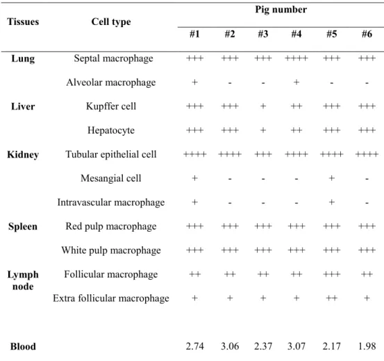
Microscopic lesions based on tissue hemorrhage and lymphoid destruction were commonly observed in all necropsied pigs. ASFV antigen loads were determined by immunohistochemistry and were antigen positive for monocyte/macrophage lineage cells as well as for hepatocytes and tubular epithelial cells. Through this pathological study, it was confirmed that naturally occurring ASF in Asia was of an acute form where ASFV antigen distribution was widely spread in virus-infected pigs.
In the third study, twenty domestic pigs were orally inoculated with ASFV genotype II Asian isolates. A temporal pathology model was established by sacrificing 4 pigs
each at 1-, 3-, 5-, 7-, and 9-days post inoculation (dpi).Gross and microscopic lesions based on hemorrhage and lymphoid destruction were observed in five major organs (lung, liver, kidney, spleen, and lymph nodes) beginning at 3 dpi. Lesions were prominent at 5 dpi and reached peak severity at 9 dpi. ASFV antigenwas observed from 3 dpi, mainly in the monocyte/macrophage lineage, but other cell types such as hepatocytes and tubular epithelial cells were antigen-positive at later stages of infection (7 dpi). These results demonstrated that ASFV circulating in Asia is highly virulent and sufficient in inducing the acute form of ASF via the oral route. It also provides evidence that the monocyte/macrophage lineage may play a major role in the acute ASF induction in early phase of viral infection.
Conclusively, this thesis suggested that ASF occurring in Asia is an acute clinical form of disease caused by genotype II ASFV, and that the monocyte/macrophage lineage plays a major role in the acute course of the disease. Through this study, an intergrated methodology was presented for the elucidation of the pathogenesis of ASF. Whole genome sequencing and comparative pathological studies of newly discovered virus isolates continue to be needed in case other ASFV genotypes enter Asia in the future.
Keywords: African swine fever virus; Pathogenesis; Genetic characterization;
Pathological investigation; Antigen distribution
Student Number: 2018-22255
IV
Table of Contents
ABSTRACT ... Ⅰ TABLE OF CONTENTS ... Ⅵ LIST OF TABLES ... Ⅷ LIST OF FIGURES ... Ⅸ LIST OF ABBREVIATIONS ... Ⅺ
GENERAL INTRODUCTION ... 1
LITERATURE REVIEW ... 3
1. History ... 3
2. Etiology ... 3
2-1. Classification ... 3
2-2. Virion Properties ... 4
2-3. Genetic structure ... 4
2-4. Virus Replication ... 5
3. Epidemiology ... 6
3-1. Sylvatic Cycle ... 6
3-2. Domestic Cycle ... 7
4. Clinical signs ... 8
4-1. Peracute form ... 8
4-2. Acute form ... 8
4-3. Subacute form ... 9
4-4. Chronic form ... 9
5. Pathology ... 10
5-1. Pathogenesis of Lymphoid lesions ... 10
5-2. Pathogenesis of Vascular change ... 11
6. Diagnosis... 12
6-1. Viral genome detection ... 12
6-2. Virus isolation ... 12
6-3. Antigen detection ... 13
6-4. Antibody detection ... 13
7. Prevention ... 14
7-1. Quarantine ... 15
7-2. Vaccination ... 15
8. References ... 16
PART Ⅰ. Genetic Characterization of Asian Isolate of African Swine Fever Virus Abstract ... 27
Introduction ... 28
VI
Materials and Methods ... 30
Results ... 34
Discussion ... 39
References ... 42
PART Ⅱ. Pathology of Naturally Occurring African Swine Fever in Asia Abstract ... 48
Introduction ... 49
Materials and Methods ... 51
Results ... 55
Discussion ... 64
References ... 67
PART Ⅲ. Pathology of Experimentally Induced African Swine Fever in Asia Abstract ... 72
Introduction ... 73
Materials and Methods ... 75
Results ... 79
Discussion ... 91
References ... 94
GENERAL CONCLUSION ... 99
ABSTRACT IN KOREAN ... 101
VIII
LIST OF TABLES
PART Ⅱ
Table 1. Gross lesions of pigs naturally infected with ASF ... 56
Table 2. Histopathology in pigs naturally infected with ASF ... 59
Table 3. Viral antigen distribution in pigs naturally infected with ASF ... 62
PART Ⅲ
Table 1. Clinical signs of pigs with experimentally induced ASF ... 80
Table 2. Gross lesions of pigs with experimentally induced ASF ... 83
Table 3. Histopathology in pigs with experimentally induced ASF ... 86
Table 4. Viral antigen distribution in pigs with experimentally
induced ASF ... 89
LIST OF FIGURES
PART Ⅰ
Figure 1. Replication kinetics of ASFV isolate in primary porcine kidney cells ... 35
Figure 2. Phylogenetic analysis of major structural protein p72 partial sequence ... 37 Figure 3a. Phylogenetic analysis of major structural protein p54 partial sequence ... 38 Figure 3b. Phylogenetic analysis of major structural protein p30 partial sequence ... 38
PART Ⅱ
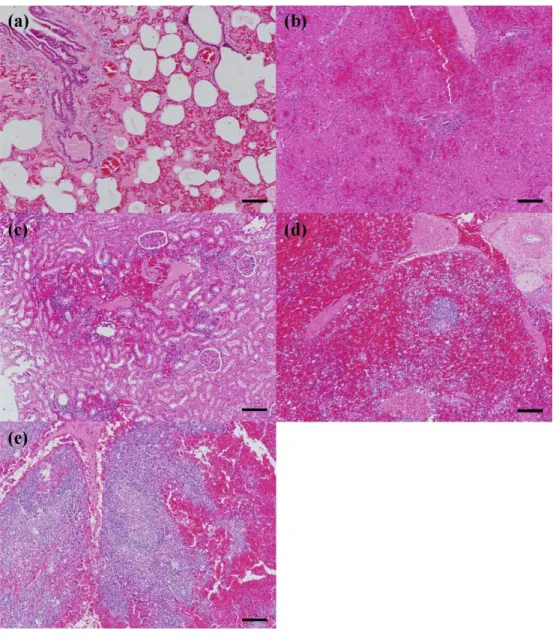
Figure 1. Gross lesions of pigs naturally infected with ASF ... 57 Figure 2. Histopathology in pigs naturally infected with ASF ... 60 Figure 3. Immunohistochemistry in pigs naturally infected with ASF ... 63
PART Ⅲ
Figure 1. Clinical signs of pigs with experimentally induced
ASF ... 81
X
Figure 2. Gross lesions of pigs with experimentally induced
ASF ... 84
Figure 3. Histopathology in pigs with experimentally induced
ASF ... 87
Figure 4. Immunohistochemistry in pigs with experimentally
induced ASF ... 90
LIST OF ABBREVIATIONS
ASF African swine fever ASFV African swine fever virus CPE Cytopathic effect
CSF Classical swine fever CSFV Classical swine fever virus CVR Central variable region DPI Days post infection FBS Fetal bovine serum
FMDV Foot-and-mouth disease virus HAD Hemadsorption reaction IHC Immunohistochemistry IPT Immunoperoxidase test MGF Multigene family ORF Open reading frames PBS Phosphate-buffered saline
PIM Pulmonary intravascular macrophages PPK Primary porcine kidney
PRRS Porcine reproductive and respiratory syndrome PRRSV Porcine reproductive and respiratory syndrome virus RT-PCR Real-time PCR
TCID50 50% Tissue culture infective dose
GENERAL INTRODUCTION
African swine fever (ASF) is a contagious hemorrhagic disease that affects domestic pigs and wild boars. African swine fever virus (ASFV) is the causative agent of ASF and is a large, icosahedral, enveloped, linear double strand DNA virus (Salas and Andrés, 2013). ASFV was first described in 1921 in Kenya (Montgomery, 1921), and the virus was transmitted intercontinentally to Portugal in 1957 (Manso‐
Ribeiro et al., 1958). After half a century, ASF entered Georgia in 2007 (Rowlands et al., 2007), with the first outbreaks occurring in China in 2018 (Zhou et al., 2018).
Currently, ASF has spread throughout Asia, including Vietnam, The Philippines, Mongolia, North Korea, South Korea, Indonesia, Myanmar, Laos, and Cambodia since 2019 (Mighell et al., 2021).
Genetic characterization of ASFV is a priority in the establishment of the heterogeneity and epidemiological links between the viruses in each ASF outbreak area (Misinzo et al., 2011). It means that possible routes and sources of ASFV can be identified through genotyping of the virus. A genotyping classification from partial sequencing of major structural protein p72 gene has been developed and widely used to define 24 ASFV genotypes from various regions (Achenbach et al., 2017; Quembo et al., 2017). Additional sequence assessments of structural proteins from the p30 and p54 genes have been used for epidemiological tracking with a higher resolution definition of virus relationships in regions where virus isolates are closely related to each other (Gallardo et al., 2009; Sanna, et al., 2017).
ASFV causes hemorrhagic disease such as Classical swine fever or erysipelas, showing no major clinical symptoms in wild boars, but various clinical symptoms in
domestic pigs. According to the course of clinical symptoms, the disease is classified into four types: peracute form, acute form, subacute form, and chronic form (Sánchez‐Vizcaíno et al., 2019). Among them, acute ASF is the most typical form and occurs when the virus is exposed to naïve pigs, resulting in high fever, loss of appetite, and inactivity. The affected animals have extensive necrosis and hemorrhage of their lymphoid tissues, splenomegaly, erythema, and pulmonary edema. (Mebus et al., 1983).
It is well established that monocyte/macrophage cell lineage is a main target for ASFV (Gomez-Villamandos et al., 2013). Viral antigens can be detected in macrophages of the spleen, liver, and lung from 3 days post infection (dpi) in case of acute ASF (Fernandez et al., 1992). Cellular necrosis resulting in lymphoid destruction starts at 3 dpi and becomes apparent at 5 dpi. Thrombocytopenia which causes hemorrhages in multiple organs under experimental conditions is also observed at 3 dpi (Zakaryan et al., 2014). The pathogenesis of ASFV can be varied depending on the virulence of the virus strain, the route of infection, and the dose of infection.
This dissertation was designed to reveal the pathogenesis of ASF occurring in Asia.
In Part Ⅰ, genetic characterization of the ASFV strain isolated from a recent Vietnam outbreak was conducted to understand the genetic and epidemiologic characteristics of ASF in Asia. In Part II, pathologic investigations consisted of observed gross and microscopic lesions were conducted in successive ASF outbreak cases in Vietnam to elucidate the pathogenesis of naturally occurring ASFV infection in Asia. In Part Ⅲ, a link between viral antigen distribution and tissue lesion development was confirmed by establishment of temporal pathology in pigs experimentally infected with Asian isolate of ASFV.
LITERATURE REVIEW
1. History
ASF, originally indigenous disease in wild African suids, was first described in 1921 in Kenya, when it transmitted to domestic pigs introduced from Europe (Montgomery, 1921). After its transcontinental transmission was reported in Portugal in 1957 (Manso‐Ribeiro et al., 1958), ASF was consecutively found in Europe including Spain (1960), France (1964), Italy (1967), Sardinia (1978), Malta (1978), Belgium (1985), and the Netherlands (1986) (Arias and Sanchez-Vizcaino, 2002). The disease also affects Central, North, and South America, including Cuba (1971), the Dominican Republic (1978), Brazil (1978), and Haiti (1979).
Subsequently ASF was eradicated from the western hemisphere with the exception of Portugal and Spain, where it became endemic for a period until 1995 (Bech- Nielsen et al., 1995; Arias et al., 2002). In 2007, ASF entered in Georgia (Rowlands et al., 2007), and spread to the Russian Federation (2007), Ukraine (2012) and Belarus (2013). The disease continued to spread to Asia through China in 2018 (Zhou et al., 2018), and has occurred sporadically in Vietnam, The Philippines, and South Korea since 2019 (Mighell et al., 2021). Currently, ASF outbreaks occur worldwide, and cause great damage to the pig industry and the economies of the affected countries.
2. Etiology
2-1 Classification
ASFV, the causing agent of ASF, is the only member of the family Asfarviridae, and is included in the genus Asfivirus (Dixon et al., 2005). ASFV is also the only known DNA arbovirus and is transmitted by soft ticks.
2-2 Virion Properties
The ASFV virion is enveloped and approximately 170 to 190 nm in diameter. The envelope contains hemagglutinin protein which is homologous to cellular CD2. The nucleocapsid core is surrounded by internal lipid layers and an icosahedral capsid.
The capsid consists of four concentric layers and an external hexagonal membrane (Salas and Andrés, 2013). ASFV has 151 to 167 viral proteins depending on the isolate (Jia et al., 2017), and more than 50 structural proteins in virions, indicating that ASFV particles are complex. Many of structural proteins are highly antigenic, including the major capsid protein p72 and the membrane proteins p30 and p54.
ASFV particles are sensitive to lipid solvents disrupting the envelope (Plowright and Parker, 1967). They are able to remain stable at a wide range of pH values (pH 4 – pH 10) but become inactive within minutes outside of the indicated range (EFSA, 2010). Virus particles found in serum or meat can remain infectious for months and even years at 4 °C. ASFV is thermolabile in that can be inactivated by heating at 60 °C for 30 minutes (Plowright and Parker, 1967) or 56 °C for 70 minutes (Mebus, 1988).
2-3 Genetic structure
ASFV is a linear double-stranded DNA virus. The viral genome of different isolates varies in length from 170 to 190 kb and encodes 151–167 open reading frames (ORF).
The Genome has covalently closed ends with inverted terminal repeats and hairpin loops. DNA consists of a conserved central region and two variable ends containing five multigene family (MGF) genes (Yañez et al., 1995). Gene Deletions and insertions occur within this MGF, generating antigenic variability which is associated with the degree of virulence and tick host range (Zsak et al., 2001; Burrage et al., 2004).
Since ASFV antigens do not sufficiently form neutralizing antibodies, a serotyping classification has not been developed. Instead, a genotyping classification with a partial sequence of structural protein from the p72 gene is used to define 24 ASFV genotypes (Achenbach et al. 2017; Quembo et al. 2017). ASFV isolates found in the Western Hemisphere before 2006 were identified as genotype I from West Africa, while a new genotype II from southeast Africa was found in the Caucasian region of Europe (EFSA, 2010). The sequence analysis of the central variable region (CVR) within the conserved central region is currently used for ASFV subtyping, which complements standard p72 genotyping (Phologane et al., 2005; Gallardo et al., 2014).
Also, an additional sequence assessment of p30 and p54 genes have been used for virus tracking. 15 complete genome sequences found from different regions are available to-date, each with a different degree of virulence and host range (wild pigs, domestic pigs, and ticks) (De Villiers et al., 2010).
2-4 Virus Replication
Field isolates of ASFV replicate in porcine monocytes and macrophages.
Replication occurs mainly in the cytoplasm, although the nucleus is needed at the early stage of viral DNA synthesis. Once the virions enter the endosomal pathway, transcription of early viral genes begins before the onset of DNA replication.
Intermediate and late genes are expressed following DNA replication. ASFV transcription follows temporal control which is relatively independent from the host cell (Rodriguez et al., 2013).
Several isolates can replicate in continuous cell lines including VERO, MS, CV-1, COS-1 and MA-104 after cell adaptation. These cell lines are currently used for ASFV research purposes, but there is a limitation in that they cause genetic and antigenic changes in the virus when subcultured for a long period of time (Sánchez‐
Vizcaíno et al., 2019).For these reasons, primary cells such as pulmonary alveolar macrophages are widely used for studying ASFV biology or diagnosis, but there are alternate problems with batch-to-batch consistency. This is one of the obstacles to the development of an ASFV vaccine.
3. Epidemiology
Although ASF has been eradicated in many countries, including Spain and Portugal, ASF has become endemic in some countries in Eastern Europe and continues to occur in Asia. Recently, the moderate virulent strains with low virulence have been found in endemic countries in Europe (Sehl et al., 2020). So far, two genetic variants have been identified, but it is necessary to monitor the emergence of new virus strains exhibiting unknown pattern of virulence. There are two conventional transmission patterns of ASF,one is the sylvatic cycle in wild boars and ticks, and the other is the epizootic and enzootic cycle in domestic swine.
3-1 Sylvatic Cycle
In southern and eastern Africa, ticks are biological vectors of ASFV. Indirect
transmission by biological vectors occurs in outdoor pig farms. The virus asymptomatically infects wild pigs (warthogs) and soft ticks (genus Ornithodoros) and is maintained in a sylvatic cycle (Maclachlan and Dubovi, 2010). ASFV replicates in the gut of ticks after the tick feeds on virus-infected pigs, and subsequently infects the reproductive system, causing transovarian transmission.
The virus is also excreted in saliva or Malpighian excrement, causing trans-stadial transmission between the ticks. Young wild boars may become infected with the virus by eating the infected ticks can develop viremia. In contrast, older wild boars do not show viremia, but the virus can cause persistent infections in the lymphoid tissues.
3-2 Domestic Cycle
In Europe, ASFV is transmitted mainly via direct contact between infected and uninfected pigs (Sánchez‐Vizcaíno et al., 2019). The primary introduction of ASFV into domestic pigs occurs through the bite of infected ticks or infected meat, or through contact with wild pigs (Maclachlan and Dubovi, 2010). Once the pigs are infected, the virus is excreted by oral fluid, feces, urine, and nasal discharge following the onset of clinical signs. The virus can be transmitted between pigs by direct contact, and within buildings by aerosol. Mechanical transmissions by worker and vehicles are possible and common, as the virus remains infectious in feces for several weeks. In recent years, there has been an increase in international transmission of ASFV reported in the process of importing and exporting uncooked pork.
4. Clinical signs
Wild boars usually do not show major clinical symptoms, whereas domestic pigs display a wide range of clinical symptoms. ASF is often confused with diseases such as classical swine fever (CSF) or erysipelas as they all cause hemorrhagic lesions.
The incubation period is 5 to 15 days, and the clinical symptoms, mortality and morbidity vary depending on the virulence of the virus strain, the route of exposure, and the dose of exposure.
4-1 Peracute form
Peracute form of ASF is induced by a highly virulent strain and is characterized by high fever, cutaneous hyperemia, hyperpnea, loss of appetite, and inactivity. The affected animals die suddenly, 1–4 days after clinical signs appear, but lesions may not be present in the organs of infected animals. The peracute form is usually reported in early outbreaks of ASF‐naïve regions (Sánchez-Vizcaíno et al., 2015).
4-2 Acute form
The acute form of ASF is induced by highly virulent or moderately virulent strains, and characterized by fever (40–42 °C), loss of appetite, inactivity, and leukopenia.
The affected animals have extensive necrosis and hemorrhage of lymphoid tissue, splenomegaly, erythema, and pulmonary edema.(Mebus et al., 1983).
Splenomegaly and petechia in the kidney are characteristic lesions of acute ASF, and affected lymph nodes (mainly the renal and gastrohepatic) display extensive signs of hemorrhage. Cutaneous hyperemia is an obvious sign, in which erythema and cyanosis appear in the skin of extremities, ears, chest, abdomen, and the perineum.
Hemorrhagic discharge from the nasal cavity or anus may be observed. Severe respiratory changes with labored breathing are often observed in the last stage of the disease. The mortality rate is almost 100% at 7–9 days after the onset of clinical signs, with abortion as a possible first clinical sign in an outbreak. Acute ASF is the most common form observed when the disease causes an outbreak in ASF‐naïve regions (Sánchez-Vizcaíno et al., 2015; Sánchez-Vizcaíno et al., 2019).
4-3 Subacute form
A subacute form of ASF is induced by moderately virulent strains. This form of the disease displays less severe clinical signs than the acute form, but hemorrhage and edema are sometimes more intense than the acute form (Gómez‐Villamandos et al., 2013). Transitory thrombocytopenia and leukopenia are observed in the early and mid-stages of the disease (Gómez‐Villamandos et al., 1997). Other symptoms include moderate to high fever, edema in several organs, hemorrhage and splenomegaly. The affected animals may die within 20 days, resulting in a 30 to 70%
range of mortality rate. Subacute ASF can be observed in endemic areas, and surviving animals usually recover after 3-4 weeks, with intermittent viremia and anti-ASFV antibodies detected (Mur et al., 2016).
4-4 Chronic form
The chronic form of ASF is induced by a low virulent strain, reported in Spain, Portugal and recently Eastern Europe (Gallardo et al., 2018). Animals infected with this form of disease have no specific clinical signs, but some animals may excrete the virus for long periods of time resulting in the persistence of the disease in those
affected regions. These animals may show necrotic lesions of the skin, arthritis (fibrinous arthritis/periarthritis), and lameness, but remain void of vascular lesions.
The problem with this form of the disease is that it causes chronic growth retardation.
5. Pathology
Pigs are initially infected with ASFV via the oronasal route, where the virus first replicates in the tonsils and regional lymph nodes then spreads rapidly through the lymph and blood into the body. Pigs suffering from ASF show a wide range of lesions in multiple organs depending on the virus virulence. Spleen enlargement, necrosis and hemorrhage in lymph nodes, petechia in kidneys, edema in the lung, and congestion in the liver are typical lesions associated with ASF. The lesions are based on lymphoid destruction and hemorrhage in the most clinical form of the disease, both of which have characteristic pathogenic mechanisms.
5-1 Pathogenesis of Lymphoid lesions
The main target of ASFV is of monocyte/macrophage lineage, and their ability to induce (or inhibit) the expression of pro-inflammatory cytokines and interferons; a critical factor in viral virulence (Maclachlan & Dubovi, 2010). The ASFV genome contain genes involved in the modulation of inflammation and host immune responses. The A238L gene encodes a protein that inhibits the activation of cellular transcription factor, and nuclear factor kB, while the 8DR gene induces T lymphocyte activation and mediation of hemadsorption by infected cells (Maclachlan & Dubovi, 2010). Also, there are genes involved in unprogrammed cell death in both inhibitory and inducing manners. In general, ASFV infection results in
severe leukopenia caused by apoptosis of lymphocytes and mononuclear cells, in which the massive destruction of lymphoid tissues including tonsil, thymus, spleen and lymph nodes is observed as disease progresses (Salguero, 2020). The upregulation of proinflammatory cytokines described as cytokine storm is a necessary mechanism for the extensive induction of apoptosis of lymphocytes (Salguero et al., 2005).
5-2 Pathogenesis of Vascular change
Hemorrhages, the typical lesion of the ASF, appear in multiple organs including kidney and visceral lymph nodes which lack fixed vascular macrophages. (Gomez- Villamandos et al., 2013). Importantly, hemorrhages may occur at earlier phase of the disease, while ASFV replicates in endothelial cells at only late phase (Gomez- Villamandos et al., 1995). This means that hemorrhage is not directly related with virus-induced cell damage. For this reason, a pathogenic mechanism has been proposed in that, after capillary endothelial cells activate phagocytosis, cell hypertrophy occurs, occludes the capillary lumen, and thus increases intravascular pressure (Gomez-Villamandos et al., 2013). The subsequent endothelial loss causes the exposure of the basal membrane where platelets can adhere resulting in the activation of the coagulation system and finally leading to disseminated intravascular coagulation (Gomez-Villamandos et al., 2013). The pathogenesis of the pulmonary edema is relevant with pulmonary intravascular macrophages (PIM), which secrete chemotactic proinflammatory cytokines such as IL-1α and TNF-α, thereafter, increasing the endothelial permeability. Increased permeability causes capillary fluid to leak into the alveolar and interalveolar space (Carrasco et al., 2002).
6. Diagnosis
Because the clinical signs and lesions of ASF are similar to those of erysipelas, salmonellosis, and especially CSF, a diagnosis cannot be made through observation or autopsy alone. There are various laboratory analyses available for ASFV diagnosis and samples including blood, oral fluid, lymphoid organs, kidney and lung are recommended for analysis (Arias and Sánchez‐Vizcaíno, 2002).
6-1 Viral genome detection
Viral genome detection using either conventional or real-time PCR is currently the golden standard because of its high sensitivity and specificity. Most PCR methods are designed to ensure detection of almost all ASFV strains by targeting the highly conserved gene which encodes for a major viral structural protein, p72 (King et al., 2003). PCR methods have the advantage of being able to detect the virus even in the early phase of infection when animals do not show clinical symptoms. Multiplex PCR methods have been developed for differential diagnosis of ASF from similar clinical disease such as Classical swine fever virus (CSFV) (Agüero et al., 2004).
Although multiplex PCR usually have lower sensitivity than single assays, it is useful for surveillance in ASF naïve regions and ASF/CSF co-circulation regions.
6-2 Virus isolation
Field strains of ASFV can be isolated in susceptible primary cell cultures of swine such as monocytes and macrophages obtained from blood or the lung. The infectious virus in the specimen replicates in cell culture and displays cytopathic effect (CPE)
and hemadsorption reaction (HAD) (Sánchez‐Vizcaíno et al., 2019). Despite these useful tools, isolation of a wild-type virus from field specimens may provide irregular results and low effectiveness (Gallardo et al., 2015). Some field isolates do not display HAD, but rather CPE (Leitão et al., 2001; Boinas et al., 2004). Isolation of these non-HAD strains requires further confirmation using PCR to produce consistent results (Oura et al., 2013). Despite these limitations, virus isolation is essential in studying the biological characterization of ASFV strains.
6-3 Antigen detection
ASFV antigen detection has been conducted with direct, indirect and sandwich ELISAs, that have been developed both commercially as well as in-house. While in- house antigen ELISAs are not typically the preferred method, commercially developed antigen ELISA kits are available for the analysis of serum samples. They produce results rapidly and methods are easy to scale up, but have relatively low sensitivity (77.2%) when compared to the routine PCR recommended by OIE, even in high-tittered samples (Oura et al., 2013; Gallardo et al., 2015). Antigen ELISAs are therefore only recommended when combined with other serological tests.
6-4 Antibody detection
Serological antibody detection tests are commonly used for ASF diagnosis due to their relatively simple methodology and low cost involved as they require only a few devices and minor facilities. Diseased animals affected with ASFV have anti-ASFV antibodies for up to several months or even years after infection (Arias and Sánchez- Vizcaíno, 2002). Since there is currently no commercially available ASF vaccine, all
animals with positive serum antibody tests can be diagnosed as having a natural infection. Likewise, detection of ASFV-specific antibody is important in the evaluation of epidemiological situations in disease affected regions. Commercial ELISA kits from various manufacturers have been developed for the detection of ASF antibodies against most antigenic viral proteins including p30, p54, p72, and pp62. The ELISA test may exhibit lower sensitivity in detection during early stage of ASFV infection (7–12 days post infection), but it is reliable for the detection of antibodies during a fulminant course of infection (12-14 days post infection) (Gallardo et al., 2015). Although it is limited in its use of serum samples, antibody detection using ELISA methodology has still advantages for large-scale serological studies as it is a fast, easy, and economical method.
Positive ASFV test results from an ELISA could be confirmed by back-up methods such as an Indirect Immunoperoxidase test (IPT), recommended by the OIE (Gallardo et al., 2015). It requires the use of a fixed monolayer culture of VERO or an MS cell line infected with adapted-grown ASFV. Although there are some drawbacks such as results can be subjectively interpreted, the tests have a high sensitivity and specificity when performed by well trained staff.This method has an advantage in that any kind of specimen such as serum, excreted discharge, and body fluids can be used for accurate diagnosis.
7. Prevention
Since there is no commercially available vaccine for ASF, epidemic countries rely on active surveillance and culling as preventative measures. Eradication of ASFV has been achieved without vaccination in certain countries by having an effective
contingency plan in place. To prepare for ASF outbreak that is spreading around the world, a plan suitable for each country is needed.
7-1 Quarantine
Countries that are free of ASF have quarantine strategies in place that ban the importation of live pigs and pork products from infected countries (EFSA, 2010). In Asia and Europe, where recent ASF outbreaks occur sporadically, vehicles, people, and products related to the swine industry are monitored while taking a preemptive culling strategy around the outbreak area. Endemic countries such as Sardinia implement monitoring and control of live pig and pork product movement as a preventive measure, and extensive serological surveillance to detect carriers that are the source of ASFV. In endemic areas of Africa, control of natural hosts, such as wild African suids or soft ticks, is important in preventing the spread of ASF to domestic pigs (Sánchez‐Vizcaíno et al., 2019).
7-2 Vaccination
Currently, a successful vaccine against ASFV is not developed or available. The first trial that used a live attenuated strain in 1963 in Portugal was unsuccessful (Manso‐Ribeiro et al., 1963). Since then, some experimental vaccines based on attenuated ASFV strains have been developed but safety and efficacy issues remain unresolved (King et al., 2011; O’Donnell et al., 2016; Reis et al., 2016). An inactivated ASF vaccine is the fastest and easiest to make, but has low immunogenicity compared to live attenuated vaccine options. Inactivated vaccines have a major drawback in that it is impossible to induce a long-term T-cell immune
response. Subunit vaccines induce specific immunity by combining the expression of specific ASFV proteins. Viral epitope analysis is important, but effective subunit candidates are difficult to select as there are more than 167 ORFs of ASFV. Previous attempts of combining p30, p54, and p72 failed to induce neutralizing antibodies.
Recombinant live vaccines have been developed by removing specific genes that related with virulence or a host immune response using CRISPR-Cas 9 or DNA homologous recombination. Also, live attenuated vaccines have been developed through unintentional genetic changes made during long subcultures. Chronic side effects accompanied by high fever have occurred from vaccination with some live strains, and catastrophic recombination may occur through recombination with wild type strains. In order to develop an effective ASF vaccine, there are many problems regarding not only efficacy, but also safety issues (Rock, 2017; Teklue et al., 2020).
8. References
Achenbach, J. E., Gallardo, C., Nieto‐Pelegrín, E., Rivera‐Arroyo, B., Degefa‐Negi, T., Arias, M., ... & Sánchez‐Vizcaíno, J. M. (2017). Identification of a new genotype of African swine fever virus in domestic pigs from Ethiopia. Transboundary and emerging diseases, 64(5), 1393-1404.
Agüero, M., Fernández, J., Romero, L. J., Zamora, M. J., Sánchez, C., Belák, S., ...
& Sánchez-Vizcaíno, J. M. (2004). A highly sensitive and specific gel-based multiplex RT-PCR assay for the simultaneous and differential diagnosis of African swine fever and Classical swine fever in clinical samples. Veterinary research, 35(5), 551-563.
Arias, M., Sánchez-Vizcaíno, J. M., Morilla, A., Yoon, K. J., & Zimmerman, J. J.
(2002). African swine fever. Trends in emerging viral infections of swine, 119-124.
Bech-Nielsen, S., Fernandez, J., Martinez-Pereda, F., Espinosa, J., Bonilla, Q. P., &
Sanchez-Vizcaino, J. M. (1995). A case study of an outbreak of African swine fever in Spain. British veterinary journal, 151(2), 203-214.
Boinas, F. S., Hutchings, G. H., Dixon, L. K., & Wilkinson, P. J. (2004).
Characterization of pathogenic and non-pathogenic African swine fever virus isolates from Ornithodoros erraticus inhabiting pig premises in Portugal. Journal of general virology, 85(8), 2177-2187.
Burrage, T. G., Lu, Z., Neilan, J. G., Rock, D. L., & Zsak, L. (2004). African swine fever virus multigene family 360 genes affect virus replication and generalization of infection in Ornithodoros porcinus ticks. Journal of virology, 78(5), 2445-2453.
Carrasco, L., Nunez, A., Salguero, F. J., San Segundo, F. D., Sanchez-Cordon, P., Gomez-Villamandos, J. C., & Sierra, M. A. (2002). African swine fever: Expression of interleukin-1 alpha and tumour necrosis factor-alpha by pulmonary intravascular macrophages. Journal of comparative pathology, 126(2-3), 194-201.
De Villiers, E. P., Gallardo, C., Arias, M., Da Silva, M., Upton, C., Martin, R., &
Bishop, R. P. (2010). Phylogenomic analysis of 11 complete African swine fever virus genome sequences. Virology, 400(1), 128-136.
Dixon LK, Escribano JM, Martins C, et al. (2005). The Asfarviridae. In Fauquet CM, Mayo MA, Maniloff J, et al., eds. Virus Taxonomy: VIII Report of the International Committee for the Taxonomy of Viruses. London: Elsevier/Academic Press, pp. 135-143.
Donald, P. K. (2010). Asfarviridae and Iridoviridae. In Maclachlan NJ, & Dubovi EJ, eds. Fenner's veterinary virology (4th ed). Burlington, MA: Elsevier/Academic press, pp. 167-172.
European Food Safety Authority (EFSA). 2010. Scientific opinion on African swine fever. EFSA J 8:1-149.
Fernandez, A., Perez, J., Carrasco, L., Bautista, M. J., Sanchez‐Vizcaino, J. M., &
Sierra, M. A. (1992). Distribution of ASFV antigens in pig tissues experimentally infected with two different Spanish virus isolates. Journal of veterinary medicine, Series B, 39(1‐10), 393-402.
Gallardo, C., Fernández-Pinero, J., Pelayo, V., Gazaev, I., Markowska-Daniel, I., Pridotkas, G., ... & Arias, M. (2014). Genetic variation among African swine fever genotype II viruses, eastern and central Europe. Emerging infectious diseases, 20(9), 1544.
Gallardo, C., Mwaengo, D. M., Macharia, J. M., Arias, M., Taracha, E. A., Soler, A., ... & Bishop, R. P. (2009). Enhanced discrimination of African swine fever virus
isolates through nucleotide sequencing of the p54, p72, and pB602L (CVR) genes.
Virus genes, 38(1), 85-95.
Gallardo, C., Nieto, R., Soler, A., Pelayo, V., Fernández-Pinero, J., Markowska- Daniel, I., ... & Arias, M. (2015). Assessment of African swine fever diagnostic techniques as a response to the epidemic outbreaks in Eastern European Union countries: how to improve surveillance and control programs. Journal of clinical microbiology, 53(8), 2555-2565.
Gallardo, C., Nurmoja, I., Soler, A., Delicado, V., Simón, A., Martin, E., ... & Arias, M. (2018). Evolution in Europe of African swine fever genotype II viruses from highly to moderately virulent. Veterinary microbiology, 219, 70-79.
Gomez-Villamandos, J. C., Bautista, M. J., Carrasco, L., Caballero, M. J., Hervas, J., Villeda, C. J., ... & Sierra, M. A. (1997). African swine fever virus infection of bone marrow: lesions and pathogenesis. Veterinary pathology, 34(2), 97-107.
Gomez-Villamandos, J. C., Bautista, M. J., Sanchez-Cordon, P. J., & Carrasco, L.
(2013). Pathology of African swine fever: the role of monocyte-macrophage. Virus research, 173(1), 140-149.
Gómez-Villamandos, J. C., Hervás, J., Méndez, A., Carrasco, L., Villeda, C. J., Wilkinson, P. J., & Sierra, M. A. (1995). Ultrastructural study of the renal tubular system in acute experimental African swine fever: virus replication in glomerular mesangial cells and in the collecting ducts. Archives of virology, 140(3), 581-589.
Jia, N., Ou, Y., Pejsak, Z., Zhang, Y., & Zhang, J. (2017). Roles of African swine fever virus structural proteins in viral infection. Journal of veterinary research, 61(2), 135.
King, D. P., Reid, S. M., Hutchings, G. H., Grierson, S. S., Wilkinson, P. J., Dixon, L. K., ... & Drew, T. W. (2003). Development of a TaqMan® PCR assay with internal amplification control for the detection of African swine fever virus. Journal of virological methods, 107(1), 53-61.
King, K., Chapman, D., Argilaguet, J. M., Fishbourne, E., Hutet, E., Cariolet, R., ...
& Takamatsu, H. H. (2011). Protection of European domestic pigs from virulent African isolates of African swine fever virus by experimental immunisation. Vaccine, 29(28), 4593-4600.
Leitão, A., Cartaxeiro, C., Coelho, R., Cruz, B., Parkhouse, R. M. E., Portugal, F.
C., ... & Martins, C. L. (2001). The non-haemadsorbing African swine fever virus isolate ASFV/NH/P68 provides a model for defining the protective anti-virus immune response. Journal of general virology, 82(3), 513-523.
Manso Ribeiro, J., Roza Azevedo, J. A., Teixeira, M. J. O., Braco Forte, M. C., Rod‑rigues Ribeiro, A. M., Oliveira, F., ... & Dias Vigario, J. (1958). Peste Porcine provoquée par une souche différente (Souche L) de la souche classique. Bulletin - Office international des epizooties, 50, 516-534.
Manso-Ribeiro, J., Nunes-Petisca, J. L., Lopez-Frazao, F., & Sobral, M. (1963).
Vaccination against ASF. Bulletin - Office international des epizooties, 60, 921-937.
Mebus, C. A. (1988). African swine fever. Advances in virus research, 35, 251-269.
Mebus, C. A., McVicar, J. W., & Dardiri, A. H. (1983). Comparison of the pathology of high and low virulence African swine fever virus infections.
Comparison of the pathology of high and low virulence African swine fever virus infections., 183-194.
Mighell, E., & Ward, M. P. (2021). African swine fever spread across Asia, 2018–
2019. Transboundary and emerging diseases, 68(5), 2722-2732.
Misinzo, G., Magambo, J., Masambu, J., Yongolo, M. G., Van Doorsselaere, J., &
Nauwynck, H. J. (2011). Genetic characterization of African swine fever viruses from a 2008 outbreak in Tanzania. Transboundary and emerging diseases, 58(1), 86- 92.
Montgomery, R. E. (1921). On a form of swine fever occurring in British East Africa (Kenya Colony). Journal of comparative pathology and therapeutics, 34, 159- 191.
Mur, L., Atzeni, M., Martínez‐López, B., Feliziani, F., Rolesu, S., & Sanchez‐
Vizcaino, J. M. (2016). Thirty‐five‐year presence of African swine fever in Sardinia:
History, evolution and risk factors for disease maintenance. Transboundary and
emerging diseases, 63(2), 165-177.
O'Donnell, V., Risatti, G. R., Holinka, L. G., Krug, P. W., Carlson, J., Velazquez- Salinas, L., ... & Borca, M. V. (2017). Simultaneous deletion of the 9GL and UK genes from the African swine fever virus Georgia 2007 isolate offers increased safety and protection against homologous challenge. Journal of virology, 91(1), e01760-16.
Oura, C. A. L., Edwards, L., & Batten, C. A. (2013). Virological diagnosis of African swine fever—comparative study of available tests. Virus research, 173(1), 150-158.
Phologane, S. B., Bastos, A. D., & Penrith, M. L. (2005). Intra-and inter-genotypic size variation in the central variable region of the 9RL open reading frame of diverse African swine fever viruses. Virus genes, 31(3), 357-360.
Plowright, W., & Parker, J. (1967). The stability of African swine fever virus with particular reference to heat and pH inactivation. Archiv für die gesamte Virusforschung, 21(3), 383-402.
Quembo, C. J., Jori, F., Vosloo, W., & Heath, L. (2018). Genetic characterization of African swine fever virus isolates from soft ticks at the wildlife/domestic interface in Mozambique and identification of a novel genotype. Transboundary and emerging diseases, 65(2), 420-431.
Reis, A. L., Abrams, C. C., Goatley, L. C., Netherton, C., Chapman, D. G., Sanchez-
Cordon, P., & Dixon, L. K. (2016). Deletion of African swine fever virus interferon inhibitors from the genome of a virulent isolate reduces virulence in domestic pigs and induces a protective response. Vaccine, 34(39), 4698-4705.
Rock, D. L. (2017). Challenges for African swine fever vaccine development—“…
perhaps the end of the beginning.”. Veterinary microbiology, 206, 52-58.
Rodríguez, J. M., & Salas, M. L. (2013). African swine fever virus transcription.
Virus research, 173(1), 15-28.
Rowlands, R. J., Michaud, V., Heath, L., Hutchings, G., Oura, C., Vosloo, W., ... &
Dixon, L. K. (2008). African swine fever virus isolate, Georgia, 2007. Emerging infectious diseases, 14(12), 1870.
Salas, M. L., & Andrés, G. (2013). African swine fever virus morphogenesis. Virus research, 173(1), 29-41.
Salguero, F. J. (2020). Comparative pathology and pathogenesis of African swine fever infection in swine. Frontiers in veterinary science, 7, 282.
Salguero, F. J., Sanchez-Cordon, P. J., Nunez, A., de Marco, M. F., & Gomez- Villamandos, J. C. (2005). Proinflammatory cytokines induce lymphocyte apoptosis in acute African swine fever infection. Journal of comparative pathology, 132(4), 289-302.
Sánchez‐Vizcaíno, J. M., Laddomada, A., & Arias, M. L. (2019). African swine fever virus. Diseases of swine, 443-452.
Sánchez-Vizcaíno, J. M., Mur, L., Gomez-Villamandos, J. C., & Carrasco, L.
(2015). An update on the epidemiology and pathology of African swine fever.
Journal of comparative pathology, 152(1), 9-21.
Sanna, G., Dei Giudici, S., Bacciu, D., Angioi, P. P., Giammarioli, M., De Mia, G.
M., & Oggiano, A. (2017). Improved strategy for molecular characterization of African swine fever viruses from Sardinia, based on analysis of p30, CD2V and I73R/I329L variable regions. Transboundary and emerging diseases, 64(4), 1280- 1286.
Sehl, J., Pikalo, J., Schäfer, A., Franzke, K., Pannhorst, K., Elnagar, A., ... &
Breithaupt, A. (2020). Comparative pathology of domestic pigs and wild boar infected with the moderately virulent African swine fever virus strain “Estonia 2014”.
Pathogens, 9(8), 662.
Teklue, T., Sun, Y., Abid, M., Luo, Y., & Qiu, H. J. (2020). Current status and evolving approaches to African swine fever vaccine development. Transboundary and emerging diseases, 67(2), 529-542.
Yanez, R. J., Rodrı́guez, J. M., Nogal, M. L., Yuste, L., Enrı́quez, C., Rodriguez, J.
F., & Vinuela, E. (1995). Analysis of the complete nucleotide sequence of African swine fever virus. Virology, 208(1), 249-278.
Zakaryan, H., Karalova, E., Voskanyan, H., Ter-Pogossyan, Z., Nersisyan, N., Hakobyan, A., ... & Karalyan, Z. (2014). Evaluation of hemostaseological status of pigs experimentally infected with African swine fever virus. Veterinary microbiology, 174(1-2), 223-228.
Zsak, L., Lu, Z., Burrage, T. G., Neilan, J. G., Kutish, G. F., Moore, D. M., & Rock, D. L. (2001). African swine fever virus multigene family 360 and 530 genes are novel macrophage host range determinants. Journal of virology, 75(7), 3066-3076.
Zhou, X., Li, N., Luo, Y., Liu, Y. E., Miao, F., Chen, T., ... & Hu, R. (2018).
Emergence of African swine fever in China, 2018. Transboundary and emerging diseases, 65(6), 1482-1484.
PART Ⅰ. Genetic Characterization of an Asian Isolate
of African Swine Fever Virus
ABSTRACT
African swine fever virus (ASFV) is a large, icosahedral, enveloped, linear double strand DNA virus, causes hemorrhagic disease in domestic pigs and wild boars, and has been present in Asia since 2018. Genetic characterization of ASFV is essential in establishing the heterogeneity and epidemiological links between the viruses in each ASF outbreak area. In this study, the genetic characterization of an ASFV isolate recently found in Vietnam was conducted to understand the genetic and epidemiologic characteristics of ASFV circulation within Asia. The wild-type virus was isolated using primary porcine kidney (PPK) cells, and successfully propagated to obtain a strain stock. Partial sequencing of viral major structural proteins p72, p54, and p30 categorized the new ASFV isolate as Genotype Ⅱ as it showed a high genetic homology with all isolates from other Asian countries. The partial sequences of these Asian isolates were also identical to those found in Georgia and Eastern Europe since 2007. These results revealed that ASF outbreaks in Asia were probably due to a single introduction of ASFV genotype Ⅱ that spread slowly eastward, and without the introduction of additional virus genotypes.
Keywords: African swine fever virus; Genetic characterization; Asian Isolate;
Primary Porcine Kidney cells
INTRODUCTION
African Swine Fever Virus (ASFV), the causing agent of ASF, is a large, icosahedral, enveloped, linear double strand DNA virus. ASFV is the only member of the family Asfarviridae, found in the genus Asfivirus (Dixon et al., 2005). After ASFV was first described in 1921 in Kenya (Montgomery, 1921), its transcontinental transmission was reported in Portugal in 1957 (Manso‐Ribeiro et al., 1958). In 2007, ASF entered in Georgia (Rowlands et al., 2007), and spread to China in 2018 (Zhou et al., 2018). Currently, ASF continues to occur throughout Asia including Vietnam, Philippines, Mongolia, North Korea, South Korea, Indonesia, Myanmar, Laos, and Cambodia (Mighell et al., 2021).
Genetic characterization of ASFV is a priority in the establishment of the heterogeneity and epidemiological links between the viruses in each ASF outbreak area (Misinzo et al., 2011). It means that possible routes and sources of ASFV can be identified through genotyping, thereby preventing further introductions of ASF.
Because ASFV antigens do not form sufficient neutralizing antibodies, a genotyping classification rather than a serotyping classification has been developed. A genotyping classification from partial sequencing of major structural protein p72 gene has been developed and widely used to define 24 ASFV genotypes from various regions (Achenbach et al., 2017; Quembo et al., 2017). Additional sequence assessments of structural proteins from the p30 and p54 genes have been used for epidemiological tracking with a higher resolution definition of virus relationships in regions where virus isolates are closely related to each other (Sanna, et al., 2017;
Gallardo et al., 2008).
In this study, the genetic characterization of the ASFV strain isolated from a recent Vietnam outbreak was conducted to understand the genetic and epidemiologic characteristics of ASFV, which is recently prevalent in Asia. As ASFV can undergo genetic and antigenic changes when extensively cultured in an existing continuous cell line (Sánchez‐Vizcaíno et al., 2019), primary porcine kidney cells were selected for isolation of the wild-type virus to reduce these risks. Thereafter, partial sequencing against p72 genes as well as p30 and p54 genes of the virus isolates were performed.
MATERIALS AND METHODS
Preparation of primary porcine kidney cell culture
Crossbred pigs that were 21-28 days old were used to harvest primary porcine kidney cells with slight modifications from the referenced methodology (Takenouchi et al., 2014). The piglets were screened where they tested seronegative for PCV2, PRRSV and M. hyopneumoniae (Kim et al., 2003; Do et al., 2016; Dubosson et al., 2004). The tissue collection process was performed by anesthetizing the animals prior to euthanasia. The kidneys were then removed during necropsy. Post-necropsy, the tissues were further processed by removing the fibrous renal capsule from the kidneys. The renal cortex was then cut into small pieces (3–5 g each), and finely minced with scissors. The prepared tissues were transferred into 50 mL tubes containing a phosphate-buffered saline (PBS) solution (0.5 mg/mL) supplemented with dispase (250 U/ml, Falcon, Corning, NY, USA) and DNase I (13 U/ml, Worthington, Lakewood, NJ, USA). Tissues were then digested by a tube rotator containing collagenase (Wako, Tokyo, Japan) while incubating for 1h at 37 °C. Once digested, the material was filtered through a nylon mesh (pore size: 100mm), and re- suspended in growth medium composed of supplemented RPMI-1640 medium (Gibco, Grand Island, NY, USA) containing 10% heat- inactivated fetal bovine serum (Gibco), 0.005 mM 2-mercaptoethanol (Sigma-Aldrich, St. Louis, MO, USA), and 10 µg/mL bovine insulin (Sigma). The media also contained antibiotics as follows: 100U/ mL penicillin (Gibco), 100 mg/ mL streptomycin (Gibco), and 0.25 ug/ mL Amphotericin B (Gibco). The cell suspension was split into 20 x T-75 tissue culture flasks (Falcon) and cultured at 37 °C in a 5% CO2 incubator for 2–3 weeks.
Full media changes occurred every 3–4 days throughout incubation. At the end of the incubation period, cells were harvested with a cell scraper and suspended in Cell Reservoir One with DMSO (Nacalai tesque, Kyoto, Japan) and immediately frozen in liquid nitrogen. This study was approved by Seoul National University Institutional Animal Care and Use Committee.
ASFV isolation using primary porcine kidney cells
After a stock of primary porcine kidney cells was established, ASFV was isolated from tissues of a pig suffering from acute ASF. A clinically suspected 10-week-old pig was selected from a farm with an ASF outbreak in Vietnam, and was confirmed through testing as positive exclusively for ASFV (King et al., 2003). Renal and gastrohepatic lymph nodes and spleen were collected from this pig post-euthanasia.
Lymph nodes and spleen tissue homogenates were prepared for ASFV isolation as follows: Approximately 2% of the tissues were suspended into RPMI-1640 medium containing antibiotic-antimycotics (100 U of penicillin G per mL, 100 g of streptomycin sulfate per mL, and 0.025 g of amphotericin B per mL; Gibco, Invitrogen-Gibco, Carlsbad, CA, USA). The suspensions were then homogenized with a laboratory blender for 3 minutes, centrifuged at 1500 g for 5minutes, and sterilized through a 0.45 μm filter. The homogenates prepared in this manner was used as the inoculum for virus isolation. Once primary porcine kidney cells reached 80% confluency in T-25 tissue culture flasks, cells were dissociated with trypsin.
Trypsin was then neutralized with, and cells were resuspended in 2mL RPMI-1640 medium containing 10% fetal bovine serum (FBS). The cell suspension was then mixed with 1mL aliquots of the inoculum (0.3 Multiplicity of Infection) described above. Three mL of infected cell aliquots was seeded into new T-25 flasks and placed
inside of a 37 °C, 5 % CO2 incubator for 2 hours for adsorption onto cell layers. After two hours, flasks were fed with 5mL of RPMI-1640 medium and returned to the incubator for 48−72 hours. Post-incubation, three freeze-thaw cycles were performed on the infected flasks. Freshly cultured cells were infected by adding 1 mL of the cell lysate obtained from the previous 3x freeze/thawed flask. Several passages were performed in the same manner to propagate the virus stock.
Replication kinetics of ASFV isolated in primary kidney cells
Replication kinetics were determined for the ASFV isolates. The viral titration of culture supernatants was performed with real-time PCR (RT-PCR) and cytopathic effect (CPE)-based assays at in 1,3,5 and 7 days post infection (dpi). A titration of cell-free viral DNA was conducted by RT-PCR targeting p72 genes (King et al., 2003). Briefly, the forward and reverse primers, and probe were 5′-
CTGCTCATGGTATCAATCTTATCGA-3′ and 5′-
GATACCACAAGATC(AG)GCCGT-3′, and 5′-(FAM)-
CCACGGGAGGAATACCAACCCAGTG-3′-(TAMRA), respectively. The RT- PCR conditions were 95 °C for 5 min, followed by 40 cycles of 95 °C for 15 s and 60 °C for 40 s. Genome copy numbers were calculated from Ct values obtained from RT-PCR using a standard curve constructed by 10-fold dilutions of quantified ASFV DNA. The results were expressed as Log10 genome copies/mL.
A titration of live-infectious virus was conducted by the CPE-based assay as previously described except for slight modifications (Hakobyan et al., 2018). Briefly, the titer was calculated by observing CPE on primary porcine kidney cells for the end-point of the 10-fold serial dilution. The results were expressed as log10
TCID50/mL according to the Reed and Muench method by tissue culture infective dose (TCID) (Reed and Muench, 1983).
Viral genome sequencing analysis
The sequencing of major structural proteins p72, p54, and p30 of the virus isolate was conducted as previously described (Bastos et al., 2003; Rowlands, et al., 2008;
Gallardo et al., 2009). Genotyping primers which amplify the C-terminal region of the p72 gene were 5′-GGCACAAGTTCGGACATGT- 3′and 5′- GTACTGTAACGCAGCACAG- 3′. The whole gene encoding the p54 protein was amplified using the primers 5′ -CGAAGTGCATGTAATAAACGTC-3′and 5′
-TGTAATTTC ATTGCGCCACAAC-3′. The primer pairs used to amplify the CP204L (p30) gene were 5′-ATGAAAATGGAGGTCATCTTCAAAAC-3′and 5′-AAGTTTAATGACCATGAGTCTTACC-3′. The obtained sequences of each of the three genes were deposited in the NCBI GenBank with the accession numbers of MW039157 (p72), MW039156 (p54) and MW039155 (p30) respectively. These sequences were further analyzed using BioEdit 7.2.5 with ClustalW for a comparison with other ASFV strains. To genotype the newly isolated strain, p72 sequences of all 24 genotype strains known to date were obtained from Genbank. In addition, p30 and p54 sequences of strains corresponding to the same genotype as the newly isolated strain were also obtained for further genetic characterization. Comparative genetic analysis was performed using BioEdit 7.2.5 with a sequence identity matrix, and the phylogenetic trees were constructed using the Neighbor-joining method with 1000 bootstrap values in Mega X software.
RESULTS
Replication kinetics of the ASFV isolate
ASFV DNA was not detected in infected cell culture supernatant at 1 dpi. Detection began in infected cell culture supernatant from 3 dpi, and the genomic copies of DNA continuously increased until 7 dpi. Infectious ASFV was also absent in infected cell culture supernatant at 1 dpi. The infectious virus was first detected in the infected cell culture supernatant from 3dpi and onward, with a continuously increasing titer until 7dpi (Figure 1).
Figure 1. Replication kinetics of ASFV isolate in primary porcine kidney cells. RT- PCR results (bar) were expressed as log10 genomic copies/mL and CPE-based assay results (line) were expressed as log10 TCID50/mL. Results of each time point represents the mean of three independent experiment of titration.
Phylogenetic analysis of the ASFV isolate
A phylogenetic analysis of the p72 partial sequence of the Vietnamese ASFV isolate categorized the isolate as part of genotype Ⅱ with high homology. The p72 partial sequence of this isolate was 100% identical to the genotype Ⅱ isolates from Georgia (2007), Armenia (2007), Russia (2012), Lithuania (2014), Poland (2014), China (2018), South Korea (2019), and Indonesia (2020) (Figure 2).
During phylogenetic analysis of the full p54 genome, the Vietnamese isolate was 100% identical with isolates from South Korea (2019), Mongolia (2019), Indonesia (2019), Timor-Letse (2019), Russia (2019), China (2018), Georgia (2007), Mozambique (2005), Madagascar (2003), but only 97.4% identical with the isolate from Zambia (2015) (Figure 3a).
During phylogenetic analysis of the partial genome of p30, the Vietnamese isolate was 100% identical with isolates from Timor-Letse (2019), China (2018), Russia (2012), Georgia (2007), and Mozambique (2002), but only 99.8% and 97.7%
identical with isolates from Madagascar (1998) and Zambia (2002) respectively (Figure 3b).
Figure 2. Phylogenetic analysis with neighbor-joining method of major structural protein p72 partial sequence (MW039157) of a causative virus strain (●) from an ASF outbreak in Vietnam, 2019. The strains used in the analysis are represented by GenBank accession numbers/country of origin/collection date/genotype. Scale bar indicates nucleotide substitutions per site.
Figure 3. Phylogenetic analysis with neighbor-joining method of major structural protein p54 (MW039156; a) and p30 (MW039155; b) partial sequence of a causative virus strain (●) from an ASF outbreak in Vietnam, 2019. The strains used in the analysis are represented by GenBank accession numbers/country of origin/collection date/genotype. Scale bar indicates nucleotide substitutions per site.
DISCUSSION
The main finding in this study confirmed that the ASFV strain isolated from a recent outbreak in Vietnam belonged to Genotype Ⅱ, the prevalent genotype found in Asia.
Partial sequencing of the major structural proteins p72, p54, and p30 of the Vietnamese strain showed 100% identity with all strains found in other Asian countries including China, which was the first Asian country to be infected. Primary porcine kidney (PPK) cells were used to obtain a stock of the ASFV strain, resulting in the successful replication of the virus.
PPK cells were selected to establish a stable stock of virus strains for this study.
Because ASFV mainly replicates in primary leukocyte lineage from pigs and can undergo genetic and antigenic changes when cultured for a long time in an existing continuous cell line, isolation of an ASFV strain is limited (Sánchez‐Vizcaíno et al., 2019). Primary porcine kidney cells are one of the target cells of ASFV, and as they have the advantages of primary cells, many passages are allowed unlike monocytes and macrophages. (Oh et al., 2020). As a result of this study, PPK cells produced cell-free viral DNA and infectious viral progeny, leading to the successful isolation and establishment of new ASFV strains in Asia.
The major structural protein p72 viral gene (B646L) is highly conserved and is mainly targeted for sequencing in differential genotyping between virus strains isolated from different regions (Achenbach et al. 2017; Quembo et al. 2017). In this study, p72 genotyping confirmed that Vietnamese ASFV isolates belonged to Genotype Ⅱ, together with other Asian isolates from China, Indonesia, and South Korea. Further phylogenetic analysis of p54 and p30 genes revealed that all Asian
strains had identical gene sequences to each other. Therefore, it can be concluded that the ASF outbreaks in Asia were probably due to a single introduction of the virus.
In addition, the partial genes of these Asian isolates were 100% identical to those of the isolates found in Georgia and Eastern Europe since 2007, indicating that ASF spread slowly eastward without the introduction of additional genotypes. These isolates were most closely related to the isolate circulating in Mozambique but were distinguishable from the Zambian isolate in p54 and p30 gene analyses. These results are consistent with reports in 2007 that the source of the ASF outbreak in Georgia was from the eastern side of Southern Africa rather than from central or west Africa (Rowlands et al., 2007).
Several causes of ASFV transmission in Asia have been postulated including movement of infected pork or contaminated fomites, wild boar populations, and ineffective biosecurity methods. Long-distance ASF transmission between continents and countries is rare but possible through infected meat products, since ASFV can survive and remain infectious for long periods in the tissues of infected pigs. The first outbreak of ASF in Timor-Leste was more than 2,000 km from the last outbreak (Lu et al., 2020). The long-distance transport of pigs has traditionally been common in Asia, so it has been assumed that this was the main route of ASFV during the early epidemic phase (Mighell et al., 2021). ASF infection cases continue to be reported in wild boars to the OIE from China and Korea, another important risk factor of ASF transmission. In the later phase of the ASF epidemic, outbreaks have been reported at higher regional densities, suggesting that the virus was spread through direct contact with wild boars then transmitted between farms with poor biosecurity (Mighell et al., 2021).
In conclusion, an Asian strain of ASFV was successfully isolated and genetically
characterized from Vietnam. The isolate had high genetic homology with other isolates found in Asia and belonged to genotype II which was prevalent in eastern Europe. These results revealed that ASF outbreaks in Asia were probably due to a single introduction of the ASFV genotype Ⅱ that spread slowly eastward and not from the introduction of additional virus genotypes. This study used partial-genome sequencing which is widely used for ASFV genetic characterization, but in the long term, whole-genome sequencing will be required to monitor the introduction of new virus genotypes into Asia.
REFERENCES
Achenbach, J. E., Gallardo, C., Nieto‐Pelegrín, E., Rivera‐Arroyo, B., Degefa‐Negi, T., Arias, M., ... & Sánchez‐Vizcaíno, J. M. (2017). Identification of a new genotype of African swine fever virus in domestic pigs from Ethiopia. Transboundary and emerging diseases, 64(5), 1393-1404.
Bastos, A. D., Penrith, M. L., Cruciere, C., Edrich, J. L., Hutchings, G., Roger, F., ...
& Thomson, G. R. (2003). Genotyping field strains of African swine fever virus by partial p72 gene characterisation. Archives of virology, 148(4), 693-706.
Dixon LK, Escribano JM, Martins C, et al. (2005). The Asfar