저작자표시-비영리-변경금지 2.0 대한민국 이용자는 아래의 조건을 따르는 경우에 한하여 자유롭게
l 이 저작물을 복제, 배포, 전송, 전시, 공연 및 방송할 수 있습니다. 다음과 같은 조건을 따라야 합니다:
l 귀하는, 이 저작물의 재이용이나 배포의 경우, 이 저작물에 적용된 이용허락조건 을 명확하게 나타내어야 합니다.
l 저작권자로부터 별도의 허가를 받으면 이러한 조건들은 적용되지 않습니다.
저작권법에 따른 이용자의 권리는 위의 내용에 의하여 영향을 받지 않습니다. 이것은 이용허락규약(Legal Code)을 이해하기 쉽게 요약한 것입니다.
Disclaimer
저작자표시. 귀하는 원저작자를 표시하여야 합니다.
비영리. 귀하는 이 저작물을 영리 목적으로 이용할 수 없습니다.
변경금지. 귀하는 이 저작물을 개작, 변형 또는 가공할 수 없습니다.
수의학석사학위논문
개 피질골 탈회가 지방 유래 중간엽 줄기세포의 골분화에 미치는 영향
Effect of Canine Cortical Bone Demineralization on Osteogenic Differentiation of Adipose-Derived Mesenchymal Stromal Cells
2017 년 8월
서울대학교 대학원
수의학과 임상수의학 전공
조 광 래
i
Effect of Canine Cortical Bone Demineralization on Osteogenic Differentiation of Adipose-Derived
Mesenchymal Stromal Cells
Supervisor: Professor Oh-Kyeong Kweon Kwangrae Jo
Major in Veterinary Clinical Sciences Department of Veterinary Medicine Graduate School of Seoul National University
ABSTRACT
Demineralized bone allografts and mesenchymal stromal cells have been used to promote bone regeneration. However, the degree to which cortical bone should be demineralized for use in combination with adipose-derived mesenchymal stromal cells (Ad-MSCs) remains to be clarified. In this study, the in vitro osteogenic
ii
ability of Ad-MSCs on allografts was investigated in relation to the extent of demineralization. Three treatment groups were established by varying exposure time to 0.6 N HCL: partially demineralized (PDB; 12 h), fully demineralized (FDB; 48 h), and non-demineralized bone (NDB; 0 h, as a control). Allografts were prepared as discs 6 mm in diameter for in vitro evaluation, and their demineralization and structure were evaluated by micro-computed tomography and scanning electron microscopy. Ad-MSC adhesion and proliferation were measured by MTS assay, and osteogenesis-related gene expression was assessed by real time polymerase chain reaction. PDB and FDB demineralization rates were 57.13 and 92.30 %, respectively.
Moreover, Ad-MSC adhesion rates on NDB, PDB, and FDB were 100, 109.71, and 110.33 %, respectively. Proliferation of these cells on FDB increased significantly after 2 days of culture compared to the other groups (P < 0.05). Furthermore, expression of the osteogenic genes ALP, BMP-7, and TGF-β in the FDB group on culture day 3 was significantly elevated in comparison to the other treatments. Given its biocompatibility and promotion of the osteogenic differentiation of Ad-MSCs, our results suggest that FDB may be a suitable scaffold for use in the repair of bone defects.
iii
___________________________________________________________________
Key words: Osteogenesis, allograft, demineralization degree, adipose-derived mesenchymal stromal cells, dogs.
Student Number: 2015-21839
iv
CONTENTS
I. INTRODUCTION…..……….1
II. MATERIALS AND METHODS………....3
1. Bone harvesting and demineralization...3
2. Isolation and culture of Ad-MSCs….…….……….4
3. Cell seeding...………...5
4. MTS assays...………..6
5. Micro-computed tomography………...7
6. Scanning electron microscopy………....……….7
7. Real time polymerase chain reaction………8
8. Statistical analysis………...10
III. RESULTS………...11
1. Demineralized bone morphology...………....11
2. Ad-MSC adherence to and proliferation on bone discs ………...13
3. Osteogenic gene expression in Ad-MSCs ………...………..16
IV. DISCUSSION………....18
V. REFERENCES………...21
VI. ABSTRACT IN KOREAN………...25
1
I. INTRODUCTION
Bone allografts have been successfully used as an alternative tissue engineering material for bone regeneration (Mankin et al. 1992, Mankin et al.
1996). Cortical bone contains bone morphogenetic proteins (BMPs) and matrix proteins. These former are osteoinductive glycoproteins that participate in bone formation (Tapp et al. 2009) and can be activated by demineralization of cortical bone. Osteoclasts or macrophages degrade an allograft at the implantation site, digesting the calcium component, before blood vessels and osteoblasts move into the demineralized construct, resulting in new bone formation (Drosse et al. 2008).
Mesenchymal stromal cells (MSCs) are an attractive therapeutic resource for bone tissue repair (Kuk et al. 2016). MSCs derived from adipose and placental tissue, bone marrow, muscle, and umbilical cord blood are capable of osteoblastic lineage differentiation under osteogenic culture conditions (Drosse et al. 2008).
Bone formation using adipose-derived MSCs (Ad-MSCs) requires suitable scaffolds for the adhesion and proliferation of these cells. Type I collagen is the major organic component of the extracellular matrix of bone and interacts with MSCs, contributing to the bone healing process (Kang et al. 2013).
Demineralization by hydrochloric acid exposes type I collagen fibers in bone through the removal of mineral components. Certain synthetic scaffolds, such as
2
those composed of tricalcium phosphate, hydroxyapatite, and poly (methyl methacrylate), exhibit a high level of mechanical stiffness but achieve relatively poor cell seeding efficiency and inadequate cell distribution (Kang et al. 2013).
Demineralized bone allografts not only consist of a natural biomaterial, but are also osteoinductive and accelerate bone regeneration by eliminating the time needed to decalcify non-demineralized bone allografts in vivo.
Demineralized cortical bone may be used as a suitable scaffold for therapy in combination with Ad-MSCs. However, the optimal degree of demineralization to promote osteogenic differentiation of these cells has not been determined. The present study was designed to evaluate the in vitro biocompatibility and osteogenic potential of canine Ad-MSCs according to the extent of bone allograft demineralization.
3
II. MATERIALS AND METHODS
1. Bone harvesting and demineralization
Cortical bone samples were harvested from adult beagles. All animal experimental procedures were approved by the Institutional Animal Care and Use Committee of Seoul National University (SNU-160831-6). Demineralization of these specimens was performed as previously described (Mauney et al. 2004, Mauney et al. 2005), with modifications. Briefly, cortical bone samples were cut with an oscillating saw into sections approximately 2 cm in length and 1 cm in diameter. These were then soaked in absolute ethanol for 8 h. Decellularization was achieved by exposure to 1 % Triton X-100 for 48 h. Degreasing was performed in anhydrous ethyl ether for 12 h, before demineralizing samples in the partially (PDB) and fully demineralized bone (FDB) groups in 0.6 N HCl (50 ml/g of bone) for 12 and 48 h, respectively, followed by soaking in sterile phosphate buffered saline (PBS) for 24 h to neutralize the acidic pH. Non-demineralized bone (NDB) control samples were not treated with HCl. The cortical bone specimens were subsequently dried at room temperature for 24 h and used to make bone discs with a 6 mm biopsy punch (Miltex GmbH, Rietheim-Weilheim, Germany; Fig. 1). The latter procedure was carried out in sterilized distilled water to minimize heat damage.
4 Figure. 1. Shape of bone discs.
Bone discs were 6 mm in diameter and collected by punch biopsy.
2. Isolation and culture of Ad-MSCs
Canine Ad-MSCs were obtained as described in our previous report (Kang et al. 2012). Adipose tissues were collected from dogs under general anesthesia from the gluteal region. The collected tissues were repeatedly washed with Dulbecco’s
PBS (DPBS; Gibco, Grand Island, NY, USA) and minced using scissors, before being digested with collagenase type I (1 mg/ml; Sigma-Aldrich, St. Louis, MO, USA) at 37 °C for 2 h. Following further washing in DPBS, the tissues were centrifuged at 980 × g for 10 min at 4 °C, and the resulting cell pellet was
5
resuspended in low-glucose Dulbecco’s modified Eagle’s medium (DMEM;
HyClone, Logan, UT, USA) containing 10 % fetal bovine serum (FBS; Gibco) and 1 % penicillin-streptomycin (HyClone). Cells were then cultured at 37 °C in 5 % CO2, with the medium being changed every 2 days to remove unattached cells.
When confluency reached 90 %, cells were subcultured. Third passage cells were used in subsequent experiments.
3. Cell seeding
Suspensions of 2 × 10⁴ Ad-MSCs in 200 μl low-glucose DMEM containing 10 % FBS, 1 U/mL penicillin and 1 μg/mL streptomycin were seeded onto each bone disc in 96-well plates, which were then incubated at 37 °C in 5% CO2 for comparison of adhesion and proliferation rates among groups. The cells were left on the disk for 24 hours to give them enough time to attach because of the different disc surface characteristics. Cultures were maintained for up to 3 days under these conditions, with the medium being replaced every 24 h. Cell-free bone discs and cell-seeded scaffolds cultured in growth medium were processed alongside the samples as controls.
6
4. MTS assays
To assess the adherence of Ad-MSCs to scaffolds, the number of viable cells remaining at the bottom of each well after removal of bone discs was measured 1 day after seeding using an MTS assay (CellTiter 96® AQueous One Solution Cell Proliferation Assay; Promega, Madison, WI, USA). Cell adherence rate was calculated with the following formula: (number of cells seeded – number of cells remaining on well bottom) / number of cells seeded. An MTS assay was also used to assess cell proliferation in each treatment group by measuring the number of viable cells. After 1, 2, and 3 days of culture, bone discs were washed with PBS and transferred to a fresh 96-well plate, before being incubated with 20 % MTS reagent in serum-free medium at 37 °C in 5% CO2. After 2 h, aliquots were pipetted into a 96-well plate. Light absorbance by the contents of each well was measured at 490 nm using a spectrophotometric plate reader (SmartSpec 3000 Spectrophotometer; Bio-Rad, Hercules, CA, USA) (Ghasemi-Mobarakeh et al.
2008). MTS assay was repeated three times with five or more bones in each group.
5. Micro-computed tomography (micro-CT)
7
The ratio of demineralized to non-demineralized bone was measured non- destructively under water using a SkyScan 1172 micro-CT scanner (Micro Photonics, Allentown, PA, USA). A custom-prepared jig was used to position specimens. Micro-CT settings were as follows: image pixel size, 9.86 µm;
exposure time, 590 ms; voltage, 70.0 kV; current, 141.0 µA; rotation range, 180°.
Estimation of the proportion of demineralized bone and porosity for each experimental group were performed using CT-Analyser software (version 1.14.4.1;
Bruker microCT, Kontich, Belgium). More than 900 sections of bone specimens were analyzed for each experimental group.
6. Scanning electron microscopy (SEM)
The surfaces of bone discs and distribution of Ad-MSCs were visualized by SEM (SUPRA 55VP; Carl Zeiss, Oberkochen, Germany) 3 days after cell seeding.
No cells were observed on the non-seeded control discs. For observation of bone disc surfaces, samples were fixed in Karnovsky fixative (2% glutaraldehyde) at 4 °C for 2 h. Subsequently, samples were washed with 0.05 M sodium cacodylate buffer and post-fixed with 2 % osmium tetroxide and 0.1 M cacodylate buffer for 2 h. Samples were then washed twice in distilled water and dehydrated using a series
8
of ethanol solutions (30, 50, 70, 80, 90, 100, 100, and 100 %) for 10 min at each step. Drying was carried out by two 10 min incubations in hexamethyldisilazane and placing discs in a drying oven overnight. After pre-treatment, samples were coated with platinum in an EM ACE200 vacuum coater (Leica Microsystems, Wetzlar, Germany) and mounted on stubs prior to observation by SEM. Three samples were photographed using the SEM for each group.
7. Real time polymerase chain reaction (RT-PCR)
For RT-PCR, Ad-MSCs were seeded on bone discs of each group and cultured for 3 days in 96-well plates. Total RNA was isolated from these cells using an RNA extraction kit (Hybrid-RTM; GeneAll Biotechnology, Seoul, Korea).
Complementary DNA (2 µl) was mixed with AMPIGENE™ qPCR Green Mix Hi- ROX (Enzo Life Sciences, Farmingdale, NY, USA) and forward and reverse primers, then amplified on an ABI StepOnePlusTM Real-Time PCR System (Applied Biosystems, Foster City, CA, USA). To assess the osteogenic potential of canine Ad-MSCs on bone discs, expression of the genes runt-related transcription factor 2 (RUNX2), alkaline phosphatase (ALP), BMP-7, and transforming growth factor β (TGF-β) was measured (Table 1). More than 10 samples were used for each group and all RT-PCR experiments were repeated 3 times.
9 Table 1. Primers used for RT-PCR.
Target Sequence (5′–3′)
GAPDH
Forward CATTGCCCTCAATGACCACT Reverse TCCTTGGAGGCCATGTAGAC
RUNX2
Forward CGCATTCCTCATCCCAGTAT Reverse GGCCACTGCTGAGGAATTT
ALP
Forward CCGAGACACAAGCACTCTCA Reverse GCTGGCCATCTGTCATAGGT
BMP-7
Forward TCGTGGAGCATGACAAAGAG Reverse AACTTGGGGTTGATGCTCTG
TGF-β
Forward CTCAGTGCCCACTGTTCCTG Reverse TCCGTGGAGCTGAAGCAGTA
TGF-β, transforming growth factor-β; Runx2, runt-related transcription factor 2;
BMP-7, bone morphogenetic protein-7; ALP, alkaline phosphatase; GAPDH, glyceraldehyde-3-phosphate dehydrogenase
8. Statistical analysis
10
Data was expressed as means ± SDs. Statistical analysis was performed using SPSS version 23.0 (SPSS Inc., Chicago, IL, USA). The Kruskal–Wallis test was used to analyze differences among groups, and the Mann–Whitney U test was carried out for post-hoc analysis. P-values < 0.05 were considered statistically significant.
11
III. RESULTS
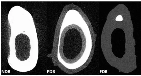
1. Demineralized bone morphology
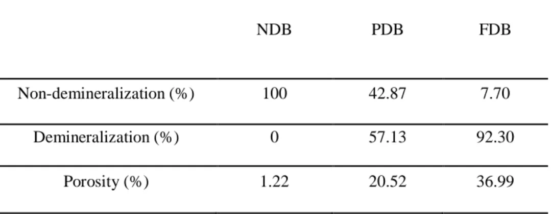
Micro-CT analysis revealed that the entire cortical bone was mineralized in NDB samples, appearing white in color (Fig. 2). Demineralization was seen to occur from the outer and inner surfaces. Demineralization rates of PDB and FDB samples were 51.3 and 92.3 %, respectively. Porosity ratios in the NDB, PDB, and FDB groups were 1.03, 19.99, and 36.93, respectively (Table 2). There were no differences in closed porosity among these groups.
Fig. 2. Micro-CT images showing bone demineralization in relation to HCl exposure time (increasing from the left panel to the right).
White and grey regions represent non-demineralized and demineralized tissue, respectively. Demineralization and porosity rates were 0 and 1.23 % for NDB, 57.13 and 20.52 % for PDB, and 92.30 and 36.99 % for FDB, respectively.
12 Table 2 Results of 3D analysis by micro-CT.
NDB PDB FDB
Non-demineralization (%) 100 42.87 7.70
Demineralization (%) 0 57.13 92.30
Porosity (%) 1.22 20.52 36.99
SEM showed the surface of NDB samples to be irregular and almost completely covered by mineral components (Fig. 3), whereas collagen fibers were observed in PDB and FDB specimens. In particular, the surface of FDB exhibited numerous collagen fibers, appearing as twisted threads. Small mineral deposits were evident on the collagen fibers in PDB; however, FDB demonstrated no such mineralization.
Fig. 3. SEM images of bone disc surfaces at 30,000× magnification.
A. NDB; the disc surface was covered by mineral components. B. PDB;
mineral components were partially removed and collagen fibers exposed. C.
FDB; collagen fibers were completely exposed and free of mineral
components.
13
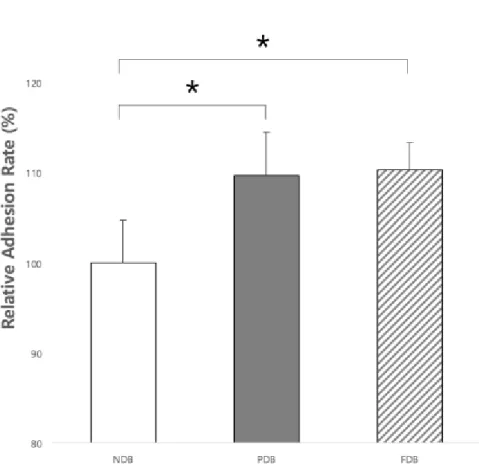
2. Ad-MSC adherence to and proliferation on bone discs
Relative adherence rates in the NDB, PDB, and FDB groups were 100, 109.71, and 110.33 %, respectively (Fig. 4). The difference between PDB and FDB samples in this respect was not significant, although adherence rates in both of these groups were significantly higher than that in the NDB group (P < 0.05; Fig.
4). Proliferation rates in the FDB group increased gradually over time, but those in the other treatment groups did not (Fig. 5). From day 2 after cell seeding, proliferation was significantly higher using FDB than NDB or PDB (P < 0.05). Ad- MSCs were able to attach to and grow on all three bone disc types; however, those cultured on FDB appeared in a stacked formation (Fig. 6).
14
Figure 4. Adherence rates of Ad-MSCs to bone discs of each treatment group.
Adherence of Ad-MSCs to PDB and FDB was greater than that to NDB; however, no significant difference was observed between PDB and FDB in this respect.
*Indicates a statistically significant difference (P < 0.05) between groups. Each bar represents the mean ± SD of 6 samples.
15
Figure. 5. Ad-MSC proliferation rates on bone discs at days 1, 2, and 3 of culture.
Day 1: Ad-MSC proliferation rates in the PDB and FDB groups were higher than that in the NDB group. Day 2: Only FDB exhibited increased Ad-MSC proliferation rates. Day 3: Proliferation on FDB was seen to have gradually increased from day 1, in contrast to the other treatment groups. *Indicates a statistically significant difference (P < 0.05) between groups. Each bar represents the mean ± SD of 6 samples.
16
Figure. 6. Ad-MSCs cultured for 3 days on bone discs in low-glucose DMEM at 800× magnification.
A. NDB; B. PDB; C. FDB; On NDB, only small fraction of the surface was covered with Ad-MSCs. Denser on PDB and the entire surface was covered with Ad-MSCs on FDB.
3. Osteogenic gene expression in Ad-MSCs
RUNX2 expression of Ad-MSCs on FDB was significantly lower than that of
those cultured on NDB (P < 0.05; Fig. 7). However, ALP, BMP-7, and TGF-β transcriptions by Ad-MSCs of the FDB group were significantly elevated compared to the other groups. Specifically, expression levels of these osteogenic genes in Ad-MSCs on FDB were more than twice those observed in the NDB group.
17
Figure. 7. Expression of RUNX2, ALP, BMP-7, and TGF-β genes 3 days after cell seeding. *Indicates a statistically significant difference (P < 0.05) between groups.
Error bar represents the mean ± SD of 6 samples.
18
IV. DISCUSSION
In the present study, SEM and micro-CT showed that HCl demineralization for 48 h successfully removed the mineral components of bone, exposing the organic constituents. In addition, demineralization increased bone porosity.
Demineralization triggers changes in the mechanical and biological properties of bone (Guo et al. 1991). Higher porosity results in a larger surface area, contributing to osteogenic protein adsorption, as well as ion exchange (Yuan et al. 1999).
Moreover, cell attachment, proliferation, and differentiation may be enhanced by increased surface roughness related to bone porosity (Yuan et al. 1999).
Here, greater bone disc demineralization was found to enhance Ad-MSC adherence more than non-demineralization of it. Organic bone components, such as type I collagen, are exposed following demineralization (Figueiredo et al. 2011).
These demonstrate chemotactic or adhesive characteristics, as they contain arginine-glycine-aspartic acid (RGD) peptides that mediate cellular adhesion (Anselme et al. 2000). The greater the degree of type I collagen exposure by demineralization, the more cell adhesion is promoted by the RGD sequence. Thus, the different adhesion rates observed among NDB, PDB, and FDB may be explained by varying accessibility of the RGD peptide. In the current work, Ad- MSCs on FDB exhibited higher proliferation rates as well. Differences in
19
proliferation between treatment groups might also be explained by the RGD sequence. As RGD-mediated cell attachment occurs, transmembrane receptors trigger signaling cascades that initiate cell proliferation (Sawyer et al. 2005).
Three days after cell seeding, we found expression of BMP-7, ALP, and TGF-β in the PDB and FDB groups to be upregulated compared with the NDB
control. However, RUNX2 transcript levels in the former two groups were downregulated compared with the latter. Bone contains osteogenic proteins, such as those of the BMP subfamily and TGF-β (Pietrzak et al. 2012). BMPs are exposed by demineralization and might contribute to Ad-MSC osteogenic differentiation through SMAD and MAPK pathway (Beederman et al. 2013). RUNX2 is a transcription factor required for bone formation and a major target of TGF-β and BMP signaling (Lee et al. 2000). BMP subfamily proteins can upregulate RUNX2 (Lee et al. 2000); however, such elevated expression is only transiently maintained and begins to decrease soon after the initiation of upregulation (Lee et al. 2000).
Therefore, the observed downregulation of RUNX2 may be the eventual consequence of promoted expression by members of the BMP subfamily.
Based on the above results, demineralization may enhance the biocompatibility of bone allografts for use in bone regeneration. Fully demineralized cortical bone might be a suitable allograft material capable of
20
stimulating the osteogenic differentiation of Ad-MSCs.
21
V. REFERENCES
H. Mankin, D. Springfield, M. Gebhardt, W. Tomford. Current status of allografting for bone tumors, Orthopedics.1992;15(10):1147-1154.
H.J. Mankin, M.C. Gebhardt, L.C. Jennings, D.S. Springfield, W.W. Tomford, Long-term results of allograft replacement in the management of bone tumors, Clinical orthopaedics and related research. 1996;324:86-97.
H. Tapp, E.N. Hanley, J.C. Patt, H.E. Gruber, Adipose-Derived Stem Cells:
Characterization and Current Application in Orthopaedic Tissue Repair, Experimental Biology and Medicine. 2009;23(4):1-9.
A. Hofmann, L. Konrad, M.H. Hessmann, R. Küchle, J. Korner, J.D. Rompe, P.M.
Rommens, The influence of bone allograft processing on osteoblast attachment and function, Journal of Orthopaedic Research.
2005;23(4):846-854.
M. Kuk, Y. Kim, S.H. Lee, W.H. Kim, O.K. Kweon, Osteogenic Ability of Canine Adipose-Derived Mesenchymal Stromal Cell Sheets in Relation to Culture Time, Cell Transplant. 2016;25(7):1415-1422.
I. Drosse, E. Volkmer, R. Capanna, P. De Biase, W. Mutschler, M. Schieker, Tissue engineering for bone defect healing: an update on a multi-component approach, Injury. 2008;39:S9-S20.
22
B.J. Kang, Y. Kim, S.H. Lee, W.H. Kim, H.M. Woo, O.K. Kweon, Collagen I gel promotes homogenous osteogenic differentiation of adipose tissue-derived mesenchymal stem cells in serum-derived albumin scaffold, J Biomater Sci Polym Ed.2013;24(10):1233-1243.
J. Mauney, S. Sjostorm, J. Blumberg, R. Horan, J. O’leary, G. Vunjak-Novakovic, V. Volloch, D. Kaplan, Mechanical stimulation promotes osteogenic differentiation of human bone marrow stromal cells on 3-D partially demineralized bone scaffolds in vitro, Calcified tissue international.
2004;74(5):458-468.
J.R. Mauney, C. Jaquiery, V. Volloch, M. Heberer, I. Martin, D.L. Kaplan, In vitro and in vivo evaluation of differentially demineralized cancellous bone scaffolds combined with human bone marrow stromal cells for tissue engineering, Biomaterials. 2005;25(16):3173-3185.
B.-J. Kang, H.-H. Ryu, S.S. Park, Y. Koyama, M. Kikuchi, H.-M. Woo, W.H. Kim, O.-K. Kweon, Comparing the osteogenic potential of canine mesenchymal stem cells derived from adipose tissues, bone marrow, umbilical cord blood, and Wharton's jelly for treating bone defects, Journal of veterinary science.
2012;13(3):299-310.
23
L. Ghasemi-Mobarakeh, M.P. Prabhakaran, M. Morshed, M.H. Nasr-Esfahani, S.
Ramakrishna, Electrospun poly(epsilon-caprolactone)/gelatin nanofibrous scaffolds for nerve tissue engineering, Biomaterials. 2008;29(34):4532-4539.
M.Z. Guo, Z.S. Xia, L.B. Lin, The mechanical and biological properties of demineralised cortical bone allografts in animals, Journal of Bone & Joint Surgery, British Volume. 1991;73(5):791-794.
H. Yuan, K. Kurashina, J.D. de Bruijn, Y. Li, K. de Groot, X. Zhang, A preliminary study on osteoinduction of two kinds of calcium phosphate ceramics, Biomaterials. 1999;20(19):1799-1806.
M. Figueiredo, S. Cunha, G. Martins, J. Freitas, F. Judas, H. Figueiredo, Influence of hydrochloric acid concentration on the demineralization of cortical bone, Chemical Engineering Research and Design. 2011;89(1):116-124.
K. Anselme, Osteoblast adhesion on biomaterials, Biomaterials. 2000;21(7):667- 681.
A.A. Sawyer, K.M. Hennessy, S.L. Bellis, Regulation of mesenchymal stem cell attachment and spreading on hydroxyapatite by RGD peptides and adsorbed serum proteins, Biomaterials. 2005;26(13):1467-1475.
24
W.S. Pietrzak, M. Dow, J. Gomez, M. Soulvie, G. Tsiagalis, The in vitro elution of BMP-7 from demineralized bone matrix, Cell Tissue Bank.
2012;13(4):653-661.
M. Beederman, J.D. Lamplot, G. Nan, J. Wang, X. Liu, L. Yin, R. Li, W. Shui, H.
Zhang, S.H. Kim, W. Zhang, J. Zhang, Y. Kong, S. Denduluri, M.R.
Rogers, A. Pratt, R.C. Haydon, H.H. Luu, J. Angeles, L.L. Shi, T.-C. He, BMP signaling in mesenchymal stem cell differentiation and bone formation, Journal of Biomedical Science and Engineering.
2013;06(08):32-52.
K.-S. Lee, H.-J. Kim, Q.-L. Li, X.-Z. Chi, C. Ueta, T. Komori, J.M. Wozney, E.-G.
Kim, J.-Y. Choi, H.-M. Ryoo, Runx2 is a common target of transforming growth factor β1 and bone morphogenetic protein 2, and cooperation between Runx2 and Smad5 induces osteoblast-specific gene expression in the pluripotent mesenchymal precursor cell line C2C12, Molecular and cellular biology. 2000;20(23):8783-8792.
25
VI. 국문 초록
개 피질골 탈회가 지방 유래 중간엽 줄기세포의 골분화에 미치는 영향
지도 교수 권 오 경
조 광 래
서울대학교 대학원
수의학과 임상수의학
골 재생을 촉진시키기 위해 동종탈회골과 줄기세포가 사용되어져 왔다.
그러나 지방유래 줄기세포와 함께 사용되어지기 위해 피질골을 얼마나 탈회시켜야 하는지에 대해서는 명백히 밝혀지지는 않았다. 본 연구에서는 동종골에서의 지방유래 줄기세포의 골분화능이 탈회의 정도와 얼마나 관련이 있는지 실험적으로 조사하였다. 세 실험군은 0.6 N HCL에 노출 시킨 시간을 달리하여 설정하였다.; 12시간 탈회시킨 그룹(PDB), 48시간
26
완전 탈회시킨 그룹(FDB), 탈회시키지 않은 그룹(NDB). 동종골은 실험적 평가를 위해 6 mm 지름의 원형으로 준비하였고 탈회의 정도와 구조는
Micro CT 와 SEM 으로 평가하였다. 지방유래 줄기세포의 부착과 증식
정도는 MTS 로 평가하였고 골분화와 관련된 유전자의 발현은 RT- PCR을 통해 평가하였다. PDB와 FDB 그룹의 탈회율은 각각 57.13% 와 92.30 % 였다. NDB, PDB, FDB 세 그룹의 지방유래 줄기세포 부착율은 각각 53.41 %, 60.65 %, 61.32 % 였다. FDB 그룹에서 배양 3 일 후 증식율은 다른 그룹과 비교하여 유의적으로 높았다. 게다가 FDB 그룹의 골분화 관련 유전자 ALP, BMP-7, TGF-베타의 활성도는 다른 그룹에 비해 유의적으로 상승하였다. 위의 결과에 따라, FDB 그룹이 골결손 치유를 위해 적합한 충진물일 것으로 생각된다.
주요어 : 골 재생, 동종골, 탈회 정도, 지방유래 줄기세포, 개 학번 : 2015-21839