저작자표시-비영리-변경금지 2.0 대한민국 이용자는 아래의 조건을 따르는 경우에 한하여 자유롭게
l 이 저작물을 복제, 배포, 전송, 전시, 공연 및 방송할 수 있습니다. 다음과 같은 조건을 따라야 합니다:
l 귀하는, 이 저작물의 재이용이나 배포의 경우, 이 저작물에 적용된 이용허락조건 을 명확하게 나타내어야 합니다.
l 저작권자로부터 별도의 허가를 받으면 이러한 조건들은 적용되지 않습니다.
저작권법에 따른 이용자의 권리는 위의 내용에 의하여 영향을 받지 않습니다. 이것은 이용허락규약(Legal Code)을 이해하기 쉽게 요약한 것입니다.
Disclaimer
저작자표시. 귀하는 원저작자를 표시하여야 합니다.
비영리. 귀하는 이 저작물을 영리 목적으로 이용할 수 없습니다.
변경금지. 귀하는 이 저작물을 개작, 변형 또는 가공할 수 없습니다.
의학박사 학위논문
Musculoskeletal assessment in patients with Duchenne muscular dystrophy
뒤셴느근디스트로피 환자에서의 근골격계 평가
2020년 8월
서울대학교 대학원 의학과 재활의학 전공
최 영 아
뒤셴느근디스트로피 환자에서의 근골격계 평가
지도 교수 신 형 익
이 논문을 의학박사 학위논문으로 제출함 2020년 5월
서울대학교 대학원 의학과 재활의학 전공
최 영 아
최영아의 의학박사 학위논문을 인준함 2020년 7월
위원장 __________________ (인)
부위원장 __________________ (인)
위 원 __________________ (인)
위 원 __________________ (인)
위 원 __________________ (인)
Musculoskeletal assessment in patients with Duchenne muscular dystrophy
By
Young-Ah Choi
(Directed by Hyungik Shin, PhD)
A thesis submitted to the Department of Medicine in partial fulfilment of the requirements for the Degree of
Doctor of Philosophy in Rehabilitation Medicine at Seoul National University College of Medicine
July 2020
Approved by Thesis Committee:
Professor __________________ Chairman Professor __________________ Vice chairman Professor __________________
Professor __________________
Professor __________________
i
Abstract
Musculoskeletal assessment in patients with Duchenne muscular dystrophy
Young-Ah Choi Department of Rehabilitation Medicine Seoul National University College of Medicine
Introduction:The aim of this study was to investigate systematic evaluation of limb and spinal musculoskeletal rehabilitation in accordance with the current clinical situation of early administration of steroids in patients with Duchenne muscular dystrophy (DMD). Three researches were conducted in this thesis. First, we investigated range of motion and contracture of lower extremity joints among male patients with DMD, based on the patients’ ambulatory status. Differences in major joint contractures, based on passive stretching exercise participation, were also investigated. Second, patients with DMD often develop scoliosis that progresses rapidly after loss of ambulation. We attempted to examine the incidence of scoliosis, flexibility of scoliosis and pelvic tilt associated with scoliosis after two years of wheelchair-bound status to identify trends over time. Finally, in persons with DMD, weakness of the upper extremity (UE) muscles has a significant impact on daily activities and body function. This problem necessitates a screening tool that can be used quickly and easily in clinical situations, such as the Upper limb short questionnaire (ULSQ). However, its validity and reliability as a clinical measure have not yet been evaluated.
Methods: In the first research, total of 128 boys with DMD, followed at the DMD
ii
clinic of a tertiary care hospital, were included in this cross-sectional study. The passive ranges-of-motion of the hip, knee, and ankle joints were measured, in the sagittal plane, using a goniometer. The Vignos Scale was used to grade ambulatory function. Boys with DMD who performed stretching exercises for more than 5 min/session, >3 sessions/week, were classified into the stretching group. In the second research, we reviewed the medical records of 273 boys who were genetically identified as having DMD, and finally, 50 boys with serial records of radiographs after loss of ambulation were finally enrolled. And among them, only 31 patients developed scoliosis. Spine radiographs in sitting and supine positions were also reviewed to obtain Cobb angle, curve flexibility, and pelvic obliquity. Flexibilities (%) were calculated by the difference in angles between the sitting and supine positions divided by the angle at the sitting position, multiplied by 100. In the third research, face to face ULSQ interviews were held, and then repeated by telephone, at least four weeks later. Lower extremity and UE body function were measured by a physician using Vignos and a modified Brooke scale, respectively.
Results: In the first research, the hip flexion (23.5 o), knee flexion (43.5 o), and ankle plantarflexion (34.5 o) contracture angles in the non-ambulatory group were more severe than those in the ambulatory group. Ankle plantarflexion contractures (41 patients, 52.6%) were more frequently observed early, even within the ambulatory period, than were hip (8 patients, 10.3%), and knee joint (17 patients, 21.8%) contractures. Passive stretching exercises >3 sessions/week were not associated with the degree of lower extremity joint contractures in the ambulatory or non-ambulatory group. In the second research, among 31 boys who had scoliosis, all but 2 boys with curves went through a sequential course of 1) no scoliosis, 2) nonstructural scoliosis, when scoliosis was only measurable in the sitting position, and 3) structural scoliosis, when scoliosis was also detectable in the supine position. Flexibility decreased each year after detection of scoliosis in those who developed scoliosis the first year, from 75.5 ± 5.0% to 57.1 ± 10.5% and to 49.1 ± 10.0% (mean ± standard deviation). Spinal
iii
flexibility was significantly correlated with curve magnitude of scoliosis in both sitting and supine position. In the third research, 160 subjects participated in the initial ULSQ interview among 167 participants, and 132 subjects completed follow- up interviews. Construct validity was confirmed by exploratory and subsequent confirmatory factor analysis. Sum scores of UE function correlated with the modified Brooke scale (Kendall’s Tau 0.64, p < .001). Total and sum scores for each ULSQ component were higher in non-ambulators than in ambulators. Reliability was acceptable, determined by internal consistency and test-retest tools.
Conclusions: There have been few studies about the range of motion and the degree of contracture of the joints (hip, knee, and ankle) of the lower limb since the 2000s when steroids began to be widely used. Knowledge of lower extremity joint contracture profiles, based on ambulatory status, may be useful for developing appropriate strategies for joint management in this patient group. Regarding to scoliosis, our result suggests that in the early stage of scoliosis, wherein flexibility is maintained without structural scoliosis, interventions such as bracings should be considered in DMD scoliosis. Also, scoliosis curve in DMD patients should be evaluated dynamically to detect the scoliosis when the curve is fully reducible. This study could be a cornerstone for further studies involving application of spinal braces for neuromuscular scoliosis. Finally, ULSQ could be a valid and reliable measurement tool for persons with DMD to screen UE function, pain, and stiffness in clinical settings
Keywords: Contracture; Duchenne muscular dystrophy; Function; Lower extremity;
Pain; Scoliosis; Steroid; Stiffness; Upper extremity Student Number: 2016-30602
iv
Table of Contents
Abstract ... i
Table of Contents ... iv
List of Tables ... vii
List of Figures ... viii
List of Abbreviations ... ix
1. Introduction ... 1
1.1. Research I: Lower extremity joint contracture according to ambulatory status ... 1
1.2. Research II: Scoliosis in Duchenne muscular dystrophy children ... 2
1.3. Research III: Upper limb short questionnaire for Duchenne muscular dystrophy ... 3
2. Methods ... 7
2.1. Research I: Lower extremity joint contracture according to ambulatory status ... 7
2.1.1. Participants... 7
2.1.2. Measures ... 8
2.1.3. Procedure ... 8
2.1.4. Data analysis ... 9
2.2. Research II: Scoliosis in Duchenne muscular dystrophy children ... 10
2.2.1. Participants... 10
v
2.2.2. Review of medical records ... 11
2.2.3. Evaluation of scoliosis and pelvic obliquity ... 11
2.2.4. Evaluation of flexibility ... 12
2.2.5. Statistical analysis ... 13
2.3. Research III: Upper limb short questionnaire for Duchenne muscular dystrophy ... 13
2.3.1. Participants... 13
2.3.2. Procedure ... 14
2.3.3. Statistical Analysis ... 18
3. Results ... 19
3.1. Research I: Lower extremity joint contracture according to ambulatory status ... 19
3.1.1. Demographic and clinical information ... 19
3.1.2. Lower extremity joint contracture comparisons .... 22
3.1.3. Differences in joint contracture angles, based on stretching participation ... 26
3.2. Research II: Scoliosis in Duchenne muscular dystrophy children ... 28
3.2.1. Clinical Characteristics ... 28
3.2.2. Development of scoliosis ... 31
3.2.3. Scoliosis curve type changes ... 33
3.2.4. Changes in curve flexibility ... 35
3.2.5. Correlation between spinal curve flexibility and
other parameters ... 35
vi
3.2.6. Development of pelvic obliquity ... 35
3.3. Research III: Upper limb short questionnaire for Duchenne muscular dystrophy ... 38
3.3.1. Clinical characteristics ... 38
3.3.2. The result of Upper limb short questionnaire survey ... 40
3.3.3. Factor analysis ... 42
3.3.4. Construct validity ... 42
3.3.5. Reliability ... 47
4. Discussion ... 49
4.1. Research I: Lower extremity joint contracture according to ambulatory status ... 49
4.2. Research II: Scoliosis in Duchenne muscular dystrophy children ... 51
4.3. Research III: Upper limb short questionnaire for Duchenne muscular dystrophy ... 55
4.4. Implication for clinical trial in Duchenne muscular dystrophy ... 58
5. Concluding Remarks ... 60
Acknowledgments ... 61
Funding ... 61
References ... 62
Appendix ... 70
국문 초록 ... 72
vii
List of Tables
Table 1. Upper limb short questionnaire (ULSQ) by Jenssen et al (2018) ... 6 Table 2. Korean version of Upper limb short questionnaire
(ULSQ) ... 15 Table 3. Patient Vignos scale score ... 21 Table 4. Severity of lower limb joint contractures in patients with DMD, based on ambulatory status ... 23 Table 5. Average lower extremity joint contracture angles,
estimated using a generalized estimating equation ... 25 Table 6. Independent-sample t-test results comparing the
stretching and non-stretching groups ... 27 Table 7. Demographic and clinical characteristics of participants
... 30 Table 8. Values are presented as mean ± standard deviation ... 32 Table 9. Pelvic obliquity of subjects ... 37 Table 10. Correlation between scores of Upper limb short
questionnaire (ULSQ) and modified Brooke scale ... 44 Table 11. Results from Kruskal-Wallis tests and post-hoc
pairwise comparisons for total scores and sum scores of
each component between different ambulatory stages ... 46
Table 12. Test-retest reliability ... 48
viii
List of Figures
Figure 1. Subject flow diagram ... 20 Figure 2. Flowchart of subject enrollment ... 29 Figure 3. Curve type changes of subjects ... 34 Figure 4. The numbers of individuals with DMD by Vignos
Scale and modified Brooke scale ... 39 Figure 5. The result of Upper limb short questionnaire (ULSQ)
survey ... 41 Figure 6. The box plot for sum scores of Upper limb short
questionnaire (ULSQ) from 1 to 5 items (Above) and total
scores of ULSQ (Below) according to the modified Brooke
scale. UE: upper extremity ... 45
ix
List of Abbreviations
AC 1 Gwet’s first-order agreement coefficient AFO Ankle-foot orthosis
ANOVA One-way analysis of variance APF Ankle plantarflexion
CFI Comparative fit index
DMD Duchenne muscular dystrophy EFA Exploratory factor analysis GFI Goodness-of-fit index
HF Hip flexion
ICF International classification of functioning, disability and health
KF Knee flexion
KMO Kaiser-Meyer-Olkin
PUL Performance of upper limb assessment RMR Root mean square residual
RMSEA Root-mean-square error of approximation ROM Range of motion
UE Upper extremity
ULSQ Upper limb short questionnaire
WHO World Health Organization
1
1. Introduction
Duchenne muscular dystrophy (DMD) is an X-linked recessive disorder caused by a lack of dystrophin, and has an overall incidence of 1 in 4700 male births.1, 2 Muscular dystrophy is characterized by progressive muscular weakness, from childhood, which eventually results in the loss of gait performance when patients are approximately 10–12-years-old.3 In recent decades, the survival of patients with DMD has improved because of interdisciplinary care, such as the inclusion of noninvasive ventilation.4 For a more fundamental therapeutic approach, novel treatments such as dystrophin-target gene therapies have been investigated in the past decade.5,6 However, even if successful dystrophin protein restoration makes it possible to improve motor function in DMD, its effect may be limited in patients with advanced musculoskeletal complications. Furthermore, efforts to reduce musculoskeletal complications are important for maintaining function and have a close impact on quality of life.
1.1. Research I: Lower extremity joint contracture according to ambulatory status
In patients with neuromuscular conditions, contractures develop due to intrinsic myotendinous structural changes and extrinsic factors.7 Specifically, in patients with DMD, joint contractures are associated with several factors, including loss of full joint range-of-motion (ROM), static positioning, muscle imbalance around a joint, and fibrotic changes (fatty tissue infiltration) within muscle tissues.8 Lower
2
extremity joint contractures negatively affect the gait of patients with ambulatory DMD.9 For example, hip joint contractures can lead to pelvic obliquity, which is associated with scoliosis development.10 In recent decades, the survival of patients with DMD has improved because of interdisciplinary care, such as the inclusion of noninvasive ventilation.4 Hence, contracture prevention is essential for maintaining a patient’s functional ability and an acceptable quality of life.
Although knowledge regarding lower extremity joint contracture profiles, based on disease progression, is necessary for the development of appropriate preventive strategies, studies is this area remain scarce. McDonald et al. reported that lower extremity contractures were rare in patients able to maintain an upright posture, but developed soon after the patients became confined to a wheelchair for the majority of the day.11 However, these authors did not describe any differences between the major lower extremity joints, i.e., the hip, knee, and ankle. This study aimed to investigate the profile of hip, knee, and ankle joint contractures, based on the ambulatory status of patients with DMD. Furthermore, differences in major joint contractures were evaluated, based on the passive stretching exercises performed by ambulatory and non-ambulatory patients.
1.2. Research II: Scoliosis in Duchenne muscular dystrophy children
Scoliosis is a frequent complication of DMD that progresses rapidly, in the non- ambulatory stage of the disease.12-17 Pelvic obliquity is also thought to be a mechanism of compensation for scoliosis.18 These deformities in the musculoskeletal system together make sitting difficult, limiting the use of upper extremities and hampering activities of daily living. When scoliosis progresses, rib
3
impingement onto the ilium may occur, causing pain and making hygiene difficult.19 It is crucial to prevent scoliosis as it affects other organ systems.
After loss of ambulation, rapid progression of spinal deformity leads to a deterioration in pulmonary function.14, 16, 17 Kurz et al. reported that with 10 degrees of thoracic curve progression, functional vital capacity decreased by 4%.20 According to Hsu et al., in DMD patients whose spinal curves exceeded 40 degrees, vital capacity diminished by 12 to 16%.21 Therefore, it is important to prevent, or delay spinal deformity as it leads to compromise of respiratory function.
Spinal orthosis attempts to prevent or delay scoliosis using spinal support at three points of the controlling mechanism; the lateral curve should be flattened by the pressure. Therefore, it is assumed that spine flexibility or reducibility is a significant influencing factor for the effectiveness of braces.22, 23 Information regarding curve flexibility helps establish a strategy for brace application to manage scoliosis. If there is sufficient flexibility, the effectiveness of bracing therapy is expected.
Nevertheless, there have been only a few reports investigating spine flexibility in this patient group.24 Therefore, this study is to investigate the curve flexibility of scoliosis for 2 years after loss of walking ability in children with DMD.
1.3. Research III: Upper limb short questionnaire for Duchenne muscular dystrophy
DMD is characterized by progressive muscular weakness. The function of the upper extremity (UE) decreases later than that of the lower extremity, usually after losing walking ability. The UE weakness appears first in the proximal and progresses to the distal region, which results in limitations performing daily activities.4, 25 In recent
4
decades, corticosteroid treatment can mitigate the progression of limb muscle weakness, and survival has improved because of interdisciplinary care, including noninvasive ventilation.4, 25 With the current life expectancy and care techniques, men with DMD will live for longer than before with their impaired UE function. The weakness of the UE muscle will affect daily life functions in different ways to weakness in the lower extremity muscle because it is more difficult to compensate body functions with assistive devices, such as wheelchairs in the lower extremities.
Janssen et al. suggested that pain and stiffness could negatively affect the UE body function in people with DMD.26 Therefore, a patient’s UE function should be evaluated in the clinic, especially after losing ambulatory function. Existing UE evaluation tools record the observational findings27-30 or use the patient's report31-33 of their ability during UE activities.
In most cases, however, they include too many evaluation items, which can be challenging to complete in the clinical setting. For example, the Performance of Upper Limb assessment in DMD (PUL) includes 22 items subdivided into the shoulder, middle, and distal levels of the upper extremities.34-36 For the PUL, scoring options varied across the scale between 0–1 and 0–6, according to performance and equipment such as weights, coins, and pencils, etc., which are required for the evaluation. Considering the progressive nature of the disease, rather than conducting detailed UE assessments at each clinic, the strategy of a simple screening assessment in each clinic and a detailed assessment when changes are detected may be more efficient. A screening assessment tool should be simple and closely related to the intervention plan.
Janssen et al. proposed the Upper limb short questionnaire (ULSQ) consisting of 14 items based on a review of existing relevant UE assessment tools for DMD.37 The questionnaire consists of upper limb function (5 items), pain (6 items), and stiffness
5
(3 items) that require yes or no answers and can be used quickly and easily in clinical situations for screening purposes (Table 1).
However, the questionnaire is not a conventional clinical measure, and its validity and reliability have not been sufficiently demonstrated. This study aimed to evaluate the construct validity of ULSQ and its reliability to investigate whether the ULSQ can be used as a clinical measure when converted to a score with 1 point per item.
6
Table 1. Upper limb short questionnaire (ULSQ) by Jenssen et al (2018)
Factor Questions Score optionsa
ULSQ-1 Heavy lifting
Do you experience problems in your arms when lifting heavy objects (>5 pounds)?
0: No 1: Yes ULSQ-2
Light or no lifting
Do you experience problems in your arms when you reach for or lift light objects such as an empty can?
0: No 1: Yes ULSQ-3
Basic hand function
Do you experience problems using your hands for basic functions like manipulating small objects or holding a key?
0: No 1: Yes ULSQ-4
Gross hand function
Do you experience problems using your hands when performing daily activities that require gross hand function like washing your hands or eating with a spoon?
0: No 1: Yes ULSQ-5
Fine hand function
Do you experience problems using your hands when performing daily activities that require fine hand function like buttoning up your shirt?
0: No 1: Yes ULSQ-6
Pain limitations
Do you experience limitations performing daily activities due to pain in your upper limb?
0: No 1: Yes ULSQ-7
Pain severity (not shoulder)
How severe is the pain you experience in your upper limb when performing daily activities?
0: No pain
1: Mild or severe pain ULSQ-8
Distal pain frequency
How often do you have pain in your hands or fingers?
0: Not more than once a month
1: More than once a month
ULSQ-9
Shoulder pain Do you experience pain in your
shoulder(s)? 0: No
1: Yes ULSQ-10
Proximal pain frequency (not shoulder)
How often do you experience pain in your upper or lower arm?
0: Not more than once a month
1: More than once a month
ULSQ-11 Elbow pain frequency
How often do you experience pain in your elbows?
0: Not more than once a month
1: More than once a month
ULSQ-12 Stiffness frequency
How often do you experience stiffness in your arms?
0: Not more than once a month
1: More than once a month
ULSQ-13 Stiffness limitations
Do you experience limitations performing daily activities due to stiffness in your upper limb?
0: No 1: Yes ULSQ-14
Stiffness severity
How severe is the stiffness you experience in your upper limb when performing daily activities?
0: No stiffness
1: Mild or severe stiffness
7
2. Methods
2.1. Research I: Lower extremity joint contracture according to ambulatory status
2.1.1. Participants
Overall, 136 boys with DMD were included in this cross-sectional study conducted at the DMD clinic of Seoul National University Hospital (Seoul, Korea). DMD diagnoses were confirmed using a dystrophin gene study. The genetic test methods used to identify dystrophin mutations were multiplex polymerase chain reaction and direct sequencing (Xp21.2-p21.1, exons 1–79). If the deletion/duplication testing results were negative, dystrophin gene sequencing was performed to search for point mutations or small deletions/insertions. All children with DMD participating in the study were prescribed alternate-day deflazacort (0.9 mg/kg), according to the international consensus, after demonstrating a partial Gower sign.38 Patients were excluded from the study due to the presence of co-morbidities (e.g., an acquired brain or spinal injury), absence of corticosteroid administration, use of ankle-foot orthosis (AFO), or use of therapeutic weight bearing (e.g., passive standing frame) to increase lower extremity ROM. The research was conducted in accordance with the Declaration of the World Medical Association, and was reviewed and approved by the Seoul National University Hospital Institutional Review Board (IRB no. 1605- 028-760). Because of the study’s retrospective design, the need for consent to participate was waived.
8
2.1.2. Measures
Demographic and medical data, including age, Gower sign results, and dystrophin gene study results, were collected. Clinical variables, such as lower extremity joint passive ROM and sagittal plane contracture angles (flexion/extension movements), were recorded. The contracture angles were measured bilaterally in the hip, knee, and ankle joints, using a goniometer, during clinical examinations performed by a physical therapist. Goniometry is the most commonly used technique for measuring joint motion limitations due to muscle contracture. With the patients in a supine position, the same physiotherapist measured and recorded all lower extremity joint passive ROMs, according to the method of Norkin and White.39 Because the same physiotherapist performed all of the joint contracture measurements, the intra-tester measurement reliability is likely high, according to Pandya et al.40 For each measurement, the protocol guided the reference points for the fulcrum and the proximal and distal arms of the goniometer. At each time point, duplicate passive ROM measurements were obtained and averaged.
A physician recorded the Vignos Scale score to grade lower limb function of the children with DMD.41 The Vignos Scale classifies patients with DMD into 10 categories, based on their ability to walk. Patients with Vignos Scale scores of 1–7 were considered ambulatory, whereas patients with scores of 8–10 were considered non-ambulatory.
2.1.3. Procedure
The severities of hip flexion (HF), knee flexion (KF), and ankle plantarflexion (APF) contractures were classified, based on a previous study,11 as mild (1°–19°), moderate
9
(20°–40°), and severe (>40°) for HF contractures; mild (1°–14°), moderate (15°–
40°), and severe (>40°) for KF contractures; and mild (1°–14°), moderate (15°–30°), and severe (>30°) for APF contractures.
We investigated whether significant differences in passive stretching exercise participation existed between ambulatory and non-ambulatory patients. All patients with DMD who participated in the stretching exercises received help from their physical therapist at hospital. Children with DMD who performed stretching exercises for >5 min/session, for >3 sessions/week, were categorized into the stretching group; the others were classified into the non-stretching group.
2.1.4. Data analysis
Means and standard deviations (SD) were used to describe the basic patient characteristics. The mean values for right and left HF, KF, and APF angles were obtained for each patient, and the HF, KF, and APF contracture angles for both legs were averaged; t-tests confirmed the absence of significant differences between the left and right legs for all measurements. Data were also stratified according to patients categorized as ambulatory or non-ambulatory.
A generalized estimating equation was used to assess the extent of the differences in the degree of lower extremity joint contractures, depending on ambulatory status, and the degree of lower extremity joint contracture, between joint sites, depending on the patients’ ambulatory status. The relationship among the HF, KF and APF contracture angles were assumed to be constant. The mean contracture angle of each lower extremity joint was compared to that of the hip joint. The p-values and 99.3%
confidence intervals, including the Bonferroni correction, are presented for all seven tests.
10
Within each group, the mean HF, KF, and APF joint contracture angles were analyzed, using an independent samples t-test, to test for differences between the stretching and non-stretching groups.
Statistical analyses were performed using SPSS version 21.0 (IBM, Armonk, NY, USA). The level of statistical significance was set at 5% for 2-tailed tests.
2.2. Research II: Scoliosis in Duchenne muscular dystrophy children
2.2.1. Participants
Medical records and radiographs of 273 boys diagnosed with DMD who visited the pediatric rehabilitation department between March of 2017 and February of 2018 were reviewed. Ethical approval was obtained from the Institutional Review Board (IRB No. 1804-169-942). DMD diagnosis had been established using a dystrophin gene study. The genetic test methods used to identify dystrophin mutations included multiplex polymerase chain reaction and direct sequencing (Xp21.2-p21.1, exons 1–
79). If deletion/duplication testing results were negative, then dystrophin gene sequencing was performed to search for point mutations or small deletions/insertions.
All enrolled DMD pediatric patients were taking deflazacort (0.9 mg/kg) every other day, according to international consensus, at the pediatric department in the same hospital after a partial Gower sign had been observed.15
Inclusion criteria were as follows: (1) time points of the ambulation loss were charted;
(2) 2-year records of whole spine radiographs both in supine and sitting positions were preserved with (3) the first follow-up radiography was performed less than 1 year after the onset of ambulation loss.
11
2.2.2. Review of medical records
Patients with DMD in our hospital had regular outpatient follow-up at 12-month intervals when ambulatory and 6 months after the loss of ambulation.38 Whole spine radiographs were taken in the sitting and supine positions, and ambulatory functions were charted in Vignos scales at each outpatient follow-up. The onset of ambulation loss was defined when the charted scale value exceeded or equaled grade 8, when patients were able to stand with long leg braces, but were unable to walk even with assistance.24
2.2.3. Evaluation of scoliosis and pelvic obliquity
It was designated the patients who had no scoliosis both in sitting and supine positions as having “no scoliosis”; those who only had scoliosis in sitting position but not in supine position were designated as having “nonstructural scoliosis”; and those who had scoliosis both in sitting and supine positions were designated as having “structural scoliosis”.22 Postero-anterior radiographs of selected patients were used in this study. To improve intraobserver reliability, the measurements were taken by a single well-trained physician.
The image field in the cranio-caudal direction ranged from the occiput to the acetabula. Cobb angle of more than 10° was considered significant.14, 42, 43 Cobb angles were retrospectively measured by a single observer. The most oblique cranial and caudal end vertebrae were marked, and lines were drawn through the endplates of each vertebra, and the angles between them were measured.44 As measurement error of Cobb angle results from errors in selecting the end vertebrae,44, 45 initially selected end vertebrae were marked to be used for serial measurements to reduce measurement error.
12
The horizontal pelvic obliquity method measures angle between the line of most proximal iliac crests and the parallel line to the bottom of the radiograph. This angle is largely influenced by the patient’s position.46 Pelvic obliquity more than 5° was considered significant.18, 47 This horizontal pelvic obliquity measurement is associated with the least interobserver and intraobserver variability.46, 48 To minimize these errors, the patients were confirmed to be in the maximal and appropriate position and were well-fitted to the frame when taking images.
2.2.4. Evaluation of flexibility
Scoliosis curve flexibility was assessed by comparing the Cobb angle values in the supine position (gravity eliminated posture) and those in the sitting position (increase in the curve with gravity). Cobb angle and pelvic obliquity in each position and flexibility were analyzed at the time when scoliosis was first detected after ambulation loss, 1 year after scoliosis detection, and 2 years after scoliosis detection.
The flexibility of the spine curve at each year was calculated as below.23, 49 Flexibility (%) = 𝐶𝑜𝑏𝑏 𝑎𝑛𝑔𝑙𝑒 𝑎𝑡 𝑠𝑖𝑡𝑡𝑖𝑛𝑔 −𝐶𝑜𝑏𝑏 𝑎𝑛𝑔𝑙𝑒 𝑎𝑡 𝑠𝑢𝑝𝑖𝑛𝑒 𝑝𝑜𝑠𝑖𝑡𝑖𝑜𝑛
𝐶𝑜𝑏𝑏 𝑎𝑛𝑔𝑙𝑒 𝑎𝑡 𝑠𝑖𝑡𝑡𝑖𝑛𝑔 ×100
For the supine position, the hands were placed by the patient’s side, and patients were instructed to lie down facing up on a scanning couch and then to straighten their trunk and legs maximally.50 In the sitting position, patients were instructed to sit on a chair with a panel on the back, with the hip to be placed appropriately on the chair.
They were asked to lie back maximally to eliminate tilts. The patients were instructed to hold handles on both sides during radiography. If they were unable to hold the handles, they were simply asked to lay their hands on the handles.
13
2.2.5. Statistical analysis
The demographic characteristics and measurements of the participants were classified by scoliosis development. To analyze changes in flexibilities in the series of radiographs, repeated measure analysis of variance was used. To evaluate the relationships among each index, Spearman’s correlation tests were used. All data were statistically analyzed using the Statistical Package for Social Sciences for Windows ver. 17.0 (SPSS Inc., Chicago, IL, USA).
2.3. Research III: Upper limb short questionnaire for Duchenne muscular dystrophy
2.3.1. Participants
We recruited patients with DMD who were followed up at the DMD clinic of Seoul National University Children’s Hospital from December 2018 to August 2019. The ULSQ was carried out with DMD patients over seven years old. All children with DMD included in the study were taking deflazacort (0.9 mg/kg) every other day, according to international consensus, at the neurologic division of the pediatric department in the same hospital after a partial Gower sign was observed.38
Exclusion criteria were the same as a previous study37 : (a) patients who were diagnosed by at least age ten and (b) ambulatory status after age 14 in steroid-naïve DMD. DMD diagnosis was confirmed using a dystrophin gene study. The genetic test methods used to identify dystrophin mutations included multiplex polymerase chain reaction and direct sequencing (Xp21.2-p21.1, exons 1–79). If deletion or duplication testing results were negative, then dystrophin gene sequencing was performed to search for point mutations or small deletions or insertions. All the study
14
participants provided their informed consent before completing the questionnaire.
The research was conducted under the Declaration of the World Medical Association and was reviewed and approved by the Seoul National University Hospital Institutional Review Board (IRB no. 1810-120-982).
2.3.2. Procedure
2.3.2.1. Translation and re-translation
Before translating the questionnaire, we obtained permission from the original author of ULSQ to translate the original English version of ULSQ into Korean via e-mail.
After a translator completed the translation into Korean, two Korean physiatrists in the pediatric division checked the Korean questionnaire. A different translator completed the backward-translation of the questionnaire into English. Afterward, both the Korean physiatrists in the pediatric division and the original author reviewed the expression and clarification of the backward-translated questionnaire compared with the original, marking the completion of the Korean version of ULSQ (Table 2).
15
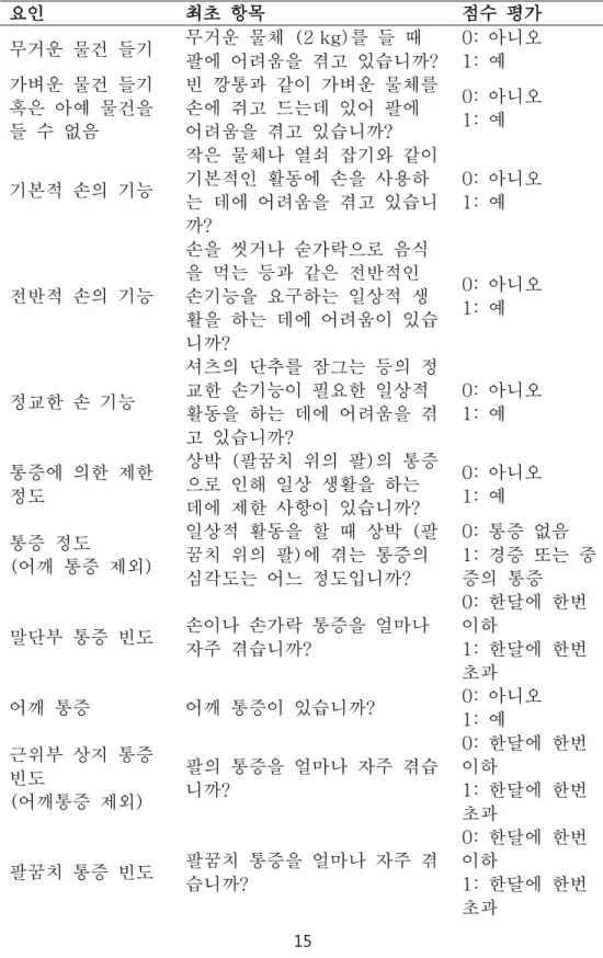
Table 2. Korean version of Upper limb short questionnaire (ULSQ)
요인 최초 항목 점수 평가
무거운 물건 들기 무거운 물체 (2 kg)를 들 때 팔에 어려움을 겪고 있습니까?
0: 아니오 1: 예 가벼운 물건 들기
혹은 아예 물건을 들 수 없음
빈 깡통과 같이 가벼운 물체를 손에 쥐고 드는데 있어 팔에 어려움을 겪고 있습니까?
0: 아니오 1: 예
기본적 손의 기능
작은 물체나 열쇠 잡기와 같이 기본적인 활동에 손을 사용하 는 데에 어려움을 겪고 있습니 까?
0: 아니오 1: 예
전반적 손의 기능
손을 씻거나 숟가락으로 음식 을 먹는 등과 같은 전반적인 손기능을 요구하는 일상적 생 활을 하는 데에 어려움이 있습 니까?
0: 아니오 1: 예
정교한 손 기능
셔츠의 단추를 잠그는 등의 정 교한 손기능이 필요한 일상적 활동을 하는 데에 어려움을 겪 고 있습니까?
0: 아니오 1: 예
통증에 의한 제한 정도
상박 (팔꿈치 위의 팔)의 통증 으로 인해 일상 생활을 하는 데에 제한 사항이 있습니까?
0: 아니오 1: 예 통증 정도
(어깨 통증 제외)
일상적 활동을 할 때 상박 (팔 꿈치 위의 팔)에 겪는 통증의 심각도는 어느 정도입니까?
0: 통증 없음 1: 경증 또는 중 증의 통증
말단부 통증 빈도 손이나 손가락 통증을 얼마나 자주 겪습니까?
0: 한달에 한번 이하
1: 한달에 한번 초과
어깨 통증 어깨 통증이 있습니까? 0: 아니오 1: 예 근위부 상지 통증
빈도
(어깨통증 제외)
팔의 통증을 얼마나 자주 겪습 니까?
0: 한달에 한번 이하
1: 한달에 한번 초과
팔꿈치 통증 빈도 팔꿈치 통증을 얼마나 자주 겪 습니까?
0: 한달에 한번 이하
1: 한달에 한번 초과
16
뻣뻣함 빈도 팔이 뻣뻣해지는 현상을 얼마 나 자주 겪습니까?
0: 한달에 한번 이하
1: 한달에 한번 초과
뻣뻣함에 의한 제 한 정도
팔의 뻣뻣함으로 인해 일상 생 활을 하는 데에 있어 한계가 있습니까?
0: 아니오 1: 예
뻣뻣함 중증도 일상적 활동 시, 팔의 뻣뻣함의 정도가 얼마나 심각합니까?
0: 경직 증상 없 음
1: 경증 혹은 중 증의 경직
17
2.3.2.2. Survey and measure
The ULSQ consisted of three-dimension regarding UE function (5 items), pain (6 items), and stiffness (3 items). All questions required a binary answer (yes or no).
The ULSQ was conducted during a face-to-face interview with the participants, their parents, and a research nurse. A physiatrist in the pediatric division recorded the Vignos scale and Brook scale in the clinic, which classifies these patients ten categories based on their walking ability. Grade 1 patients could walk and climb stairs without assistance, while grade 10 patients were confined to a bed.41 Based on the Vignos scale, the ability of ambulation was classified into three categories:
independent ambulation (Vignos scale 1-5), ambulation with assist or aids (Vignos scale 6-7), and loss of ambulation (Vignos scale 8-10). The original Brookes scales graded patients with DMD into six categories based on their ability to use their arms.27 Grade 1 patients start with their arms at their sides and abduct them in a full circle until they reached above their head, while grade 6 patients had no functional ability to use their hands. We added an intermediate stage (grade 5.5) between grades 5 (cannot raise hands to the mouth, but can use hands to hold a pen or pick up pennies from the table) and grade 6 (Cannot raise hands to the mouth and has no useful function of hands). Grade 5.5 patients cannot hold a pen; however, they can move their fingers (e.g. press on a mobile or other electronic device), which modified the Brooke scale into a seven-step scale for this study. Landfeldt et al. also used an intermediate step in their DMD functional ability self-assessment tool in the same way as in our study to adjust for the information technology (IT) devices with a touchpad that were commonly used by DMD patients.51 Four weeks after the initial interview, a physiotherapist conducted a second telephone ULSQ interview. ULSQ interview times, both by face to face and telephone, were within ten minutes.
Demographic and medical data, including age, gender, prescribed medication, and dystrophin gene study results, were collected and tabulated.
18
2.3.3. Statistical Analysis
The mean and SD described essential patient characteristics. Exploratory factor analysis (EFA) of principal factors investigated the latent constructs of the questionnaires. For EFA, Bartlett’s test of sphericity tested whether the correlation matrix of the sample was sufficient for factor analysis. The value of the Kaiser- Meyer-Olkin (KMO) measure of sampling was also measured to evaluate sample adequacy of the factor analysis. The eigenvalues-greater-than-one rule was applied to find common factors.52 Factor rotation using orthogonal rotation (the varimax method) extracted the relevant factors. Confirmatory factor analysis using maximum likelihood estimation confirmed the factor structure obtained using EFA. Model for goodness of fit was evaluated using the Chi-square test, Goodness-of-Fit Index (GFI), Root Mean Square Residual (RMR), Comparative Fit Index (CFI), and Root-Mean- Square Error of Approximation (RMSEA). GFI and CFI values usually ranged from 0 to 1.0, with values of 0.90 or higher considered evidence of a good fit.53 RMR and RMSEA values of less than 0.08 indicated an acceptable fit for the models.53, 54 To measure internal consistency, we calculated the relevant Cronbach’s α coefficients.
Inter-rater reliability analysis employed the Gwet’s AC1 (first-order agreement coefficient), which provided a reasonable chance-corrected coefficient in line with the percentage level of agreement.55
Furthermore, correlations between ULSQ and the Vignos/modified Brooke scale scores were calculated based on the Kendall rank correlation coefficient.56 To evaluate the discriminative capability of the ULSQ, the total scores, as well as sum of each component on the ULSQ of patients with DMD, were compared according to the ambulatory disease stage using the Kruskal-Wallis one-way analysis of variance (ANOVA) by ranks.57 If the difference between groups were significant, we conducted a post-hoc Tamhane’s comparison. Descriptive statistics were expressed as means and SD. Statistical analysis was performed using SPSS version
19
23.0, and confirmatory factor analysis was performed with AMOS, ver. 6 (SPSS Science, Chicago, IL) to statistically confirm the hypothesized structures.
3. Results
3.1. Research I: Lower extremity joint contracture according to ambulatory status
3.1.1. Demographic and clinical information
A total of 136 boys with DMD were evaluated for inclusion in the study; eight were excluded due to not meeting the study criteria (Figure 1). Of the 128 eligible patients, 78 (61%) were included in the ambulatory group and 50 (39%) were in the non- ambulatory group. The mean age in the ambulatory group was 9.1 ± 2.2 years (range, 4–15 years) and that in the non-ambulatory group was 13.3 ± 3.0 years (range, 8–23 years); none of the patients withdrew from corticosteroid therapy. The number of patients based on ambulatory ability, is shown in Table 3.
20 Figure 1. Subject flow diagram
21 Table 3. Patient Vignos scale score
Vignos score
Number (%)
1 19 (14.8)
2 12 (9.4)
3 22 (17.2)
4 11 (8.6)
5 4 (3.1)
6 5 (3.9)
7 5 (3.9)
8 50 (39.1)
Total 128 (100)
22
3.1.2. Lower extremity joint contracture comparisons
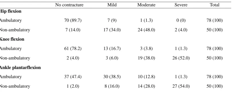
Table 4 shows the number of patients exhibiting each lower extremity joint contracture angle severity. In the ambulatory group, HF and KF contractures were observed infrequently, whereas APF contractures were observed more frequently in ambulatory; 41 patients (52.6%) in the ambulatory group showed APF contractures
23
Table 4. Severity of lower limb joint contractures in patients with DMD, based on ambulatory status
No contracture Mild Moderate Severe Total
Hip flexion
Ambulatory 70 (89.7) 7 (9) 1 (1.3) 0 (0) 78 (100)
Non-ambulatory 7 (14.0) 17 (34.0) 24 (48.0) 2 (4.0) 50 (100)
Knee flexion
Ambulatory 61 (78.2) 13 (16.7) 3 (3.8) 1 (1.3) 78 (100)
Non-ambulatory 2 (4.0) 3 (6.0) 19 (38.0) 26 (52.0) 50 (100)
Ankle plantarflexion
Ambulatory 37 (47.4) 30 (38.5) 10 (12.8) 1 (1.3) 78 (100)
Non-ambulatory 1 (2.0) 8 (16.0) 14 (28.0) 27 (54.0) 50 (100)
DMD, Duchenne muscular dystrophy; values are presented as n (%).
24
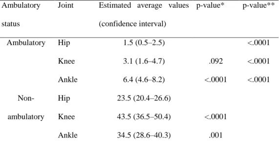
The interactions were significant, based on the use of the generalized estimating equation analysis. Consequently, the contracture angles of each lower extremity joint were compared based on ambulatory status. The mean KF and APF contracture angles were compared with the mean HF contracture angle in both the ambulatory and non-ambulatory groups. The mean APF contracture angle was 4.9° greater (99.7%
confidence interval, 2.6–7.3) than the mean HF contracture angle (adjusted p
< .0001), in the ambulatory group. Comparing the ambulatory and non-ambulatory groups, the mean HF angle was 22.0° (99.7% confidence interval, 17.5–26.5) greater, the mean KF contracture angle was 40.4° (99.7% confidence interval, 30.6–50.2) greater, and the mean APF contracture angle was 28.1° greater (99.7% confidence interval, 19.7–36.5) in the non-ambulatory group than in the ambulatory group (Table 5).
25
Table 5. Average lower extremity joint contracture angles, estimated using a generalized estimating equation
Ambulatory status
Joint Estimated average values (confidence interval)
p-value* p-value**
Ambulatory Hip 1.5 (0.5–2.5) <.0001
Knee 3.1 (1.6–4.7) .092 <.0001
Ankle 6.4 (4.6–8.2) <.0001 <.0001
Non- ambulatory
Hip 23.5 (20.4–26.6)
Knee 43.5 (36.5–50.4) <.0001
Ankle 34.5 (28.6–40.3) .001
* Corrected p-values for comparisons of other ankle and knee joint contracture angles with that of the hip joint.
** Corrected p-value for comparisons of the joint contracture angles, depending on ambulatory status.
26
3.1.3. Differences in joint contracture angles, based on stretching participation
The mean age of the ambulatory boys with DMD in the stretching group was 8.8 ± 2.2 years and 9.4 ± 2.2 years in the non-stretching group. Among the non-ambulatory boys with DMD, the mean age was 13.6 ± 3.8 years in the stretching group and 13.1
± 2.4 years in the non-stretching group. There were no significant differences in the mean ages of the boys, based on ambulatory ability, in the stretching and non- stretching groups. The mean duration of each stretching session was 16.2 ± 8.8 min (range, 5–30 min) for the ambulatory group and 16.8 ± 13.4 min (range, 5–60 min) in the non-ambulatory group; 31 (81.6%) patients in the ambulatory group and 17 (81.0%) in the non-ambulatory group had performed stretching exercises for >1 year.
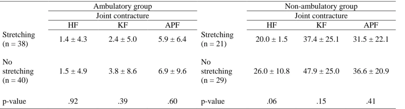
No differences were seen in the severities of the HF, KF, and APF joint contractures between the stretching and non-stretching groups, regardless of ambulatory status (Table 6).
27
Table 6. Independent-sample t-test results comparing the stretching and non-stretching groups
Ambulatory group Non-ambulatory group
Joint contracture Joint contracture
HF KF APF HF KF APF
Stretching
(n = 38) 1.4 ± 4.3 2.4 ± 5.0 5.9 ± 6.4 Stretching
(n = 21) 20.0 ± 1.5 37.4 ± 25.1 31.5 ± 22.1 No
stretching (n = 40)
1.5 ± 4.9 3.8 ± 8.6 6.9 ± 9.6 No stretching (n = 29)
26.0 ± 10.8 47.9 ± 25.0 36.6 ± 20.9
p-value .92 .39 .60 p-value .06 .15 .41
Values are presented as means ± standard deviations.
HF: Hip flexion; KF: Knee flexion; APF: Ankle plantarflexion
28
3.2. Research II: Scoliosis in Duchenne muscular dystrophy children
3.2.1. Clinical Characteristics
Among 273 boys, 146 boys who were still ambulatory were excluded. In the remaining boys, 4 who had undergone spine surgeries during the follow-up period were also excluded. After excluding those without relevant medical information and radiologic records, 50 boys remained for analysis (Figure 2). There were 31 boys who developed scoliosis during follow up period. Characteristics of participants according to scoliosis development is shown in Table 7. Age of boys at ambulation loss was 13.1 ± 2.6 years. There were no significant difference between two groups except pulmonary function (p < .05). Among those who were included for analysis, no patient was prescribed for spinal orthosis.
29 Figure 2. Flowchart of subject enrollment
30 Table 7. Demographic and clinical characteristics of participants
Values are presented as mean ± standard deviation. Comparison between groups was by Mann-Whitney test. FVC ; Functional vital capacity.
Scoliosis development No scoliosis p-value Total
Number of boys 31 19 - 50
Age (year) 13.6 ± 2.5 12.4 ± 2.6 .08 13.14 ± 2.6
Height (cm) 149.4 ± 25.5 142.9 ± 34.3 .66 146.9 ± 25.6
Body weight (kg) 49.1±16.5 45.9 ± 12.1 .36 47.9 ± 14.9
Functional assessment
Brooke scale 2.5 ± 1.7 2.0 ± 1.0 .31 2.3 ± 1.5
Vignos scale 8.4 ± 0.6 8.3 ± 0.5 .43 8.3 ± 0.5
Pulmonary function FVC % of
predicted (%) 74.2 ± 12.5 84.5 ± 15.2 .03 78.2 ± 14.7
31
3.2.2. Development of scoliosis
The 50 boys had 2 years of annual follow-up radiographs available from the onset of ambulation loss. During this follow-up period, 19 boys fell under the category of no scoliosis, and 31 boys developed either nonstructural or structural scoliosis. At the first follow-up after ambulation loss, 12 boys developed scoliosis, and another 13 boys developed scoliosis the following year. At the last year of follow-up, 6 more patients were found to have scoliosis. Cobb angle increased each year after the ambulation loss both in sitting and supine positions (Table 8).
32 Table 8. Values are presented as mean ± standard deviation
Time 1 represents the time when scoliosis was first detected after loss of walking ability Time 2 represents the time 1 year after the detection of scoliosis
Time 3 represents the time 2 years after the detection of scoliosis
Time 1 Time 2 Time 3
Number of boys
Cobb angle (°) Flexibility (%)
Cobb angle (°) Flexibility (%)
Cobb angle (°) Flexibility (%)
sitting supine sitting supine sitting supine
12 23.0±5.6 10.8±6.6 75.5±5.0 30.9 ± 5.9 18.2±6.6 57.1±10.5 34.4±4.4 22.2±6.3 49.1±10.0 25 11.1±3.5 5.2±3.3 60.7±6.5 27.2 ± 3.2 13.7±3.6 36.2±9.2
31 22.4±2.8 8.2±3.0 86.2±5.0
33
3.2.3. Scoliosis curve type changes
Among the 31 boys who developed scoliosis, except for the 6 boys who developed scoliosis at the last year of follow-up and the 4 boys who already had structural scoliosis at the first year of follow-up, only 2 boys had a course of “no scoliosis” that progressed directly to “structural scoliosis.” The remaining 19 boys went through the sequence of (1) no scoliosis, (2) nonstructural scoliosis, and (3) structural scoliosis (Figure 3).
34 Figure 3. Curve type changes of subjects
Values are presented as number of patients. Note that only 2 boys at the first year had a course of no scoliosis to structural scoliosis
Time 1 represents the time when scoliosis was first detected after loss of walking ability Time 2 represents the time 1 year after the detection of scoliosis
Time 3 represents the time 2 years after the detection of scoliosis
35
3.2.4. Changes in curve flexibility
Scoliosis was detected in the sitting position at the first visit after ambulation loss in 12 boys. In these 12 patients, consecutive follow-ups of supine and sitting radiographs were available for 2 years. Flexibility decreased over the follow-up period (p = .011). Mean values for flexibility were 75.5% at the first follow-up, 57.1%
the next year, and 49.1% at the last follow-up. Cobb angle in this population increased over time. In the same context, for 25 patients whose scoliosis developed after 1 year follow up, consecutive follow-ups of supine and sitting radiographs were available for 2 time points. Flexibility decreased during follow-up period (p = .02).
Mean values of flexibility were 60.7% at the first follow-up, 36.2% the next year.
Cobb angle also increased over time in this group (Table 7).
3.2.5. Correlation between spinal curve flexibility and other parameters
Spinal flexibility of 31 patients was inversely correlated with scoliosis curve angle in sitting and supine position (r = -0.504 and r = -0.77, respectively, p < .05 in both).
Pulmonary function, forced vital capacity of % predicted was not correlated with spinal curve flexibility.
3.2.6. Development of pelvic obliquity
There was no pelvic obliquity in the 19 boys who had not developed scoliosis. In the remaining 31 boys with scoliosis, 18 developed pelvic obliquity. In these boys, pelvic obliquity had an increasing tendency after ambulation loss both in the sitting and
36
supine positions (Table 9). The Cobb angle in the sitting position had a significant correlation with pelvic obliquity both in sitting (r = 0.758, p < .001) and supine positions (r = 0.639, p < .001). The Cobb angle in the supine position also significantly correlated with pelvic obliquity both in the sitting (r = 0.844, p < .001) and supine positions (r = 0.810, p < .001). Pelvic obliquity in sitting and supine position did not show correlations with spinal curve flexibility.
37 Table 9. Pelvic obliquity of subjects
Values are presented as mean ± standard deviation (SD).
Time 1 represents the time when scoliosis was first detected after loss of walking ability Time 2 represents the time 1 year after the detection of scoliosis
Time 3 represents the time 2 years after the detection of scoliosis
Time 1 Time 2 Time 3
Number of boys
Pelvic obliquity (°) Flexibility (%)
Pelvic obliquity (°) Flexibility (%)
Pelvic obliquity (°) Flexibility (%)
sitting supine sitting supine sitting supine
4 12.0±8.0 5.3±4.1 100.0±40.0 11.4±3.7 9.1±3.5 27.3±18.7 18.4±7.3 12.6±5.8 32.5±12.7 10 9.1±3.5 4.2±4.9 61.5±42.5 11.4±1.1 6.9±1.5 42.4±10.7
18 8.6±1.0 2.5±0.8 71.9±9.2
38
3.3. Research III: Upper limb short questionnaire for Duchenne muscular dystrophy
3.3.1. Clinical characteristics
Among 167 participants, 160 subjects participated in face-to-face ULSQ interviews and 132 subjects completed follow-up telephone interviews. The mean age was 15.2
± 4.4 years old (range 7-25 years). The number of patients with DMD by the Vignos scale and modified Brooke scale are represented in Figure 4. Forty-nine patients (30.6%), fifty-seven patients (35.6%), and fifty-four patients (33.8%) were independent ambulators, ambulators with assist or aids, and non-ambulators, respectively. The average age of diagnosis was 4.0 ± 2.8 years. All analyses included patients who used corticosteroid currently or in the past.
39
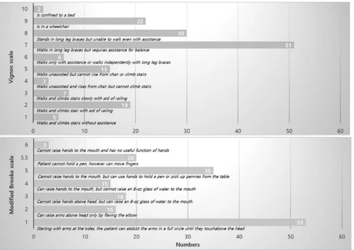
Figure 4. The numbers of individuals with DMD by Vignos Scale and modified Brooke scale
40
3.3.2. The result of Upper limb short questionnaire survey
The participants had answered either “yes” or “no” for fourteen items of ULSQ. The results for each question’s responses are found in Figure 5. Among the questionnaire regarding UE function, lifting heavy objects over 5 pounds was the most difficult UE function. Among the questionnaire related to UE pain, the most common pain area was the shoulder followed by proximal UE, elbow, and distal UE. For other questions, including ULSQ 3, 6, 8, and 10-14, less than 10% of respondents reported problems.
41 Figure 5. The result of Upper limb short questionnaire (ULSQ) survey
42
3.3.3. Factor analysis
The result of Bartlett’s test of sphericity (df = 762.77, p < .001) indicated that the correlation matrix of the sample was worthy of factor analysis. The value of the KMO Measure of Sampling was 0.74, which was a middling value in sample adequacy. Three components had the possibility of an eigenvalue greater than 1.00.
The eigenvalues of the first, second, and third components were 3.84, 2.19, and 1.87, respectively, and the first component explained 27.46% of the total variance. Factor rotation using orthogonal rotation was applied, and the result demonstrated three distinct factors comprised of UE function, pain, and stiffness in line with the previous study.35 Subsequent confirmatory factor analysis showed that the sample size- dependent chi-square was significant (χ2= 138.77, p < .001). The GFI value was 0.90, and the CFI value was 0.91. The RMSR value was 0.006, and the RMSEA value was 0.074. The confirmatory factor analysis indicated that the three-factor model could be acceptable in statistical terms as well as conceptual terms.
3.3.4. Construct validity
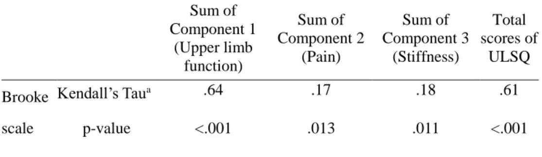
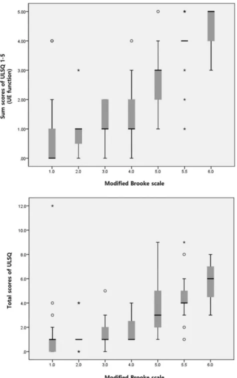
Kendall rank correlation coefficient (Kendall’s Tau) and the p-value are summarized in Table 10. Among the three components, the sum scores of the first component, UE function was the most highly related to Brook scale (Kendall’s Tau 0.64, p < .001) (Figure 6). Although the sum scores of the second component regarding pain and the third component regarding stiffness also showed that it statistically significantly correlated with the modified Brooke scale (Kendall’s Tau 0.17, p = .013; Kendall’s Tau 0.18, p = .011), the strength of the relationship was very weak. The total scores of fourteen items of ULSQ had a significant correlation with the modified Brooke scale (Kendall’s Tau 0.61, p < .001), respectively. The total scores and sum scores of
43
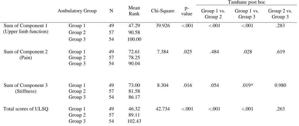
each component of differences between ambulatory groups were compared to assess the discriminative capability of ULSQ using the Kruskal-Wallis test (Table 11). For total scores and each sum score regarding upper limb dysfunction, pain, and stiffness, there was a significant difference between ambulatory groups. Post-hoc Tamhane’s test revealed a significant difference in total scores and sum scores of upper limb function between the independent ambulatory and assisted ambulatory groups (p
< .001) as well as between the independent ambulatory and non-ambulatory group (p < .001). However, in the case of pain and stiffness, the difference of sum scores were only between the independent ambulatory and the non-ambulatory groups (pain:
p = .028, stiffness: p = .019).
44
Table 10. Correlation between scores of Upper limb short questionnaire (ULSQ) and modified Brooke scale
Sum of Component 1
(Upper limb function)
Sum of Component 2
(Pain)
Sum of Component 3
(Stiffness)
Total scores of
ULSQ Brooke
scale
Kendall’s Taua .64 .17 .18 .61
p-value <.001 .013 .011 <.001
a: Kendall Rank Correlation Coefficient
45
Figure 6. The box plot for sum scores of Upper limb short questionnaire (ULSQ) from 1 to 5 items (Above) and total scores of ULSQ (Below) according to the modified Brooke scale. UE: upper extremity
46
Table 11. Results from Kruskal-Wallis tests and post-hoc pairwise comparisons for total scores and sum scores of each component between different ambulatory stages
Ambulatory Group N Mean
Rank Chi-Square p- value
Tamhane post hoc Group 1 vs.
Group 2
Group 1 vs.
Group 3
Group 2 vs.
Group 3 Sum of Component 1
(Upper limb function)
Group 1 49 47.29 39.926 <.001 <.001 <.001 .283
Group 2 57 90.58
Group 3 54 100.00
Sum of Component 2 (Pain)
Group 1 49 72.61 7.384 .025 .484 .028 .619
Group 2 57 78.25
Group 3 54 90.04
Sum of Component 3 (Stiffness)
Group 1 49 73.00 8.304 .016 .054 .019* 0.980
Group 2 57 81.58
Group 3 54 86.17
Total scores of ULSQ Group 1 49 46.32 42.734 <.001 <.001 <.001 .263
Group 2 57 89.11
Group 3 54 102.43
N: number; Group 1: independent ambulator; Group 2: ambulator with assist or aid; Group 3: non-ambulator.
47
3.3.5. Reliability
Cronbach’s alpha values were 0.71, 0.72, and 0.86 for UE function, pain, and stiffness, respectively, indicating adequate internal consistency for all factors. The test-retest reliability of the ULSQ using mean Gwet's AC1 coefficients and their 95%
confidence intervals with the percentage level of agreement summarized in Table 12.
The first and second-rater tend to conduct the ULSQ consistently, as all measured items lie within the 95% confidence interval.
48 Table 12. Test-retest reliability
Items Agreement (%) AC1 (95% CI)
ULSQ_1 73.74 0.63 (0.48, 0.78)
ULSQ_2 85.86 0.78 (0.66, 0.9)
ULSQ_3 78.79 0.69 (0.55, 0.83)
ULSQ_4 90.91 0.86 (0.76, 0.95)
ULSQ_5 81.82 0.64 (0.49, 0.79)
ULSQ_6 94.95 0.94 (0.9, 0.99)
ULSQ_7 89.9 0.88 (0.8, 0.96)
ULSQ_8 97.98 0.98 (0.95, 1)
ULSQ_9 90.91 0.89 (0.82, 0.96)
ULSQ_10 92.93 0.92 (0.86, 0.98)
ULSQ_11 94.95 0.94 (0.9, 0.99)
ULSQ_12 98.99 0.99 (0.96, 1)
ULSQ_13 100 NA
ULSQ_14 100 NA
AC1: Gwet’s first-order agreement coefficient NA: Not applicable
49
4. Discussion
4.1. Research I: Lower extremity joint contracture according to ambulatory status
This study examined HF, KF, and APF contractures among male patients with DMD, based on the patients’ ambulatory status, and investigated the differences in major joint contractures, based on passive stretching exercise participation. Our findings indicated that HF, KF, and APF contractures are more common and severe when there is deterioration of ambulatory function. Moreover, stretching exercises alone are unlikely to prevent lower extremity joint contractures.
The frequency and severity of lower extremity joint contractures rapidly increases after the loss of ambulatory function in children with DMD. This finding is consistent with the results of a previous study by McDonald et al.11 However, unlike their results, lower extremity