Collagenous fibroma is a recently defined entity that was first described by Evans in 1995 as desmoplatic fi- broblastoma and this tumor was renamed 1 year later by Nielsen et al. (1, 2). Since its initial description in 1995, fewer than 100 cases have been reported with the largest series having been published by Miettinen and Fetsch (3). Collagenous fibroma is a benign fibrous soft tissue tumor that typically arises in the subcutaneous tis- sue or skeletal muscle of adults. This tumor typically presents as a slowly growing, painless mass. This tumor is found in a people of all ages, but it is most common in the fifth and sixth decades of life. Men are affected four times more commonly than women. This entity has been reported in various locations, including the upper
extremities, lower extremities, posterior neck, upper back, abdominal wall, hip joint and head. Only a few case reports have described the MRI features of this tu- mor. We herein report on a case of a collagenous fibro- ma in the finger and we place emphasis on the MRI findings with the pathological correlation.
Case Report
A 54-year-old man was admitted to our hospital with a slowly growing, painless mass in the dorsoradial aspect of the proximal phalanx of the right 3rd finger. He had a history of fracture of the distal phalanx of the 3rd finger 5 years ago. Two year later, he had felt the slowly grow- ing mass. Physical examination showed an approxi- mately 4 × 6 cm
2firm, fixed mass without tenderness (Fig. 1A).
An MRI study was done. On the T1-weighted images, the mass was well-circumscribed, and it had iso-signal intensity compared with muscle (Fig. 1B). On the T2- weighted images, the mass had diffuse low signal inten-
J Korean Soc Radiol 2011;64:67-70
─ 67 ─
Collagenous Fibroma (Desmoplastic Fibroblastoma) of the Finger: A Case Report 1
Young Jae Sung, M.D., Joon Bum Koo, M.D.
1
Department of Radiology, Dongguk University Ilsan Hospital, Dongguk University College of Medicine
Received April 22, 2010 ; Accepted September 26, 2010
Address reprint requests to : Joon Bum Koo, M.D., Department of Radiology, Dongguk University Ilsan Hospital, 814, Siksa-dong, Ilsandong-gu, Goyang-si, Gyeonggi-do 410-773, Korea.
Tel. 82-31-961-7826 Fax. 82-31-961-8281 E-mail: [email protected]
Collagenous fibroma is a recently described, rare, benign, soft tissue tumor that aris- es in the subcutaneous tissue or muscle. We report here on a case of a collagenous fi- broma of the finger. A 54-year-old man was admitted to our hospital with a painless, slow-growing mass in the finger. On magnetic resonance imaging (MRI), the mass showed iso-signal intensity on the T1-weighted image, low signal intensity on the T2- weighted image and focal non-enhancing areas on the contrast-enhanced T1-weighted image. The lesion was totally removed by surgical excision and it was pathologically confirmed to collagenous fibroma.
Index words : Fibroma, Desmoplastic Fingers
Magnetic Resonance Imaging
Soft Tissue Neoplasm
sity with focal high signal intensity (Fig. 1C). The con- trast-enhanced T1-weighted fat-saturated images showed mild heterogeneous enhancement with focal ir- regular areas of non-enhancement (Fig. 1D).
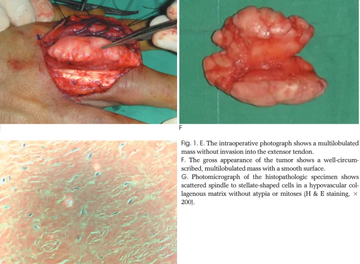
The tumor was removed by surgical excision. The tu- mor was located in the subcutaneous tissue of the 3rd finger. The tumor was loosely attached to the tendon sheath, but it had not invaded the extensor and flexor tendons (Fig. 1E). Macroscopically, the tumor appeared as a multilobulated, firm, whitish mass without gross necrosis, and it measured 5.5 5.0 2.5 cm
3(Fig. 1F).
Microscopically, the lesion was hypocellular and com- posed of fibroblast/myofibroblast cells dispersed in a collagenous-rich stroma (Fig. 1G). Cytological atypia, mitotic figures, areas of necrosis, calcifications and in- flammatory cells were not seen. Immunohistochemical staining showed the tumor cells to be focally and weak-
ly positive for smooth muscle actin and they were nega- tive for S100, CD 34 and CD 68. Thus, fibroma of the tendon sheath and nerve sheath tumor were excluded from the differential diagnosis.
There has been no local tumor recurrence at the 6 month postoperative follow-up examination.
Discussion
Collagenous fibroma characteristically presents as slowly growing, non-tender masses that have developed over a long duration. The tumor has a male predomi- nance and a peak incidence in the fifth and sixth decades of life. This tumor usually occurs in the subcu- taneous tissue, but approximately 25% of the tumors in- volve skeletal muscle. The tumor has a wide anatomic distribution, but it mainly affects the extremities. The
Young Jae Sung, et al : Collagenous Fibroma (Desmoplastic Fibroblastoma) of the Finger
─ 68 ─
A B
C D
Fig. 1. A. Clinical appearance of the tumor.
B-D. The axial T1- weighted (B) and T2-weighted (C) MR images show a well-circumscribed soft-tissue mass (arrows) of low signal
intensity on both sequences in the radial aspect of the proximal phalanx of the right 3rd finger. The axial contrast-enhanced T1-
weighted fat-saturated image (D) shows mild heterogeneous enhancement (arrow) with focal irregular areas of non-enhancement
(open arrow).
tumor typically infiltrates fat and skeletal muscle, and this has been observed in up to 51% of cases (3). The tu- mors range in size from 1 to 20 cm. Histologically, the tumor is hypocellular and it consists of stellate and spin- dle-shaped fibroblastic cells that are widely separated by a collagenous to fibromyxoid matrix (1, 3, 4).
Treatment of collagenous fibroma is surgical excision and there have been no reported incidences of local re- currence or metastases (3, 5).
Evans postulated that collagenous fibromas might be either neoplasm or a reactive condition (1). A history of antecedent trauma has been noted in only three cases of collagenous fibroma (3, 5). Our report reinforces the possibility that fibrous proliferation subsequent to trau- ma may contribute to the development of these tumors.
There have been seven previous case reports regard- ing the MRI features of collagenous fibroma. Among these cases, four case reports had the contrast-enhanced
T1-weighted images (5-8). On MRI, collagenous fibroma has a low signal on both the T1-weighted and T2-weight- ed images. The low signal intensity of the mass on both the T1-weighted and T2-weighted images is attributed to the low cellularity of the mass in a background of abun- dant collagen. The areas of persistently low signal inten- sity on the contrast-enhanced T1-weighted images corre- spond to regions of dense, relatively acellular collagen matrix (6, 7). Our case was consistent with the previous case reports.
Most soft-tissue masses have high signal intensity on T2-weighted images. The soft-tissue masses with low SI on the T2-weighted images include fibroma of the ten- don sheath, neurofibroma, cicatrical fibroma, malignant fibrous histiocytoma, aggressive fibromatosis and calci- fying fibrous pseudotumor (5, 9). Collagenous fibroma may be misdiagnosed as one of these soft tissue tumors.
In summary, we report here on a collagenous fibroma
J Korean Soc Radiol 2011;64:67-70
─ 69 ─
E F
G
Fig. 1. E. The intraoperative photograph shows a multilobulated mass without invasion into the extensor tendon.
F. The gross appearance of the tumor shows a well-circum- scribed, multilobulated mass with a smooth surface.
G. Photomicrograph of the histopathologic specimen shows
scattered spindle to stellate-shaped cells in a hypovascular col-
lagenous matrix without atypia or mitoses (H & E staining, ×
200).
of the finger that presented with a history of antecedent trauma to the finger. Collagenous fibroma should be in- cluded in the differential diagnosis of a well-circum- scribed lesion with low signal intensity seen on both the T1-weighted and T2-weighted images and the lesion shows minimal enhancement.
References
1. Evans HL. Desmoplastic fibroblastoma: a report of seven cases.
Am J Surg Pathol 1995;19:1077-1081
2. Nielsen GP, O’Connell JX, Dickersin GR, Rosenberg AE, Collagenous fibroma (desmoplastic fibroblastoma): a report of sev- en cases. Mod Pathol 1996;9:781-785
3. Miettinen M, Fetsch JF. Collagenous fibroma (desmoplastic fibrob- lastoma): a clinicopathologic analysis of 63 cases of distinctive soft tissue lesion with stellate-shaped fibroblasts. Hum Pathol 1998;29:
676-682
4. Hasegawa T, Shimoda T, Hirohashi S, Hizawa K, Sano T.
Collagenous fibroma (desmoplastic fibroblastoma): report of four cases and review of the literature. Arch Pathol Lab Med 1998;
12:455-460
5. Farina Fong, Edward Odell, Ricard Simo. Collagenous fibroma (desmoplastic fibroblastoma) of the neck presenting with neurolog- ical symptoms. Head and Neck Pathol 2009;3:47-50
6. Shuto R, Kiyosue H, Hori Y, Miryake H, Kawano K, Mori H. CT and MR imaging of desmoplastic fibroblastoma. Eur Radiol 2002;
12:2474-2476
7. Osipov V, Carrera GF. Collagenous fibroma (desmoplastic fibrob- lastoma) with vertebral body erosion. Sarcoma 2009;2009:682-687 8. Ahn M, Osipov V, Harris GJ. Collagenous fibroma (desmoplastic
fibroblastoma) of the lacrimal gland. Opthal Plast Reconstr Surg 2009;25:250-252
9. Walker KR, Bui-Mansfield LT, Gering SA, Ranlett RD.
Collagenous fibroma (desmoplastic fibroblastoma) of the shoulder.
AJR Am J Roentgenol 2004;183:1766
Young Jae Sung, et al : Collagenous Fibroma (Desmoplastic Fibroblastoma) of the Finger
─ 70 ─
대한영상의학회지 2011;64:67-70
손가락에 발생한 아교질섬유종:
증례 보고1
1